Reverse saphenous conduit flap in small animals: Clinical applications and outcomes
←
→
Page content transcription
If your browser does not render page correctly, please read the page content below
Page 1 of 4      Original Research

         Reverse saphenous conduit flap in small animals:
                Clinical applications and outcomes
Author:                               Due to the lack of skin elasticity defects of the distal hind limb can be a challenge to close. This
Ross C. Elliott1                      article assesses a well-described, but completely under-used technique for closure of wounds
Affiliation:                          on the distal tarsus. The technique was used with good success in six cases presenting to
1
 Department of Small                  the Bryanston Veterinary Hospital with a wide range of underlying pathology ranging from
Animal Surgery, Bryanston             trauma to neoplastic disease of the tarsus. All six cases were treated with a reverse saphenous
Veterinary Hospital,                  conduit flap and two of them underwent radiation therapy with no adverse side effects. All
South Africa
                                      cases showed excellent results with a very low degree of flap necrosis that never exceeded
Correspondence to:                    15% of the total flap area. This skin flap provides an excellent treatment method that is reliable
Ross Elliott                          in closure of defects of the distal tarsus with few adverse effects. To the author’s knowledge
                                      there has been only one previously published report on the clinical use of this type of skin flap,
Email:
rosselliott_2@hotmail.com             even though the flap is well described in most texts.

Postal address:
PO Box 130905, Bryanston
2021, South Africa
                                     Introduction
                                     The reverse saphenous conduit flap is a type of axial pattern flap. This type of skin flap is used for
Dates:                               one-stage reconstruction of wounds. It uses skin from surrounding areas where there is abundance,
Received: 19 Apr. 2013
                                     unlike the recipient site (Cornell et al. 1995). Axial pattern flaps are durable, full-thickness pedicle
Accepted: 14 Sept. 2013
Published: 20 Aug. 2014              flaps that contain a direct cutaneous artery and vein to supply and drain the entire length of the
                                     flap. A pedicle flap without a known direct cutaneous artery is known as a sub-dermal plexus
How to cite this article:            flap (Cornell et al. 1995; Degner, Bauer & Cozen 1993). The assurance of a blood supply allows a
Elliott, R.C., 2014, ‘Reverse        longer flap to be harvested, and an axial pattern flap shows a 50% increased graft survival area
saphenous conduit flap
in small animals: Clinical           compared with a routine sub-dermal plexus flap (Moores 2009). Axial pattern flaps can be used
applications and outcomes’,          in areas of poor vascularity, as they are not dependent on local blood supply from the wound
Journal of the South African         bed. Axial pattern flaps are more resilient to movement because they do not rely completely on
Veterinary Association 85(1),        vascularisation from the wound bed, thus making them an excellent choice when wounds extend
Art #1038, 4 pages. http://
dx.doi.org/10.4102/jsava.
                                     over, or are close to, a high-motion joint. The scar formation seen in these animals is considered
v85i1.1038                           to be acceptable by most of the pet owners (Degner et al. 1993).

Copyright:                           There are, however, some disadvantages to the axial pattern flaps. Their use is restricted to the
© 2014. The Authors.
                                     anatomical area in which the direct cutaneous vessels are situated. Therefore, there are areas in
Licensee: AOSIS
OpenJournals. This work              which they may not be suitable for the closure of wounds. Even in the face of these limitations
is licensed under the                most wounds in the body can be treated, as there are described direct cutaneous arteries and
Creative Commons                     veins throughout the body. Hair growth is reported to be normal to near normal, but given the
Attribution License.                 rotational nature of the flaps it may be in the opposite direction (Figure 1) to normal hair growth
                                     (Degner et al. 1993).

                                     There is little information published on the clinical use of the reverse saphenous flap, however,
                                     it remains a versatile and robust method of closure for wounds over the tarsus and metatarsus
                                     (Pavletic 1991). Flaps of 12 cm − 20 cm have been harvested in experimental canine patients
                                     (Pavletic 1999) and 8 cm long by 4 cm wide in feline experimental patients, both with 100%
                                     survival rates (Pavletic et al. 1983). These authors recommended the use of this flap in the feline
                                     patient on account of the well-developed medial saphenous vein, but it can also be very useful in
                                     the canine patient, as is shown in this article and indicated by Pavletic et al. (1983).

                                     The purpose of this article is to demonstrate the clinical versatility of the reverse saphenous
                                     conduit flap, the outcome of the flap, and associated complications.

                                     Methods
 Read online:
                   Scan this QR      Animal selection
                   code with your
                   smart phone or    Animals were selected from patients that presented at the Bryanston Veterinary Hospital for
                   mobile device     pathology of the hind limb. This pathology ranged from trauma, non-healing wounds and
                   to read online.
                                     oncological surgery. All patients had a serum biochemistry analysis performed, which included

                                               http://www.jsava.co.za      doi:10.4102/jsava.v85i1.1038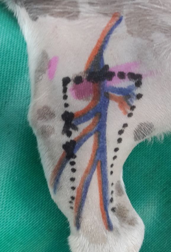
Page 2 of 4 Original Research
albumin, total solids, urea, creatinine and electrolytes, as well
as a full haematological examination prior to surgery. The
feline patients were tested for factors that may potentially
affect wound healing, such as feline immunodeficiency or
AIDS virus and feline leukaemia virus. All patients presenting
for oncological intervention had a fine needle aspirate
taken from the regional lymph nodes, ultrasonographic
examination of the abdomen and thoracic radiographs. A
biopsy of the neoplasm to be removed was performed prior
to surgery in order to determine the type of neoplasm by
histological examination.
Anaesthesia
All patients were pre-medicated with diazapam 0.2 mg/kg
(Valium 5 mg/mL, Roche, New Jersey, USA) and intravenous
buprenorphine 0.02 mg/kg (Temgesic® 0.3 mg/mL,
Schering-Plough, Woodmead, South Africa). Induction Source: Taken by Dr R.C. Elliott
was performed with intravenous diprivan 6.6 mg/kg FIGURE 1: Hair growing in the opposite direction to the surrounding hair.
(Propofol 1% 10 mg/mL, Fresenius Kabi, Midrand, South
Africa). All patients were intubated and placed on isoflorane
(Isofor 250 mL, Safeline, Pharmaceuticals, Johannesburg,
South Africa). They were also all placed on a Ringers lactate
drip (Sabax) at 10 mL/kg/h for the duration of the surgery.
A dose of intravenous amoxicillin clavulanic acid
20 mg/kg (Augmentin 600 mg, SmithKline, Wynberg
A V
Ext 6, Johannesburg) was given prior to surgery and one
hour later, which coincided with the end of the surgery. P S
No further antibiotic cover was given. Buprenorphine was
used at 0.01 mg/kg every eight hours through the night a v
after the surgery. The Ringers lactate drip was continued at
maintenance dose rates according to the weight of the patient.
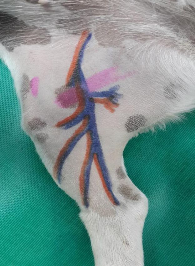
Anatomical background to surgical procedure
The saphenous artery is not a true, direct cutaneous artery
but it supplies the skin with multiple, small direct cutaneous
arteries; it therefore acts as a conduit. This flap is interesting
in that it actually relies on reversing the blood flow in the
D
saphenous artery to enable it to function and survive. The
medial saphenous vein is responsible for the vascular
drainage of the flap (Pavletic 1999).
These vessels are ligated just distal to the femoral artery and
vein, where they originate from the femoral artery and vein.
The blood supply in the saphenous artery is maintained and Source: Taken by Dr R.C. Elliott
reversed as a result of the connections between: FIGURE 2: The anatomy of the saphenous artery and vein in relation to the
reverse saphenous conduit flap, (A) femoral artery, (V) femoral vein, (P) patella,
• the cranial branch of the saphenous artery and the (S) Sartorius muscle, (a) saphenous artery branching from the femoral artery,
(v) saphenous vein branching from the femoral vein, (D) distal communications
perforating metatarsal artery by way of the medial and where the blood flow will be reversed.
lateral plantar arteries
• the cranial branch of the medial saphenous vein and the measured because of the need to ligate the flap just proximal
cranial branch of the lateral saphenous vein to the blood vessels coming off the femoral artery. There is
• other connections with the cranial and caudal branches of a maximum length to all reverse saphenous conduit flaps. A
the medial saphenous vein distal to the tibio-tarsal joint
skin incision was made across the central third of the inner
(Degner et al. 1993; Pavletic 1999).
thigh perpendicular to the long axis of the tibia. Another
useful landmark described is the patella; this incision can be
Surgical procedure made just proximal to the patella. In cats, it was possible to
All surgical procedures were performed as described below visualise the vasculature on the medial aspect of the tibia once
(Degner et al. 1993). The size of the defect to be covered was clipping, shaving and cleaning was completed. To harvest
http://www.jsava.co.za doi:10.4102/jsava.v85i1.1038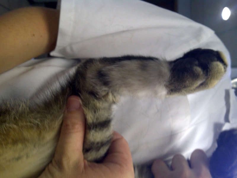
Page 3 of 4      Original Research

the skin flap two incisions were made parallel to the long                                   The donor site was closed with a subcutaneous continuous
axis of the tibia on either side of the transverse incision. These                           suture of polydioxanone 4–0 and the skin was closed with
two incisions extended distally just proximal to the medial                                  simple interrupted nylon 4–0. The flap was sutured onto the
malleolus. They tended to converge distally according to the                                 wound edges with nylon 4–0 simple interrupted sutures.
availability of skin in the distal limb.
                                                                                             Post-operative care
The saphenous artery and medial saphenous vein were                                          A light dressing and an Elizabethan collar were placed on
exposed at the transverse incision where they branch                                         all patients to prevent patient interference and/or damage
off the femoral artery and vein. Both these vessels were                                     to the skin flap. The dressing was changed after 48 h and the
double ligated and the flap was then undermined deep to                                      flap was clinically evaluated. A large number of the flaps
the vasculature. A portion of the medial gastrocnemius                                       took on a dark purple colour at 48 h. This was thought to be
fascia was included in the pedicle of the flap. This has been                                due to mild post-operative venous congestion of the flap post
recommended in order to prevent damage to the caudal                                         surgery, which had disappeared completely at four days post
branch of the saphenous artery and medial saphenous vein                                     surgery, when the flap took on a normal pink colouration.
(Pavletic 1999).                                                                             This venous congestion did not affect flap survival or increase
                                                                                             its dehiscence. The animals were sent home with strict
The epithelialised wound edges were surgically debrided                                      instructions to maintain rest and pain control for four days.
and all the skin flaps were joined to their recipient beds by                                The skin flaps were checked at five days post surgery and
a bridging incision. No tubed pedicles were performed. As                                    the skin sutures removed at 14 days post surgery. Radiation
                                                                                             therapy was started at day 10 after the operation for those
all of the flaps were rotated nearly 180° in order to cover the
                                                                                             who required it.
skin defects distal to the tarsal-crural joint, it was essential to
make sure that there was no kinking of the vasculature in the
pedicle of the flap. It is always best to allow a gradual curve                              Results
of the pedicle over the bridging incision.                                                   For ease of comparison, the results are presented in Table 1.
                                                                                             All of the patients who were presented for radiation therapy
                                                                                             had a full course of radiation and there were no adverse
                                                                                             effects detected on flap survival. All patients tolerated the
                                                                                             flap well and showed no signs of pain or discomfort during
                                                                                             the recovery period. Some patients showed mild erythema
                                                                                             and pruritus of the flap after radiation, but this was managed
                                                                                             with a short course of an anti-inflammatory medication.

                                                                                             Discussion
                                                                                             The cases demonstrated that the reverse saphenous conduit
                                                                                             flap achieved excellent results in two different species under
                                                                                             different sets of circumstances. More studies are needed to
                                                                                             evaluate the effects of radiation therapy on this type of skin
                                                                                             flap, as there is great potential to use this skin flap in cats
                                                                                             and dogs to close skin defects on the tarsus and metatarsus
                                                                                             when removing malignant or large benign neoplasms. In
                                                                                             the author’s experience, wounds resulting from attempts
                                                                                             to remove masses (mainly benign) in the tarsal area, which
                                                                                             have been surgically closed by primary closure of the wound
                                                                                             edges, have usually shown a high rate of dehiscence. A
                                                                                             more severe complication in the metatarsal area is vascular
                                                                                             compromise of the distal limb. This is due to swelling caused
                                                                                             by the surgical procedure to remove these masses, the high
                                                                                             degree of skin tension on the wound edges and the practice
                                                                                             of placing a bandage post-operatively. This excessive skin
                                                                                             tension produces a natural tourniquet due to the minimal
                                                                                             soft tissue surrounding this area and subsequently causes
                                                                                             constriction of the vascular supply, leading to severe swelling
                                                                                             of the distal pes and possible avascular necrosis of the skin of
                                                                                             the paw. This complication can be avoided by planning the
Source: Taken by Dr R.C. Elliott                                                             surgery to include a reverse saphenous conduit flap to cover
Note: The black crosses represent the sites of ligation and the dotted lines represent the
extent of the skin flap.                                                                     the skin defect. This technique prevents high tension across
FIGURE 3: Site of the flap.                                                                  the wound and all of the above complications.

                                                             http://www.jsava.co.za          doi:10.4102/jsava.v85i1.1038
Page 4 of 4 Original Research
TABLE 1: Results of treatment of wound with reverse saphenous conduit flap.
Species Age Clinical application Complicating factors Flap survival (%) Follow up
Feline 2 Traumatic non-healing wound None 100 > 12 months (Figure 1)
Feline 10 Surgical wound to remove fibrosarcoma Radiation therapy for 4 weeks. No adverse 100 11 months metastasis to regional lymph node
effects on flap survival
Canine 9 Degloving wound post road traffic 15% superficial slough, however sloughed 85 > 12 months
accident area epithelialised rapidly covering the
defect
Canine 10 Surgical wound post removal of a Mild swelling post opperation 100 > 12 months
schwannoma No radiation therapy done due to owners
consent. Mild purple discolouration day 2
post surgery
Canine 11 Surgical wound post removal of a Mild superficial slough 5% of the distal end 95 12 months at time of publication
schwannoma of the flap. Radiation therapy delayed for
2 weeks
Canine 5 Surgical removal of a spindle cell Mild purple discolouration of the flap at day 100 8 months at time of publication
sarcoma two post surgery. Normal colour returned
2 days later with no treatment. Pet
owners declined radiation therapy
Deep abrasive wounds of the tarsus occur commonly after clinical setting. The cases described above indicate how this
road traffic accidents due to a shearing force being applied to axial pattern skin flap was used in canine and feline patients
the skin. They may result in loss of relatively vast amounts in a clinical setting for closure of distal pelvic limb wounds.
of skin, connective tissue and even bone. Once granulated, A limitation of the study was that the sample size was small,
providing the distal vascular connections are not damaged but given the high success rate of this type of skin flap, this
by the initial trauma, they become excellent candidates for the report may provide important clinical information about
use of a reverse saphenous conduit flap. All ligament damage using the flap.
and other orthopaedic damage should also be accounted for
and managed prior to closure of the skin defect. Acknowledgement
Chronic non-healing wounds may present a different
Competing interests
problem in that they can have an underlying cause affecting The author declares that he has no financial or personal
wound healing. All of these wounds should first be biopsied relationship(s) which may have inappropriately influenced
to rule out any neoplastic condition as being a cause of the him in writing this article.
non-healing wound. A culture should also be done to treat
any underlying bacterial infection. All animals should be References
assessed for systemic diseases that can affect wound healing.
Cornell, K., Salisbury, K., Jacovljevic, S., Bauer, M. & Petryk, D., 1995, ‘Reverse
saphenous conduit flap in cats: An anatomic study’, Veterinary Surgery 24,
202–206. http://dx.doi.org/10.1111/j.1532-950X.1995.tb01319.x
As the reverse saphenous conduit flap brings its own blood
Degner, D.A., Bauer, M.S. & Cozen, S.M., 1993, ‘Reverse saphenous conduit flap:
supply with it, its use in wounds with a poor blood supply A case report in a cat’, Veterinary Comparative Orthopaedics and Traumatology
allows for excellent healing, as it is not dependent on a health 6, 175.
Moores, A., 2009, ‘Axial pattern flaps’, in J. Willams & A. Moores (eds.), BSAVA manual
granulation bed. This factor makes it ideal for use in cases of canine and feline wound management and reconstruction, pp. 100–143,
where post-operative radiation is planned and in this study BSAVA, Gloucester.
was used with no adverse complications arising from the Pavletic, M.M., 1991, ‘Anatomy and circulation of the canine skin’, Microsurgery 12,
103–112. http://dx.doi.org/10.1002/micr.1920120210
radiation. Pavletic, M.M., 1999 ‘Axial pattern flaps’, in M.M. Palvetic (ed.), Atlas of small animal
reconstructive surgery, n.p., Saunders, Philadelphia.
This technique has been proven in cats, but it has only been Pavletic, M.M., Watters, J., Henry, R.W. & Nafe, LA., 1983, ‘Reverse saphenous conduit
flap in the dog’, Journal of the American Veterinary Medical Association 182,
reported in dogs as an anatomical study and not in any 380–389.
http://www.jsava.co.za doi:10.4102/jsava.v85i1.1038You can also read