Oral Alterations in a COVID-19 Patient: Case Report and Update of Main Findings
←
→
Page content transcription
If your browser does not render page correctly, please read the page content below
Int. J. Odontostomat.,
15(2):315-319, 2021.

                 Oral Alterations in a COVID-19 Patient:
               Case Report and Update of Main Findings

                         Alteraciones Orales en un Paciente con COVID-19:
                 Reporte de un Caso y Actualización de los Principales Hallazgos

                     Amanda Claudino Gomes1; Diogo da Silva Ferreira1; Michelly de Melo Silva1;
              Caio César da Silva Barros2; Nicássio Silva Menezes3 & Hellen Bandeira de Pontes Santos4

GOMES, A. C.; FERREIRA, D. S.; SILVA, M. M.; BARROS, C. C. S.; MENEZES, N. S. & SANTOS, H. B. P. Oral alterations
in a COVID-19 patient: case report and update of main findings. Int. J. Odontostomat., 15(2):315-319, 2021.

        ABSTRACT: COVID-19 was characterized as a pandemic due to the worldwide dissemination and the severity with
which the disease attacks the human organism. Some oral lesions have been observed in COVID-19 patients. However,
there is still no concrete evidence of the real influence of SARS-CoV-2 on the human body, especially in the oral region. In
this context, the present report discusses a case of a COVID-19 patient with oral alterations. The male patient presented
ulcerative lesions of painful symptomatology and petechiae in the oral mucosa. This study also performed a literature review
of the main oral alterations reported in the literature. Although more studies with a larger number of cases should be performed,
the present clinical case may have manifested signs of this pathology in the oral cavity since the epithelial cells of the oral
mucosa have ACE2 receptors.

         KEY WORDS: coronavirus, oral manifestations, oral health.

INTRODUCTION

      Changing the global life scenario, the new                    literature reports some of the complications that
coronavirus originated in East Asia spread rapidly                  SARS-CoV-2 may manifest when coming into contact
around the world. On February 11th, the World Health                with the human organism. The WHO highlights that
Organization (WHO) termed it as COVID-19, caused                    cough, fever, tiredness, runny nose, sore throat, and
by the severe acute respiratory syndrome                            difficulty breathing are still the most common
coronavirus 2 (SARS-CoV-2). A month after, COVID-                   symptoms of current infection (Hamed; Guan et al.).
19 was characterized as a pandemic due to the
worldwide spread and the severity with which the                            In this new scenario, complications of this vi-
disease attacks the human body (Hamed, 2020).                       rus to the human being have been studied vehemently
                                                                    by several scientific groups, which dedicate
       Coronaviruses, belonging to the Coronaviridae                themselves to research on the symptoms,
family, are classified as simple, highly diverse RNA                immunization, and medications that help in reducing
viruses, which mutate easily (Weiss & Navas-Martin,                 the effects of this virus on the organism. In some
2005). In humans, COVID-19 infections mainly affect                 studies, oral alterations have been reported in patients
the respiratory and gastrointestinal tracts, with                   with COVID-19 (Amorim Dos Santos et al., 2020;
manifestations ranging from common colds to acute                   Carreras-Presas et al., 2020; Ciccarese et al., 2020;
pneumonia (Hamed; Guan et al., 2020). Recent                        Lechien et al., 2020; Jimenez-Cauhe et al., 2020;

1
  Undergraduate student, Department of Dentistry, Nova Esperança School (FACENE), João Pessoa, PB, Brazil.
2
  Oral Pathology and Medicine, Postgraduate Program in Dental Sciences, Federal University of Rio Grande do Norte, Natal, RN, Brazil.
3
  Physician, Nova Esperança Medical School (FAMENE), João Pessoa, PB, Brazil.
4
  Professor, Postgraduate Program in Health Family, Nova Esperança School (FACENE), João Pessoa, PB, Brazil.

    Received: 2021-01-06    Accepted: 2021-02-13

                                                                                                                                315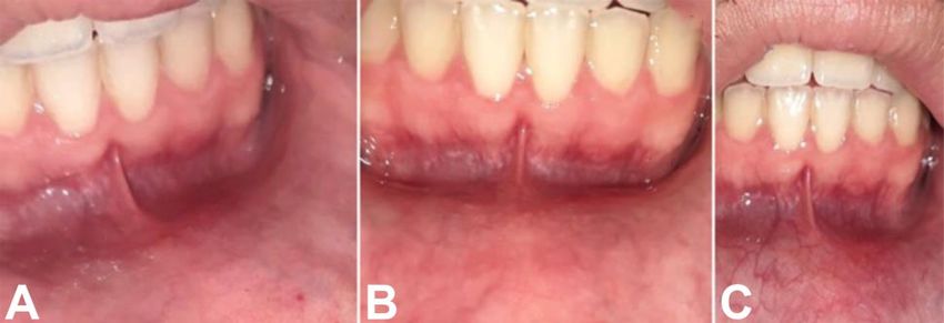
GOMES, A. C.; FERREIRA, D. S.; SILVA, M. M.; BARROS, C. C. S.; MENEZES, N. S. & SANTOS, H. B. P. Oral alterations in a COVID-19 patient: case report and update of main
findings. Int. J. Odontostomat., 15(2):315-319, 2021.
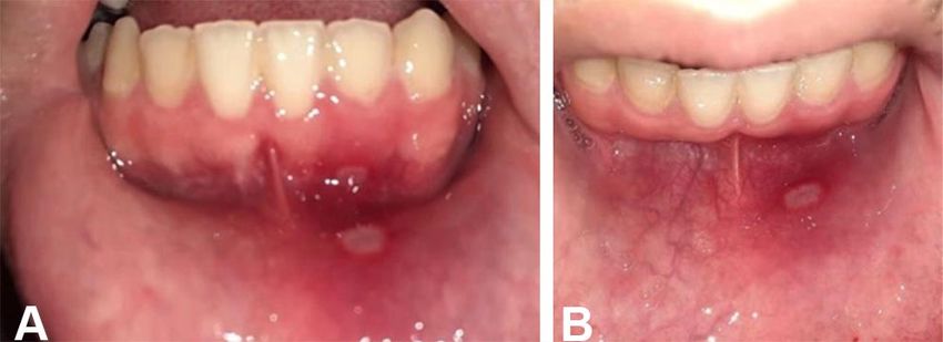
Kahraman & Çaskurlu, 2020; Soares et al., 2020). In The patient reported that on the 2nd day after the
this context, this study reported a COVID-19 patient onset of symptoms, he felt discomfort in the lower gum
who presented oral alterations and assessed his and the inner surface of the lower lip. On intraoral
clinical characteristics in comparison to cases in the physical examination, it was observed two shallow
literature. ulcerative lesions with painful symptoms and strong
burning (Fig. 1A) and the presence of petechiae, which
were not causing pain or discomfort, in the anterior region
CASE REPORT of the inner mucosa of the lower lip (Fig. 1B). The patient
stated that there was no previous trauma in the area.
A 22-year-old man, with a history of chronic The patient reported that on the 3rd day after the
asthma and recent contact with COVID-19 patients, onset of symptoms he used oral azithromycin (500 mg)
presented with remarkable respiratory symptoms, once a day for seven days, oral dexchlorpheniramine
severe headache, fever, odynophagia, cough, and (2 mg) once a day for ten days, oral ivermectin (200
runny nose. Associated with this condition, there were mcg/kg) in a single dose, and budesonide nasal spray
hyposmia and hypogeusia, as well as lack of appetite. once a day for five days. On the 9th day after the first
Furthermore, there were no cutaneous changes in the symptoms, the patient reported a gradual return of taste
patient. In the medical history, the patient reported and, without the use of any topical medication,
allergic rhinitis and drug hypersensitivities, such as presented a decrease in the number of petechiae as
acetylsalicylic acid, non-steroidal anti-inflammatory, well as involution of ulcerative lesions (Fig. 2A). On
dipyrone, and penicillin. It was performed the SARS- the 11th and 15th day after the appearance of the first
COV-2 test by the method of immunochromatography symptoms, it was possible to verify a smaller amount
with a whole blood sample, and a reagent sample for of petechiae and the complete healing, respectively, in
positive IgM and IgG was found, thus confirming the the mucosa of the lower gum and the lower lip (Figs.
patient's exposure to the virus. 2B and C).
Fig. 1. (A) Two shallow ulcerative lesions
located in the gum region and in the internal
mucosa of the lower lip. (B) Ulcerative lesions
located in the internal mucosa of the lower lip
and ulcerative lesion located in the inner
surface of the lower lip and petechiae in the
anterior region of the lower lip mucosa.
Fig. 2. Involution of ulcerative lesions and petechiae in the lower gum and the inner mucosa of the lower lip on the 9th (A) and
11th (B) day. (C) Complete healing of the lower gum and the inner surface of the lower lip after 15 days after the onset of
symptoms.
316GOMES, A. C.; FERREIRA, D. S.; SILVA, M. M.; BARROS, C. C. S.; MENEZES, N. S. & SANTOS, H. B. P. Oral alterations in a COVID-19 patient: case report and update of main
findings. Int. J. Odontostomat., 15(2):315-319, 2021.
DISCUSSION
Current research shows that SARS-CoV-2 platelets since viral infections can promote systemic
enters the host cell by targeting the angiotensin- inflammatory responses and cause an imbalance
converting enzyme 2 (ACE2), the same receptor from between procoagulant and anticoagulant homeostatic
SARS-CoV. Cells that have receptors for the ACE2 mechanisms (Giannis et al., 2020). Therefore, we
enzyme have the potential to become hosts for the suggest the possibility that petechiae located on the
virus, causing inflammation in the associated organs lower lip may be caused by changes in the
and tissues (Zhou et al., 2020; Zou et al., 2020). coagulation cascade.
According to Xu et al. (2020), the oral mucosa may
be a high-risk location for the potential development Similar to the present case, Jimenez-Cauhe
of COVID-19 infection. Although some clinical findings et al. observed the presence of macules on the hard
have suggested an association between oral palate and petechiae in the oral cavity in three of the
alterations and SARS-CoV-2 infection, the data four reported cases. These three patients were
should be analyzed with caution due to the lack of treated with systemic corticosteroids and exhibited
information. In this way, it was analyzed six articles progressive resolution of the lesions within 2 to 3 days,
that have reported oral alterations, such as ulcers while in our case, the patient showed clinical
and petechiae in patients with COVID-19 (Amorim improvement of oral lesions over 2 to 3 weeks.
Dos Santos et al.; Carreras-Presas et al.; Ciccarese Additionally, Kahraman & Çaskurlu also reported the
et al.; Jimenez-Cauhe et al.; Kahraman & Çaskurlu; presence of a largely erythematous surface in the
Soares et al.). oropharynx and oral petechiae and numerous
prominent pustular enanthem, ranging from 1-3 mm
A European study with 417 patients with mild in diameter, in the palate of a 51-year-old male patient
to moderate COVID-19 symptoms showed that the with COVID-19. Besides, this patient also stated
most prevalent general symptoms consisted of cough, anosmia and sore throat and showed regression of
myalgia, and loss of appetite. Of these, 85.6 % and oral lesions after a few days of antibiotic therapy.
88.8 % of patients had olfactory dysfunction related These clinical findings reported in the literature
to the infection and reported taste disorders with suggest a possible association between these oral
impairment of the four taste modalities, respectively, alterations and infection by the SARS-CoV-2 virus.
which appeared before the other symptoms (Lechien However, there are still doubts as to whether these
et al.). As shown in the study by Lechien et al., the lesions are caused due to coronavirus infection or
patient presented here also had olfactory dysfunction whether they represent secondary manifestations
and impairedtaste as well as general symptoms such resulting from the systemic condition of these patients
as cough, myalgia, and loss of appetite. or pharmacological therapy performed.
Ciccarese et al. reported a case of a 19-year- Some authors have reported the occurrence
old female patient with COVID-19, who presented of oral alterations associated with COVID-19, such
erythematous macules, papules, and petechiae on as ulcers, blisters on the mucosa, and scaly gingivi-
the lower extremities on physical examination. It also tis. Carreras-Presas et al. described oral lesions in
was observed erosions, ulcers, and blood scabs on one confirmed and two suspected COVID-19 patients.
the inner surface of the lips, as well as petechiae on The confirmed patient had blisters on the internal
the palate and gums. For this patient, the initial mucosa of the lip, scaly gingivitis, and generalized
antibiotic therapy was discontinued, and intravenous skin rash, while the other two cases had painful palatal
immunoglobulins (400 mg/kg) and ulcers similar to herpetic lesions. Similarly, in the case
methylprednisolone (1 mg/kg) were administered for reported by Amorim Dos Santos et al., it was observed
5 days. These oral alterations are similar to those as oral changes the presence of persistent white
presented in the present clinical case, regarding the plaque and several ulcers on the dorsum of the tongue
existence of petechiae and ulcerations in the oral resembling the late stage of recurrent herpetic oral
mucosa region. It has been reported that patients with lesions.
COVID-19 may develop thrombocytopenia and
exhibit high D-dimer, which can be explained by the Some authors have discussed the possibility
excessive activation of the coagulation cascade and of the relationship between these oral alterations and
317GOMES, A. C.; FERREIRA, D. S.; SILVA, M. M.; BARROS, C. C. S.; MENEZES, N. S. & SANTOS, H. B. P. Oral alterations in a COVID-19 patient: case report and update of main
findings. Int. J. Odontostomat., 15(2):315-319, 2021.
SARS-CoV-2 infection (Abu-Hammad et al., 2020; Al- GOMES, A. C.; FERREIRA, D. S.; SILVA, M. M.; BARROS,
Khatib, 2020; Amorim Dos Santos et al.; Petrescu et C. C. S.; MENEZES, N. S. & SANTOS, H. B. P. Alteraciones
al., 2020; Ponce & Tjioe, 2020; Rocha et al., 2020). orales en un paciente con COVID-19: reporte de un caso y
Abu-Hammad et al. emphasize the possibility of these actualización de los principales hallazgos. Int. J.
oral changes being associated with other infections of Odontostomat., 15(2):315-319,2021.
viral or fungal etiology. Besides, these authors point
RESUMEN: El COVID-19 se caracterizó como una
out that many drugs have adverse effects, as
pandemia debido a la diseminación mundial y la gravedad
impairment of the immune system, which increases the con la que la enfermedad ataca al organismo humano. Se
susceptibility of opportunistic infections to affect the han observado algunas lesiones orales en pacientes con
oral mucosa. Also, the emotional stress associated with COVID-19. Sin embargo, todavía no hay evidencia concreta
periods of social-life restrictions may promote changes de la influencia del SARS-CoV-2 en el cuerpo humano, es-
in the health condition. pecialmente en la cavidad oral. En este contexto, el presen-
te reporte analiza un caso de un paciente con COVID-19
In the case reported by Soares et al., the patient con alteraciones orales. El paciente de sexo masculino pre-
exhibited an ulcerated lesion on the buccal mucosa sentó lesiones ulcerativas de sintomatología dolorosa y
petequias en la mucosa oral. Este estudio también realizó
and multiple reddish macules on the hard palate,
una revisión de la literatura de las principales alteraciones
tongue, and lips. Analysis through orales reportadas en la literatura. Si bien se deben realizar
immunohistochemistry and in-situ hybridization más estudios con un mayor número de pacientes, el pre-
revealed negativity for herpes simplex virus 1 and 2, sente reporte de caso puede haber manifestado signos de
cytomegalovirus, treponema pallidum, and Epstein- esta patología en la cavidad oral ya que las células epiteliales
Barr virus. It also was reported that the patient had a de la mucosa oral tienen receptores ACE2.
history of diabetes and hypertension and, when
admitted to the hospital, presented petechiae and PALABRAS CLAVE: coronavirus, manifestacio-
vesiculobullous lesions of unknown etiology on the skin, nes bucales, salud oral.
which was treated with dexamethasone and dipyrone
for one week. In this way, we believe that the presence
of ulcers in different locations of the oral cavity may be REFERENCES
a relevant condition, but we also highlight that caution
is advised when associating the appearance of ulcers
Abu-Hammad, S.; Dar-Odeh, N. & Abu-Hammad, O. SARS-CoV-2
or other oral alterations with COVID-19. and oral ulcers: A causative agent or a predisposing factor? Oral
Dis., 2020. DOI: https://www.doi.org/10.1111/odi.13498
Al-Khatib, A. Oral manifestations in COVID-19 patients. Oral Dis.,
CONCLUSION 27 Suppl. 3:779-80, 2020.
Amorim Dos Santos, J.; Normando, A. G. C.; Carvalho da Silva, R.
L.; De Paula, R. M.; Cembranel, A. C.; Santos-Silva, A. R. &
Silva Guerra, E. N. Oral mucosal lesions in a COVID-19 patient:
Due to the COVID-19 be a recent disease and New signs or secondary manifestations? Int. J. Infect. Dis.,
the lack of information related to it, there is still no con- 97:326-8, 2020.
Carreras-Presas, C. M.; Amaro Sánchez, J.; López-Sánchez, A. F.;
crete evidence of the real influence of SARS-CoV-2 Jané-Salas, E. & Somacarrera Pérez, M. L. Oral vesiculobullous
on the human body, especially in the oral region. lesions associated with SARS-CoV-2 infection. Oral Dis., 27
However, given the mentioned clinical findings, the Suppl. 3:710-2, 2020.
possibility of the patient of the present clinical case Ciccarese, G.; Drago, F.; Boatti, M.; Porro, A.; Muzic, S. I. & Parodi,
A. Oral erosions and petechiae during SARS-CoV-2 infection. J.
having manifested signs of this pathology in the oral Med. Virol., 93(1):129-32, 2020.
cavity is suggested, since the epithelial cells of the oral Giannis, D.; Ziogas, I. A. & Gianni, P. Coagulation disorders in
mucosa have ACE2 receptors. coronavirus infected patients: COVID-19, SARS-CoV-1, MERS-
CoV and lessons from the past. J. Clin. Virol., 127:104362, 2020.
Guan, W.; Ni, Z.; Hu, Y.; Liang, W.; Ou, C.; He, J.; Liu, L.; Shan, H.;
We highlight the importance of longitudinal Lei, C.; Hui, D. S. C.; et al. Clinical characteristics of Coronavirus
studies aimed at oral implications due to the new Disease 2019 in China. N. Engl. J. Med., 382:1708-20, 2020.
coronavirus, which can elucidate the mechanisms Hamed, M. A. An overview on COVID-19: reality and expectation.
underlying the development of possible oral lesions Bull. Natl. Res. Cent., 44(1):86, 2020.
Jimenez-Cauhe, J.; Ortega-Quijano, D.; Carretero-Barrio, I.; Suarez-
since there is no consensus on whether the reported Valle, A.; Saceda-Corralo, D.; Moreno-Garcia del Real, C. &
ulcers, petechiae, and vesiculobullous lesions Fernandez-Nieto, D. Erythema multiforme-like eruption in patients
represent intraoral manifestations of SARS-CoV-2 with COVID-19 infection: clinical and histological findings. Clin.
infection. Exp. Dermatol., 45(7):892-5, 2020.
318GOMES, A. C.; FERREIRA, D. S.; SILVA, M. M.; BARROS, C. C. S.; MENEZES, N. S. & SANTOS, H. B. P. Oral alterations in a COVID-19 patient: case report and update of main
findings. Int. J. Odontostomat., 15(2):315-319, 2021.
Kahraman, F. C. & Ças¸kurlu, H. Mucosal involvement in a COVID- Corresponding author:
19-positive patient: A case report. Dermatol. Ther., 33(4):e13797, Hellen Bandeira de Pontes Santos
2020. Av. Frei Galvão, 12 - Gramame
Lechien, J. R.; Chiesa-Estomba, C. M.; De Siati, D. R.; Horoi, M.; Le
João Pessoa - PB
Bon, S. D.; Rodriguez, A.; Dequanter, D.; Blecic, S.; El Afia, F.;
Distinguin, L.; et al. Olfactory and gustatory dysfunctions as a 58067-698
clinical presentation of mild-to-moderate forms of the coronavirus BRAZIL
disease (COVID-19): a multicenter European study. Eur. Arch.
Otorhinolaryngol., 277(8):2251-61, 2020.
Petrescu, N.; Lucaciu, O. & Roman, A. Oral mucosa lesions in E-mail: hellenbps@hotmail.com
COVID-19. Oral Dis., 2020. DOI: https://www.doi.org/10.1111/
odi.13499.
Ponce, J. B. & Tjioe, K. C. Overlapping findings or oral manifestations
in new SARS-CoV-2 infection. Oral Dis., 27 Suppl. 3:781-2, 2020.
Rocha, B. A.; Souto, G. R.; Grossmann, S. M. C.; de Aguiar, M. C.
F.; de Andrade, B. A. B.; Romañach, M. J. & Rebello Horta, M.
C. Viral enanthema in oral mucosa: A possible diagnostic
challenge in the COVID-19 pandemic. Oral Dis., 27 Suppl.
d3:776-8, 2020.
Soares, C. D.; de Carvalho, R. A.; de Carvalho, K. A.; de Carvalho,
M. G. F. & de Almeida, O. P. Letter to Editor: Oral lesions in a
patient with Covid-19. Med. Oral Patol. Oral Cir. Bucal, 25(4):
e563-4, 2020.
Weiss, S. R. & Navas-Martin, S. Coronavirus pathogenesis and the
emerging pathogen severe acute respiratory syndrome
coronavirus. Microbiol. Mol. Biol. Rev., 69(4):635-64, 2005.
Xu, H.; Zhong, L.; Deng, J.; Peng, J.; Dan, H.; Zeng, X.; Li, T. &
Chen, Q. High expression of ACE2 receptor of 2019-nCoV on
the epithelial cells of oral mucosa. Int. J. Oral Sci., 12:8, 2020.
DOI: https://www.doi.org/10.1038/s41368-020-0074-x.
Zhou, P.; Yang, X. L.; Wang, X. G.; Hu, B.; Zhang, L.; Zhang, W.; Si,
H. R.; Zhu, Y.; Li, B.; Huang, C. L.; et al. A pneumonia outbreak
associated with a new coronavirus of probable bat origin. Nature,
579:270-3, 2020.
Zou, X.; Chen, K.; Zou, J.; Han, P.; Hao, J. & Han, Z. Single-cell
RNA-seq data analysis on the receptor ACE2 expression reveals
the potential risk of different human organs vulnerable to 2019-
nCoV infection. Front. Med., 14(2):185-192, 2020.
319You can also read