Skin diseases accompanying COVID-19 infection - literature review - Aesthetic ...
←
→
Page content transcription
If your browser does not render page correctly, please read the page content below
Skin diseases accompanying
COVID-19 infection
- literature review
Dermatozy skórne towarzyszące zakażeniu COVID-19
– przegląd piśmiennictwa

INTRODUCTION
The basic clinical manifestations of SARS-CoV-2           of skin lesions in patients infected with corona-               Sara Winkler 1
                                                                                                                          ORCID:
infection come from the respiratory system.               virus may have many causes and is related to                    0000-0001-6271-4277
Along with the spread of the virus and in more            the severity of SARS-CoV-2 (severe acute respira-               Alicja Derkacz 2
                                                                                                                          ORCID:
and more patients, symptoms of the digestive              tory syndrome CoV-2) infection. Dermatoses take
                                                                                                                          0000-0001-7685-2910
system, smell and taste disturbances and skin             various forms - from vesicular lesions, urticaria,              1. Department

lesions, which are characterized by a rather dy-          maculopapular rash to covid fingers (pseudo-fro-                   of Technology
                                                                                                                             of Medicinal
namic course, were also noticed. They gradually           stbite lesions) or net cyanosis.                                   Measures,
disappear as viral load decreases. The problem                                                                               Medical University
                                                                                                                             of Silesia in Katowice,
                                                                                                             »    432        Poniatowskiego 15,
                                                                                                                             40-055 Katowice
ABSTRACT                                                  STRESZCZENIE                                                       P: +48 506 809 982
                                                                                                                             E: sarawin@op.pl
Coronavirus disease 2019 (COVID-19) is a new dis-         Choroba koronawirusowa COVID-19 to nowa jed-
                                                                                                              2. Chair and Department
ease entity caused by SARS-CoV-2 coronavirus. It          nostka chorobowa wywołana przez koronawirusa           of Clinical Chemistry
was first diagnosed in Wuhan, China. Its high rate of     SARS-CoV-2. Po raz pierwszy została zdiagnozo- and Laboratory
                                                                                                                 Diagnostics,
infectivity, low virulence, and asymptomatic trans-       wana w Wuhan w Chinach. Wysoka zakaźność,
                                                                                                                 Medical University
mission have caused it to spread rapidly beyond           niska wirulencja i bezobjawowe przenoszenie            of Silesia in Katowice,
geographic boundaries, leading to a pandemic. The         spowodowało szybkie rozprzestrzenienie się poza        Poniatowskiego 15,
                                                                                                                 40-055 Katowice
COVID-19 outbreak was identified as a public health       granice geograficzne prowadząc do pandemii. Wy- P: +48 517 251 023
emergency of international concern following a de-        buch COVID-19 został uznany za stan zagrożenia         E: derkacz.alicja@
                                                                                                                 gmail.com
clared global pandemic. SARS-CoV-2 is an enveloped        zdrowia publicznego o zasięgu międzynarodo-
virus composed of single-stranded RNA and belongs         wym, a w następstwie ogłoszony jako pandemia
to the coronavirus family. The virus enters cells         o zasięgu światowym. SARS-CoV-2 to wirus otocz-
through the angiotensin-converting enzyme recep-          kowy złożony z jednoniciowego RNA, należy do
tor 2 (ACE2) on the surface of the cells. The lungs are   rodziny koronawirusów. Wirus wnika do komórek
the main site of COVID-19 infection, with symptoms        poprzez znajdujący się na powierzchni komórek
ranging from mild flu-like, to fulminant pneumo-          receptor enzymu konwertującego angiotensynę
nia in patients. Patients have also been diagnosed        2 (ACE2). Płuca są głównym miejscem zakażenia
with multiple cutaneous manifestations during the         COVID-19, wśród pacjentów objawy zaliczane są
course of COVID-19.                                       od łagodnych grypopodobnych, po ostre zapalenie received / otrzymano
   The aim of the article was to present, based on the    płuc. Podczas przebiegu COVID-19 zdiagnozowa- 17.08.2020
available literature, selected skin disorders in pa-      no również wiele objawów skórnych.                  corrected / poprawiono

tients with COVID-19. The basis of these changes is         Celem artykułu było przedstawienie na podstawie 27.08.2020
not fully understood and requires further research.       dostępnej literatury wybranych dermatoz skórnych accepted / zaakceptowano
                                                          u pacjentów z COVID-19. Podłoże tych zmian nie jest 05.09.2020
                                                          do końca poznane i wymaga dalszych badań.

Keywords: coronavirus, skin diseases, COVID-19            Słowa kluczowe: koronawirus, dermatozy
                                                          skórne, COVID-19
                                                                                                      5 / 2020 / vol. 9
                                                                                  Aesthetic Cosmetology and Medicine      431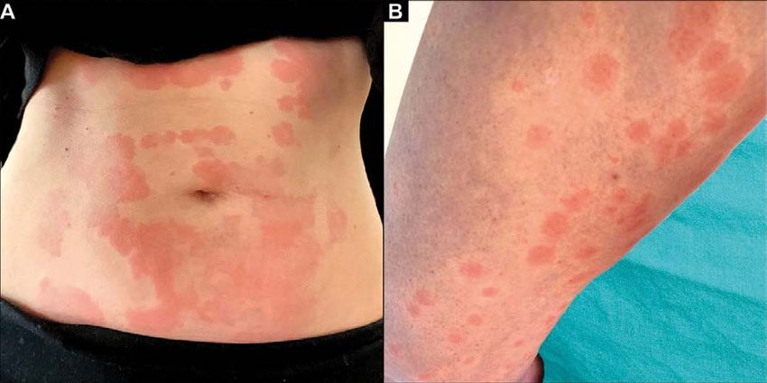
THE INFLAMMATORY PROCESS                                             proteins NLR (NOD-like receptors) responsible for the detec-
IN THE COURSE OF COVID-19                                            tion of DAMPs (Damage-associated molecular patterns) that
Fever, dry cough, shortness of breath, muscle aches, fatigue,        are expressed inside cells. Binding of DAMP activates NRLs,
a tendency to leukopenia and radiological symptoms of                causing a cascade of processes leading to the formation of
progressive pneumonia that may cause multi-organ failure             protein complexes called inflammasomes, which convert
are symptoms observed in COVID-19 (coronavirus disease               procaspase-1 to caspase-1 (Fig. 1). As a result of these events,
2019), SARS-CoV (severe acute respiratory syndrome CoV-2)            IL-1β is activated [9, 11]. If the signaling activation processes
and MERS (Middle East respiratory syndrome coronavirus).             are controlled, they are used to fight viruses and maintain
The pathogenesis of these diseases seems to be similar [1].          the body’s homeostasis. Under normal conditions, virus-in-
In the course of COVID-19, very high levels of ferritin and          fected cells are destroyed by NK cells and CD8 + cytolytic
D-dimers in the serum are observed, disproportionate to              T cells. Recognition is followed by apoptosis of the antigen
the severity of the infection, as well as the ability to mono-       presenting cells and the corresponding cytotoxic T cells in
cytosis and small amounts of NK cells (Natural Killers) and          order to avoid unnecessary antigenic activity. With acquired
cytotoxic T lymphocytes. Spikes composed of glycoproteins            diseases, a defect in the cytolytic activity of lymphocytes
on the surface of the virus belong to the most immunogenic           may occur. NK cells and CD8 + T cells become unable to lyse
parts of coronaviruses and have the ability to bind to angio-        infected and activated antigen presenting cells. Interactions
tensin-converting enzyme-2 ACE-2 (Angiotensin-convert-               between innate and adaptive immune cells take longer and
ing enzyme 2) receptors to enter the host cell. A similarity         many pro-inflammatory cytokines, including TNF (Tumor
has been demonstrated between SARS-CoV and SARS-CoV-2                necrosis factor), interferon γ, IL-1, IL-6, IL-18 and IL-33, are
spike glycoproteins. The distribution of ACE-2 receptor ex-          continuously secreted. The entire pathological process,
pression on the surface of type II follicular epithelial cells,      starting with defects in the cytolytic activity of lymphocytes,
heart, kidney, intestine and endothelial cells is consistent         through the increased activity of macrophages and the acti-
with target organs and the clinical picture in COVID-19              vation of the entire immune system, provokes a continuous
infected [1, 2]. SARS-CoV-2 is spread mainly through di-             release of cytokines. It may lead to multi-organ failure [4,
rect contact with saliva or secretions from the respiratory          12, 13]. This life-threatening condition is one of the leading
tract, when an infected person sneezes or coughs [3]. After          causes of death in COVID-19 patients.
binding to ACE-2 receptors on the cell surface through a gly-
coprotein spike, the virus enters the cytoplasm of the cell,
releases the RNA and replicates, resulting in the formation
of new viral particles. As a result, the cell breaks down and
the virus spreads to other cells. When the immune system
recognizes the antigens, they are presented to NK cells and
CD8 + cytotoxic T cells. This presentation activates both in-
nate and acquired immunity, provoking the production of
large amounts of pro-inflammatory cytokines and chemo-
kines. After binding to ACE-2 receptors on the cell surface
through a glycoprotein spike, the virus enters the cytoplasm
of the cell, releases the RNA genome and replicates, result-
ing in the formation of new viral particles. As a result, the cell
breaks down and the virus spreads to other cells. When the           Fig. 1 A cascade of pro-inflammatory events following virus entry.
                                                                     PRR – Pattern recognition receptor, DAMP – Damage-associated molecular pattern,
immune system recognizes the antigens, they are present-             PAMP – Pathogen-associated molecular pattern, ASC – Apoptosis-associated speck-like protein
                                                                     containing a CARD, CARD – Caspase Activation and Recruitment Domain,
ed to NK cells and CD8 + cytotoxic T cells. This presentation        NRLP3 – NLR family pyrin domain containing 3 (NLRP3),
activates both innate and acquired immunity, provoking the           PSTPIP1 – Proline-serine-threonine phosphatase-interacting protein 1
                                                                     Source: [13]
production of large amounts of pro-inflammatory cytokines
and chemokines. PRR receptors identify PAMP molecular                SKIN DERMATOSES ACCOMPANYING COVID-19
patterns mainly in the extracellular environment and to              Skin changes in COVID-19 are related to the body’s on-
a lesser extent in the intracellular environment. A signaling        going process of fighting the virus. The best known case
system is triggered that leads to the expression of pro-in-          stages of cutaneous COVID-19 symptoms as published by
flammatory transcription factors inducing cytokines such             Recalcati et al., in Lombardy, Italy, included 88 patients.
as NF-kβ and interferon regulatory factors that mediate the          This study reported 20.4% of confirmed COVID-19 cases,
type I interferon-mediated antiviral response [9, 10]. Anoth-        of which 19 had skin symptoms. It was shown that most of
er pathogen recognition system is in the cytosol and involves        the skin lesions were manifested by an erythematous rash
the NLRP1, NLRP3, NLRP7, NLRC4 family of leucine-rich                (77.8%). There were also several cases of urticaria (16.7%)

            432       5 / 2020 / vol. 9
                      Aesthetic Cosmetology and Medicine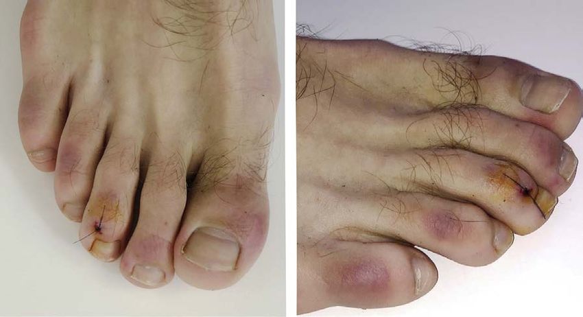
Fig. 2 A 39-year-old female with hives on the abdomen and thigh. The hives started the day before the fever appeared. Shortly
thereafter, she was diagnosed with Covid-19 positive Source: [15]
Fig. 3 Pseudo-frostbite changes, the so-called “covid fingers” Source: [16]
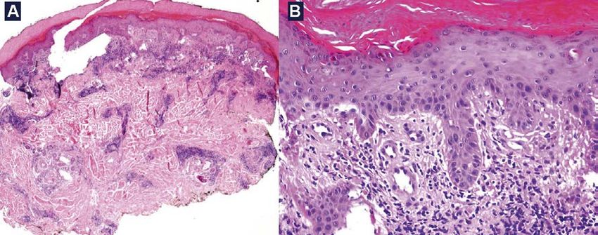
Fig. 4 Histopathological and pseudo-frostbite changes as a result of COVID-19 infection.
a) visible deep lymphoplasmic infiltrate, b) image with visible necrotic keratinocytes Source: [16]
maculopapular rash (15.3%) and the formation of vesicu- • Vesicular lesions
lar lesions on the skin (34.7%). The time of development of They are characterized by the occurrence of small vesicles
skin lesions among patients varied, ranging from 3 days located mainly in the area of the trunk, subcutaneous folds
before COVID-19 diagnosis to 13 days after diagnosis. The and legs, and they mainly affect middle-aged people. The
article describes a few selected dermatoses associated vesicular lesions accompanying COVID-19 are of medium
with coronavirus infection in the world [14]. to high intensity. The duration of the rash is 8 to 10 days.
Recalcati et al., in Lombardy reported vesicular lesions in
34.7% of patients with a positive COVID-19 result [14].
5 / 2020 / vol. 9
Aesthetic Cosmetology and Medicine 433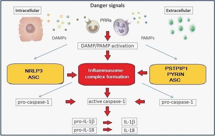
• Urticaria lesions                                                 REFERENCES
                                                                        Huang C, Wang Y, Li X, et al. Clinical features of patients infected with novel
Urticaria-like lesions are less common and last about               1.
                                                                        coronavirus in Wuhan, China. Lancet. 2019; 395(10223):497-506.
7 days. According to Recalcati et al., urticaria occurred in        2. Ding Y, He L, Zhang Q, et al. Organ distribution of severe acute respirato-

9.7% of the reported patients (Fig. 2). The accompanyng                 ry syndrome (SARS) associated coronavirus (SARS-CoV) in SARS patients:
                                                                        implications for pathogenesis and virus transmission pathways. J Pathol.
symptom of urticaria, which occurs mainly on the but-                   2004;203:622-630. doi:10.1002/2004/1560. Accessed 13.08.2020.
tocks and around the lower extremities, is itching [14].            3. Paules CI, Marston HD, Fauci AS. Coronavirus infections – more than just the
                                                                        common cold. JAMA. 2020;323(8):707-708.
                                                                    4. Crayne CB, Albeituni S, Nichols KE, Cron RQ. The immunology of macropha-
• Maculopapular rash                                                    ge activation syndrome. Front Immunol. 2019;10:119. doi:10.3389/2019/00119.
According to Italian scientists, the manifestation accom-               Accessed 13.08.2020.
                                                                    5. Sarzi-Puttini P, Giorgi V, Sirotti S, et al. COVID-19 cytokines and immunosup-
panying COVID-19 infection was observed in 15.3% of                     pression: what can we learn from severe acute respiratory syndrome? Clin Exp
patients. It lasts about 8 days, 60% of it may be pruritic. It          Rheumatol. 2020;38(2):337-342.
occurs in both children and adults. It is characterized by          6. Li X, Geng M, Peng Y, et al. Molecular immune pathogenesis and diagnosis of
                                                                        COVID-19. J Pharm Anal. 2020;4(10):102-108. doi:10.1016/2020.03.001. Acces-
infiltrating papular changes [14, 15].                                  sed 15.08.2020.
                                                                    7. Read R. Flawed methods in “COVID-19: Attacks the 1-Beta Chain of Hemoglobin
                                                                        and Captures the Porphyrin to Inhibit Human Heme Metabolism”. ChemRxiv.
• Covid fingers
                                                                        Preprint. 2020. doi:10.26434/chemrxiv.12120912.v2. Accessed 14.08.2020.
Experts say that they are increasingly noticing characteristic      8. Kawai T, Akira S. The role of pattern-recognition receptors in innateimmuni-

pseudo-frostbite changes called covid fingers in people infect-         ty: update on toll-like receptors. Nat Immunol. 2010;11:373-384. doi:10.1038/
                                                                        ni.1863. Accessed 12.08.2020.
ed with COVID-19. Changes located asymmetrically on the fin-        9. Schnappauf O, Chae JJ, Kastner DL, Aksentijevich I. The Pyrin inflammasome
gers and toes, accompanied by slight swelling (Fig. 3). Patients        in health and disease. Front Immunol. 2019;10:1745.
                                                                    10. Crow MK, Ronnblom L. Type I interferons in host defence and inflammatory
have discoloration on the hands and fingers, on which painful
                                                                        diseases. Lupus Sci Med. 2019;6(1):1-10. doi:10.1136/2019/000336. Accessed
blisters and ulcers appear. Noticeable changes are painful              15.08.2020.
red-purple bumps that tend to be on the tips of the fingers or      11. Lucherini OM, Rigante D, Sota J, et al. Updated overview of molecular pathways
                                                                        involved in the most common monogenic autoinflammatory diseases. Clin Exp
toes. The blue discoloration of the fingers may be indicated by         Rheumatol. 2018;36(1):3-9.
small blockages in the vessels, as COVID-19 is believed to cause    12. Al-Samkari H, Berliner N. Hemophagocytic lymphohistiocytosis. Annual review
                                                                        of pathology: mechanisms of disease. 2018;13:27-49. doi:10.1146/020117/043625.
an increased tendency to blood clots [14, 15]. The obtained his-
                                                                        Accessed 15.08.2020.
topathological image of the altered sites showed that there were    13. Mehmet S, Gökhan K, Pamir A, et al. Cytokine storm in COVID-19: pathogene-

pseudofreezing lesions (chilblains) with visible single, necrotic       sis and overview of anti-inflammatory agents used in treatment. International
                                                                        League of Associations for Rheumatology. 2020;39(7):2085-2094. doi:10.1007/
keratinocytes and a deep lymphoplasmic infiltrate (Fig. 4) [16].        s10067-020-05190-5. Accessed 16.08.2020.
                                                                    14. Sachdeva M, et al. Cutaneous manifestations of COVID-19: Report of three
                                                                        cases and a review of literature. J Dermatol Sci. 2020;5:98(2):75-81. doi:
• Reticular sinus (Lat. Livedo reticularis)
                                                                        10.1016/2020.04.011. Accessed 15.08.2020.
This is a blood vessel disorder. These are characteristic le-       15. Young S, Fernandez AP. Skin manifestations of COVID-19. Cleve Clin J Med.

sions forming a network of red-blue, marbled, mosaic-like               2020;5:1-4. doi:10.3949/ccjm.87a.ccc031. Accessed 15.08.2020.
                                                                    16. Athanassios K, Florence D, Delplace D, et al. Coronavirus (COVID-19) infection
lesions on the patient’s skin. Vessels in COVID-19 infected           – introduced chilblains: A case report with histophatologic findings. JAAD Case
patients can lead to lymphocytic vasculitis, similar symp-              Rep. 2020;6(6):489-492. doi:10.1016/2020.04.011. Accessed 15.08.2020.
toms are observed in thrombosis [14, 15].

SUMMARY
The underlying cause of skin lesions accompanying CO-
VID-19 is not fully recognized and requires further research.
The efficiency of the immune system determines the cour-
se of SARS-CoV-2 infection, and dermatological treatment
in many cases is immunosuppressive, which may result in
a potentially more severe course of COVID-19. In such case, if
the patient has been diagnosed with COVID-19 disease, im-
munosuppressive treatment should be discontinued. As the
pandemic unfolds and the number of cases increases, the
number of studies showing the association of COVID-19 in-
fection with skin lesions is also growing. Early detection of
the disease, especially in asymptomatic, poorly symptoma-
tic people or with atypical symptoms, may help to inhibit the             CITE / SPOSÓB CYTOWANIA
                                                                          Winkler S, Derkacz A. Skin diseases accompanying COVID-19 infection
spread of the virus, therefore disturbing, atypical skin lesions
                                                                         – literature review. Aesth Cosmetol Med. 2020;9(5):431-434.
cannot be underestimated. As COVID-19 may be asymptoma-
tic for 14 days after infection, skin symptoms can be used as            DOI: https://doi.org/10.6084/m9.figshare.13150961

an indicator of infection to aid prompt diagnosis.

            434       5 / 2020 / vol. 9
                      Aesthetic Cosmetology and Medicine
You can also read