A pilot study of estrogen receptor (ER) expression in pancreatic ductal adenocarcinoma (PDAC)
←
→
Page content transcription
If your browser does not render page correctly, please read the page content below
Original Article

A pilot study of estrogen receptor (ER) expression in pancreatic
ductal adenocarcinoma (PDAC)
Kai Siang Chan1, Bernard Chi Shern Ho2, Vishal G. Shelat3
1
Lee Kong Chian School of Medicine, Nanyang Technological University, Singapore, Singapore; 2Department of Pathology, 3Department of General
Surgery, Tan Tock Seng Hospital, Singapore, Singapore
Contributions: (I) Conception and design: VG Shelat; (II) Administrative support: None; (III) Provision of study materials or patients: BCS Ho, VG
Shelat; (IV) Collection and assembly of data: BCS Ho; (V) Data analysis and interpretation: All authors; (VI) Manuscript writing: All authors; (VII)
Final approval of manuscript: All authors.
Correspondence to: Vishal G. Shelat. Department of General Surgery, Tan Tock Seng Hospital, Singapore 308433, Singapore.
Email: vishal_g_shelat@ttsh.com.sg.

                 Background: Pancreatic ductal adenocarcinoma (PDAC) is the most common pancreatic neoplasm with
                 5-year survival as low as 6%. It is therefore imperative to explore potential treatment avenues to improve
                 survival in these groups of patients. Anti-estrogenic hormone therapy (AEHT) is well-tolerated and has been
                 used in estrogen receptor (ER) subgroups of breast cancer. ER is a type of sex hormone receptor which have
                 been reported to be expressed inconsistently in pancreatic cancer. This study aims to identify the presence of
                 ER in PDAC specimens to guide potential use of AEHT in the management of unresectable PDAC.
                 Methods: This is a retrospective case control study of 10 patients (5 males, 5 females) who underwent
                 pancreatic resections for PDAC from 2011 to 2012. Sections of the post-operative specimens were prepared
                 and sent for ER staining. Pancreatic tissue specimens that were analysed included (I) ductal epithelial cells; (II)
                 acinar cells; (III) islet cells; (IV) intralobular stromal cells; and (V) adenocarcinoma cells.
                 Results: Intralobular stromal cells were positively stained for ER in 7/10 (70%) of the cases, but were of
                 weak intensity and patchy in distribution. Islet cells (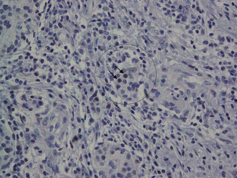
Page 2 of 5 Translational Gastroenterology and Hepatology, 2021
study. Upon completion of the patient selection, sections
of the post-operative specimens were prepared and sent
for ER staining. This study was approved by the hospital
institutional review board (reference number: 2012/00919)
and funded by the National Research Foundation
Scholarship Grant.
Specimen preparation
Sections of pancreatic cancer including adjacent normal
pancreatic tissue were cut from formalin fixed, paraffin
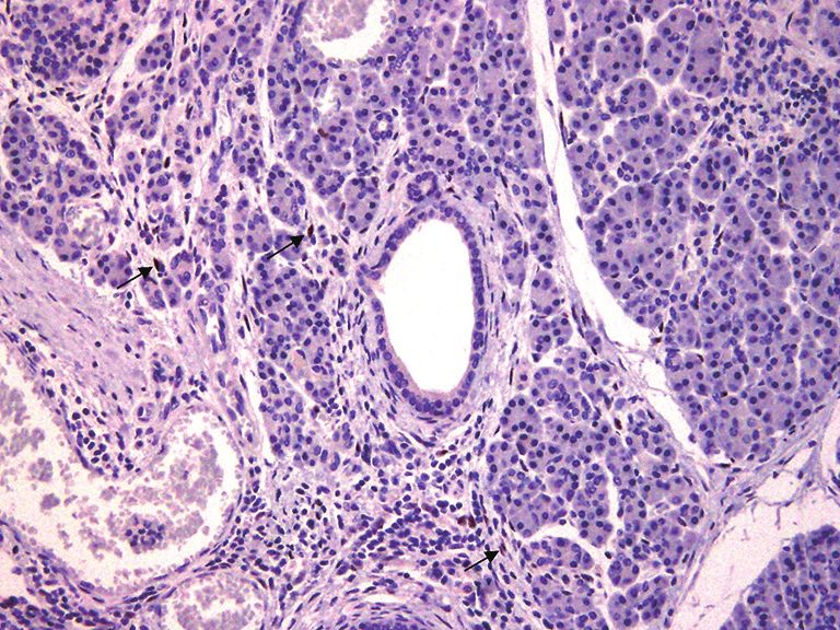
Figure 1 Intralobular cells showing weak expression of estrogen embedded tissue blocks and stained for ER using
receptor (ER) (black arrows), ×200 magnification Novocastra Liquid Mouse Monoclonal Antibody Estrogen
Receptor TM (Product code: NCL-L-ER-6F11) (Leica
Biosystems, Newcastle Upon Tyne, UK) and Ventana
cancers, including pancreatic cancer (6). ER is a type of OptiView DAB IHC Detection KitTM (Ventana Medical
sex hormone receptor which have been reported to be Systems, Arizona, USA) with appropriate tissue controls.
expressed in pancreatic cancer, though there have been Pancreatic tissue specimens that were analysed included (I)
inconsistencies in their detection (7,8). Presence of ER ductal epithelial cells; (II) acinar cells; (III) islet cells; (IV)
expression in PDAC may potentially guide treatment with intralobular stromal cells; and (V) adenocarcinoma cells.
AEHT. Several studies have been conducted to explore Reporting of ER staining was performed in accordance to
the role of AEHT (mainly tamoxifen) in the management ER staining for breast cancer specimens (22): (I) proportion
of PDAC; however, results are equivocal with varying of cells (0–100%) with ER positive nuclear staining and (II)
treatment response across different studies (9-16). It intensity of staining (weak, moderate and strong). Specimen
has been postulated that the inconsistent results may be preparation and histopathological analysis were performed
attributed to the lack of assessment of estrogen dependence by a single, blinded pathologist.
in PDAC (17). Hence, this study aims to identify the
presence of ER in PDAC specimens to guide potential use Results
of AEHT in the management of unresectable PDAC.
Intralobular stromal cells were positively stained for ER in
7/10 (70%) of the cases; these were stromal spindly cells,
Methods all of which had nuclear ER-positive staining were of weak
This is a retrospective case control study of 10 patients intensity and were patchy in distribution (Figure 1). Islet cells
(5 males, 5 females) who underwent pancreatic resections (Translational Gastroenterology and Hepatology, 2021 Page 3 of 5
immunohistochemistry analysis: adequate tissue controls
were used to identify problems with tissue fixation with
formalin; this is of important significance as it is widely
documented that ER is labile and good fixation is required
to identify its presence (25). Use of immunohistochemistry
for ER performed formalin fixed paraffin embedded tissue
has also been documented to be a reliable and convenient
method to study ER expression (26).
The role of AEHT in PDAC is still uncertain. Existing
studies thus far show equivocal evidence on its use in the
management of unresectable PDAC. A study by Wong et al.
Figure 2 Islet cells showing occasional nuclear estrogen receptor demonstrated that tamoxifen prolongs survival in post-
(ER) positivity (black arrow), ×400 magnification. menopausal women (n=6/15, 3 with minimum 2-year
survival) with unresectable PDAC (12). However, other
randomized studies fail to demonstrate the survival benefit
of tamoxifen in unresectable pancreatic adenocarcinoma
(15,24). The lack of survival benefit in patients treated with
tamoxifen could be attributed to the lack of ER expression,
as demonstrated in the study by Taylor et al. (ER expression
positive in n=0/25) (24). It was traditionally postulated that
the role of tamoxifen in breast cancer is due to its effect
on nuclear ERs (27). However, a recent translational study
by Cortes et al. in 2019 on mice demonstrated that a new
mechanism of action of tamoxifen which is independent of
its effect on nuclear ERs (28); this involves a mechanical
downregulation of hypoxia-inducible factor-1 alpha
Figure 3 Pancreatic ductal adenocarcinoma cells showing lack of (HIF-1A) which reduces the adaptive response of PDAC to
estrogen receptor (ER) staining, ×100 magnification. hypoxia and may be responsible for reprogramming of the
tumour microenvironment and its therapeutic role in some
of studies which documented survival benefit of tamoxifen
to lack of confirmatory staining with anti-insulin or anti- in PDAC. Other mechanisms of action on the role of
glucagon antibodies. However, it has described in literature tamoxifen in cancer treatment have also been described (29).
that the use of antibody-staining techniques may not be The presence of ER expression in other organs have
necessary (23); islet cells can be easily identified with also guided several investigations on the role of anti-
background counter stain by a trained pathologist. Negative hormonal therapy (6). However, these results have been
staining for ER in ductal epithelial and acinar cells may dismal. A randomized control trial on the use of tamoxifen
be due to the true absence of ER or very low quantities of in hepatocellular carcinoma (HCC) by the Cancer of the
ER which is undetectable by the immunohistochemical Liver Italian Programme Group in 1998 (30), and a multi-
technique. centre regional randomized control trial by Chow et al. in
It is still uncertain whether the ER expression is present 2002 demonstrated the lack of survival benefits in HCC (31);
in all patients with PDAC. A study by Greenway et al. failed the latter involved the use of high-dose tamoxifen which
to demonstrate presence of ER receptors at five different inhibits HCC via ER-independent mechanisms.
sites in a patient with underlying PDAC (7). Our pilot One of the limitations of this study is that only a single
study is also supported by the studies by Singh et al. (8), and pathologist was involved in the histopathological analysis
Taylor et al. (24), which detected little to no ER expression of the specimens. However, it has been demonstrated
by immunohistochemistry. The lack of ER staining in our that a second opinion surgical pathology is unlikely to
pilot study is unlikely due to the specimen preparation and change diagnosis with an overall incidence of 1.4% of
© Translational Gastroenterology and Hepatology. All rights reserved. Transl Gastroenterol Hepatol 2021;6:9 | http://dx.doi.org/10.21037/tgh.2020.02.16Page 4 of 5 Translational Gastroenterology and Hepatology, 2021
changed diagnoses, and an even lower incidence of changed distributed in accordance with the Creative Commons
diagnosis for gastrointestinal (1.2%) and endocrine (0%) Attribution-NonCommercial-NoDerivs 4.0 International
pathologies (32). Due to the small sample size in this pilot License (CC BY-NC-ND 4.0), which permits the non-
study, this is likely to be unremarkable. In addition, this is commercial replication and distribution of the article with
a pilot study with a small sample. A larger sample would the strict proviso that no changes or edits are made and the
be ideal for detecting the presence of ER staining but this original work is properly cited (including links to both the
is limited by inadequate funding to permit ER staining for formal publication through the relevant DOI and the license).
all the patients who underwent pancreatic resections for See: https://creativecommons.org/licenses/by-nc-nd/4.0/.
PDAC. Furthermore, a sample size of ten was determined
to be acceptable for a pilot study (19-21).
References
In conclusion, this pilot study did not detect the presence
of ER expression in PDAC. Hence, we were unable to 1. Kamisawa T, Wood LD, Itoi T, et al. Pancreatic cancer.
procced to explore AEHT role in management of ER- Lancet 2016;388:73-85.
positive PDAC. The role of AEHT in pancreatic cancer 2. Gillen S, Schuster T, Zum Büschenfelde CM, et al.
remains uncertain and does not appear to be of value Preoperative/neoadjuvant therapy in pancreatic cancer:
at present. Our study demonstrates the presence of ER a systematic review and meta-analysis of response and
expression in intralobular stromal and islet cells, which resection percentages. PLoS Med 2010;7:e1000267.
was previously unreported. Further studies are required 3. Alexakis N, Halloran C, Raraty M, et al. Current
to determine the significance of weak and patchy ER standards of surgery for pancreatic cancer. Br J Surg
expression in intralobular stromal and islet cells. 2004;91:1410-27.
4. Rose C, Mouridsen HT, Thorpe SM, et al. Anti-estrogen
treatment of postmenopausal breast cancer patients with
Acknowledgments
high risk of recurrence: 72 months of life-table analysis
Funding: This study is funded by the National Research and steroid hormone receptor status. World J Surg
Foundation Scholarship Grant. 1985;9:765-74.
5. Hortobagyi GN. Treatment of breast cancer. N Engl J
Med 1998;339:974-84.
Footnote
6. Stedman KE, Moore GE, Morgan RT. Estrogen
Provenance and Peer Review: This article was a free submission receptor proteins in diverse human tumors. Arch Surg
to the journal. The article has undergone external peer 1980;115:244-8.
review. 7. Greenway B, Iqbal M, Johnson P, et al. Oestrogen receptor
proteins in malignant and fetal pancreas. Br Med J (Clin
Conflicts of Interest: All authors have completed the ICMJE Res Ed) 1981;283:751-3.
uniform disclosure form (available at http://dx.doi. 8. Singh S, Baker P, Poulsom R, et al. Expression of
org/10.21037/tgh.2020.02.16). The authors have no oestrogen receptor and oestrogen-inducible genes in
conflicts of interest to declare. pancreatic cancer. Br J Surg 1997;84:1085-9.
9. Theve NO, Pousette A, Carlström K. Adenocarcinoma of
Ethical Statement: The authors are accountable for all the pancreas--a hormone sensitive tumor? A preliminary
aspects of the work in ensuring that questions related report on Nolvadex treatment. Clin Oncol 1983;9:193-7.
to the accuracy or integrity of any part of the work are 10. Tønnesen K, Kamp-Jensen M. Antiestrogen therapy in
appropriately investigated and resolved. The study was pancreatic carcinoma: a preliminary report. Eur J Surg
conducted in accordance with the Declaration of Helsinki (as Oncol 1986;12:69-70.
revised in 2013). This study was approved by the hospital 11. Crowson MC, Dorrell A, Rolfe EB, et al. A phase II study
institutional review board(reference number: 2012/00919). to evaluate tamoxifen in pancreatic adenocarcinoma. Eur J
The written informed consent was waived due to the Surg Oncol 1986;12:335-6.
retrospective nature of the study. 12. Wong A, Chan A, Arthur K. Tamoxifen therapy in
unresectable adenocarcinoma of the pancreas. Cancer
Open Access Statement: This is an Open Access article Treat Rep 1987;71:749-50.
© Translational Gastroenterology and Hepatology. All rights reserved. Transl Gastroenterol Hepatol 2021;6:9 | http://dx.doi.org/10.21037/tgh.2020.02.16Translational Gastroenterology and Hepatology, 2021 Page 5 of 5
13. Keating JJ, Johnson PJ, Cochrane AM, et al. A prospective 2015;63:543-58.
randomised controlled trial of tamoxifen and cyproterone 24. Taylor O, Benson E, McMahon M. Clinical trial of
acetate in pancreatic carcinoma. Br J Cancer tamoxifen in patients with irresectable pancreatic
1989;60:789-92. adenocarcinoma. Br J Surg 1993;80:384-6.
14. Scheithauer W, Kornek G, Haider K, et al. 25. King WJ, DeSombre ER, Jensen EV, et al. Comparison
Unresponsiveness of pancreatic adenocarcinoma to of immunocytochemical and steroid-binding assays for
antioestrogen therapy. Eur J Cancer 1990;26:851-2. estrogen receptor in human breast tumors. Cancer Res
15. Bakkevold KE, Pettersen A, Arnesjø B, et al. Tamoxifen 1985;45:293-304.
therapy in unresectable adenocarcinoma of the pancreas 26. Taylor CR, Shi SR, Chaiwun B, et al. Strategies for
and the papilla of Vater. Br J Surg 1990;77:725-30. improving the immunohistochemical staining of
16. Wong A, Chan A. Survival benefit of tamoxifen therapy in various intranuclear prognostic markers in formalin-
adenocarcinoma of pancreas. A case-control study. Cancer paraffin sections: androgen receptor, estrogen receptor,
1993;71:2200-3. progesterone receptor, p53 protein, proliferating cell
17. Yamashita J, Abe M, Ogawa M. Endocrine therapy in nuclear antigen, and Ki-67 antigen revealed by antigen
pancreatic carcinoma. Oncology 1998;55:17-22. retrieval techniques. Hum Pathol 1994;25:263-70.
18. World Health Organization. ICD-10: international 27. Hall JM, Couse JF, Korach KS. The multifaceted
statistical classification of diseases and related mechanisms of estradiol and estrogen receptor signaling. J
health problems: tenth revision. 2004. Available Biol Chem 2001;276:36869-72.
online: https://apps.who.int/iris/bitstream/ 28. Cortes E, Lachowski D, Robinson B, et al. Tamoxifen
handle/10665/42980/9241546530_eng.pdf mechanically reprograms the tumor microenvironment
19. Isaac S, Michael WB. Handbook in research and via HIF-1A and reduces cancer cell survival. EMBO Rep
evaluation: A collection of principles, methods, and 2019. doi: 10.15252/embr.201846557.
strategies useful in the planning, design, and evaluation 29. Daurio NA, Tuttle SW, Worth AJ, et al. AMPK Activation
of studies in education and the behavioral sciences. Edits and Metabolic Reprogramming by Tamoxifen through
publishers; 1995. Estrogen Receptor–Independent Mechanisms Suggests
20. Julious SA. Sample size of 12 per group rule of thumb for New Uses for This Therapeutic Modality in Cancer
a pilot study. Pharmaceut Statist 2005;4:287-91. Treatment. Cancer Res 2016;76:3295-306.
21. Van Belle G. Statistical rules of thumb. John Wiley & 30. Group C. Tamoxifen in treatment of hepatocellular
Sons; 2011. carcinoma: a randomised controlled trial. Lancet
22. Barnes DM, Harris WH, Smith P, et al. 1998;352:17-20.
Immunohistochemical determination of oestrogen 31. Chow PK, Tai BC, Tan CK, et al. High-dose tamoxifen
receptor: comparison of different methods of assessment in the treatment of inoperable hepatocellular carcinoma:
of staining and correlation with clinical outcome of breast a multicenter randomized controlled trial. Hepatology
cancer patients. Br J Cancer 1996;74:1445-51. 2002;36:1221-6.
23. Baskin DG. A historical perspective on the identification 32. Kronz JD, Westra WH, Epstein JI. Mandatory second
of cell types in pancreatic islets of Langerhans by staining opinion surgical pathology at a large referral hospital.
and histochemical techniques. J Histochem Cytochem Cancer 1999;86:2426-35.
doi: 10.21037/tgh.2020.02.16
Cite this article as: Chan KS, Ho BCS, Shelat VG. A pilot
study of estrogen receptor (ER) expression in pancreatic ductal
adenocarcinoma (PDAC). Transl Gastroenterol Hepatol
2021;6:9.
© Translational Gastroenterology and Hepatology. All rights reserved. Transl Gastroenterol Hepatol 2021;6:9 | http://dx.doi.org/10.21037/tgh.2020.02.16You can also read