Serum hypoxia inducible factor 1α and uterine artery Doppler ultrasound during the first trimester for prediction of preeclampsia - Nature
←
→
Page content transcription
If your browser does not render page correctly, please read the page content below
www.nature.com/scientificreports
OPEN Serum hypoxia‑inducible factor‑1α
and uterine artery Doppler
ultrasound during the first
trimester for prediction
of preeclampsia
Wasinee Tianthong & Vorapong Phupong*
The objective of this study was to determine the predictive value of serum hypoxia-inducible factor-1α
(HIF-1α) combined with uterine artery Doppler in singleton pregnancy during 11–13+6 weeks of
gestation for preeclampsia. This prospective observational study was conducted in singleton pregnant
women at 11–13+6 weeks of gestation who visited the King Chulalongkorn Memorial Hospital, Faculty
of Medicine, Chulalongkorn University for antenatal care between February 2019 and May 2020.
Serum HIF-1α levels and uterine artery Doppler ultrasound were performed. Pregnancy outcomes
were recorded. The sensitivity, specificity, positive predictive value (PPV), and negative predictive
value (NPV) of these tests at the optimal cut-off values were determined to predict preeclampsia.
A total of 385 participants were analyzed. Of these, 31 cases had preeclampsia (8.1%), and 6 cases
of them had early-onset preeclampsia (1.6%). Preeclamptic women had significantly higher serum
HIF-1α levels than normal pregnant women (median 1315.2 pg/ml vs. 699.5 pg/ml, p < 0.001). There
was no difference in the mean pulsatility (PI) of the uterine artery. Serum HIF-1α levels were higher
than 1.45 multiple of median for the gestational age as a cut-off value for predicting preeclampsia;
the sensitivity, specificity, PPV, and NPV were 66.7%, 71.5%, 17.2%, and 96.2%, respectively. When
a combination of abnormal serum HIF-1α levels and abnormal uterine artery Doppler PI (above the
95th percentile) were used as a predictive value to predict preeclampsia, the sensitivity, specificity,
PPV, and NPV were 74.2%, 67.2%, 16.6%, and 96.8%, respectively. This study showed that the serum
HIF-1α levels with or without uterine artery Doppler at 11–13+6 weeks of gestation were effective in
predicting preeclampsia.
Hypertensive disease of pregnancy is considered one of the most frequent obstetric complications that affects up
to 10% of pregnant w omen1. Preeclampsia, a serious type of pregnancy-induced hypertension, is a member of the
deadly obstetrical triad along with infection and hemorrhage as well as a leading cause of maternal and neonatal
morbidities2,3. The global incidence of preeclampsia is 2–8%. Furthermore, there are estimated 50,000–60,000
women worldwide who die of preeclampsia each y ear1.
The etiology of preeclampsia remains unknown, but it is thought to be the consequence of impaired tropho-
blastic invasion of the maternal spiral arteries due to an imbalance between levels of angiogenic factors and
hypoxia-induced oxidative s tress4,5. According to previous studies, a defective placental invasion results in utero-
placental insufficiency and is associated with adverse pregnancy outcomes such as fetal growth restriction and
preeclampsia, especially during the early and severe form of this condition4,5.
The American College of Obstetricians and Gynecologists (ACOG) and the National Institute for Care and
Health Excellence (NICE) recommend identifying patients who are at high risk of developing preeclampsia based
on their medical history. Pregnant women who are at high risk of developing preeclampsia are prescribed low-
dose aspirin to minimize the incidence and severity of the d isease6–8. Although this traditional approach using
clinical risk factors has limited predictive ability and can identify only 30% of pregnant women who develop
preeclampsia, it is the only currently recommended screening method6,9,10. Nowadays, researches are focused on
Placental Related Diseases Research Unit, Department of Obstetrics and Gynecology, Faculty of Medicine,
Chulalongkorn University, Rama IV Road, Pathumwan, Bangkok 10330, Thailand. *email: vorapong.p@chula.ac.th
Scientific Reports | (2021) 11:6674 | https://doi.org/10.1038/s41598-021-86073-w 1
Vol.:(0123456789)www.nature.com/scientificreports/
finding new markers that have high predictive value to provide early intervention in order to reduce morbidity
and mortality. Many screening tests, such as those using biochemical markers and ultrasound markers, were
investigated whether it can predict preeclampsia or not, but none of them have been found to be predictably
reliable, valid, and suitable for routine clinical use10.
Hypoxia-inducible factor-1 (HIF-1) is a heterodimer consisting of two subunits, α and β. It is a key transcrip-
tion factor that mediates cellular response to low oxygen tension. HIF-1α levels are rapidly reduced under normal
oxygen condition. But they are increased in hypoxic e nvironments11,12. Placentation develops in a low-oxygen
environment during early pregnancy before 10 weeks of gestation when there is limited blood flow into the
intervillous space. HIF-1α is upregulated to maintain trophoblasts in a proliferative, noninvasive, and immature
phenotype. After that, intervillous blood flow is increased at 10–12 weeks of gestation leading to normoxic condi-
tions and lower levels of HIF-1α. The differentiation of extravillous trophoblasts occurs and they physiologically
invade the myometrial segment of the uterus. For pregnancies complicated by preeclampsia, HIF-1α expression
remains abnormally elevated and the trophoblasts development remains arrested at an immature stage, causing
a shallow trophoblast invasion11,13–18. Previous studies found that pregnant women with preeclampsia had high
blood levels of HIF-1α compared to normal healthy controls19,20. Galbiati et al. found that HIF-1α was signifi-
cantly higher in women who later developed preeclampsia compared to women who did not21.
Serum HIF-1α levels have never been used to predict preeclampsia. Therefore, this study assessed the predic-
tive value of serum HIF-1α combined with uterine artery Doppler in singleton pregnancy during 11–13+6 weeks
of gestation for preeclampsia and other pregnancy complications, such as fetal growth restriction, preterm
delivery, and perinatal death.
Materials and methods
This prospective observational study was performed at the Department of Obstetrics and Gynecology, King
Chulalongkorn Memorial Hospital, Faculty of Medicine, Chulalongkorn University, Bangkok, Thailand, between
February 2019 and May 2020. This study was approved by the Research Ethics Committee of the Faculty of Medi-
cine, Chulalongkorn University. All procedures were performed in accordance with the relevant guidelines and
regulations of the Institutional Review Board. This study has been performed in accordance with the Declaration
of Helsinki. All subjects gave written informed consent.
Singleton pregnant women at a gestational age of 11–13+6 weeks, aged between 20 and 45 years who came to
the antenatal clinic were invited to participate in this study. Gestational age was determined by the last menstrual
period and confirmed by the measurement of the fetal crown-rump length at the first-trimester ultrasound.
Women who used aspirin as a prophylaxis for preeclampsia were excluded from the study. In addition, pregnant
women who were diagnosed to have fetal structural or chromosomal abnormalities were excluded.
Since there is a lack of data on the predictive value of HIF-1α for the prediction of preeclampsia, the sample
size was calculated based upon the hypothesis that the sensitivity of serum HIF-1α combined with uterine artery
PI in preeclampsia prediction was 80% with 20% allowable error. This indicated that the study needed 15 cases
of preeclampsia. The incidence of preeclampsia at our institute was 4.9%. After we adjusted the calculation using
our institute’s incidence of preeclampsia and a loss to follow-up rate of 20%, a minimum of 368 women were
required for this study.
Written informed consent was obtained from all eligible participants. The questionnaire on maternal age,
medical history, parity, and obstetric history was completed by interviewing each participant. Maternal weight
and height were measured to calculate the body mass index (BMI).
Blood pressure was measured by validated automated devices (Microlife AG, 9443 Widnau, Switzerland) after
at least 5 min of rest in the seated position. The mean arterial pressure was recorded.
Uterine artery Doppler was assessed transabdominally by a single sonographer using ultrasonographic
machines (GE Voluson E10, GE Medical Systems, Zipf, Austria) with a convex probe AB 2–7 MHz. Initially, a
mid-sagittal section of the uterus and cervix was obtained, and the probe was then tilted sideways to identify
the uterine artery blood flow along both sides. Two-millimeter gate pulse-wave Doppler was positioned on the
branch of the uterine artery close to the internal cervix, with an insonation angle < 30°. At least three identi-
cal waveforms with the peak systolic velocity more than 60 cm/s were obtained. The pulsatility index (PI) was
recorded for each side22. An abnormal Doppler of the uterine artery result was defined as having a mean PI more
than the 95th percentile for each gestational a ge22.
Venous blood samples (10 ml) were drawn from each participant into a non-heparinized tube. Serum was
separated by centrifugation at 2500 rounds per minute for 10 min and frozen at − 80 °C until assay. After recruit-
ment was completed, HIF-1α concentration was measured using the HIF 1-α enzyme-linked immunosorbent
assay (ELISA) (Cloud-Clone Corp, Massachusetts, TX, USA)23.
SPSS software version 22.0 (IBM, New York, USA) was used for statistical analysis. Results were presented as
mean with standard deviation, median with interquartile range (IQR), sensitivity, specificity, positive predictive
value (PPV), negative predictive value (NPV), and relative risk, with a 95% confidence interval. The optimal
cut-off values for HIF-1α levels were calculated using the receiver operator characteristic curve. A Chi-square
test, Fisher’s exact test, unpaired t test, and Mann–Whitney U test were used when appropriate. A p value < 0.05
was considered to be statistically significant.
Results
Four hundred and seven pregnant women were enrolled into this study. Twenty-two women were excluded
due to spontaneous miscarriage prior to 20 weeks (7 cases), lost to follow-up (13 cases), or fetal anomalies were
diagnosed during the second trimester (2 cases). The data from a total of 385 women were analyzed. Thirty-one
participants were diagnosed with preeclampsia (8.1%), and 6 of them had early-onset preeclampsia (1.6%).
Scientific Reports | (2021) 11:6674 | https://doi.org/10.1038/s41598-021-86073-w 2
Vol:.(1234567890)www.nature.com/scientificreports/
Controls (n = 354) Preeclampsia (n = 31) p value
Maternal age (years) 32.8 ± 4.9 33.3 ± 4.6 0.612
Advanced maternal age (≥ 35 years old) 135 (38.1) 15 (48.4) 0.262
Primigravida 173 (48.9) 17 (54.8) 0.523
Parity 0.537
0 202 (57.1) 20 (64.5)
≥1 152 (42.9) 11 (35.5)
Prepregnancy BMI (kg/m2) 22.6 ± 4.5 23.6 ± 3.6 0.197
Obesity (BMI ≥ 30 kg/m2) 26 (7.3) 0 (0) 0.25
Total weight gain (kg) 13.2 ± 4.7 14.6 ± 4.3 0.106
GA at measurement (weeks) 12.3 ± 0.7 12.4 ± 0.6 0.241
Mean arterial pressure (mmHg) 83.1 ± 8.4 90.8 ± 9.7 < 0.001
Gestational diabetes 22 (6.2) 5 (16.1) 0.088
Fetal growth restriction 2 (0.6) 2 (6.5) 0.034
GA at delivery (weeks) 38.2 ± 1.5 36.6 ± 2.5 < 0.001
Delivery at GA < 37 weeks 20 (5.6) 7 (22.6) 0.003
Delivery at GA < 34 weeks 5 (1.4) 2 (6.5) 0.102
Mode of delivery 0.499
Vaginal delivery 154 (43.5) 11 (35.5)
Cesarean section 200 (56.5) 20 (64.5)
Birth weight (g) 3116 ± 440.8 2747.3 ± 631.6 < 0.001
Low birth weight (< 2500 g) 20 (5.6) 7 (22.6) 0.001
Apgar score
1 min 8.9 ± 0.5 8.1 ± 1.8 < 0.001
5 min 9.9 ± 0.3 9.3 ± 1.8 < 0.001
Neonatal respiratory distress syndrome 6 (1.7) 6 (19.4) < 0.001
Perinatal death 1 (0.3) 1 (3.2) 0.155
Length of hospital stay 4.2 ± 2.8 6.7 ± 8.3 < 0.001
Table 1. Baseline characteristics and pregnancy outcomes of women with preeclampsia and controls. Data are
presented as mean ± SD or n (%). BMI body mass index, GA gestational age.
The demographic data and pregnancy outcomes among women with and without preeclampsia are shown
in Table 1. The baseline characteristics including maternal age, proportion of women who were at an advanced
maternal age (35 years old or more at the estimated date of confinement), parity, prepregnancy body mass index
(BMI), proportion of obese women (defined as prepregnancy BMI 30 kg/m2 or more), total weight gain and gesta-
tional age at measurement were not significantly different between the two groups. However, preeclamptic women
had a significantly higher mean arterial blood pressure at the first trimester than non-preeclamptic women
(90.8 ± 9.7 vs. 83.1 ± 8.4 mmHg, p < 0.001). On the other hand, for pregnancy and neonatal outcomes, pregnant
women who developed preeclampsia had a lower gestational age at delivery (36.6 ± 2.5 vs. 38.2 ± 1.5 weeks,
p < 0.001), higher preterm delivery rate (22.6% vs. 5.6%, p = 0.003), lower neonatal birth weight (2747.3 ± 631.6
vs. 3116 ± 440.8 g, p < 0.001), higher rate of low birth weight (< 2500 g) (22.6% vs. 5.6%, p = 0.001), lower Apgar
scores at the 1st and 5th-minute after birth (8.1 ± 1.8 vs. 8.9 ± 0.5, p < 0.001 and 9.3 ± 1.8 vs. 9.9 ± 0.3, p < 0.001,
respectively), higher rate of neonatal respiratory distress syndrome (RDS) (19.4% vs. 1.7%, p < 0.001), and longer
length of hospital stay (6.7 ± 8.3 vs. 4.2 ± 2.8 days, p < 0.001) than non-preeclamptic women.
The median (IQR) serum HIF-1α levels of overall preeclampsia and late-onset preeclampsia were 1315.2
(645.9, 5128.7), and 1715.5 (852, 5134) pg/ml, respectively, which were significantly higher than that of normal
pregnant women (699.5 (426.8, 1107.1) pg/ml) (p < 0.001, and < 0.001, respectively). However, the median (IQR)
serum HIF-1α level of early-onset preeclampsia was 646.7 (438.9, 5151.9) pg/ml which was not different com-
pared to the controls (p = 0.629). There was no differences in the mean PI of the uterine artery Doppler between
women with preeclampsia and control women (p = 0.775). However, the mean PI of the uterine artery Doppler
was significantly higher in women with early-onset preeclampsia than healthy control women (2.21 ± 0.47 vs.
1.72 ± 0.48, p = 0.013). The mean PI of the uterine artery Doppler was comparable between late-onset preeclamp-
sia women and the healthy control women (Table 2).
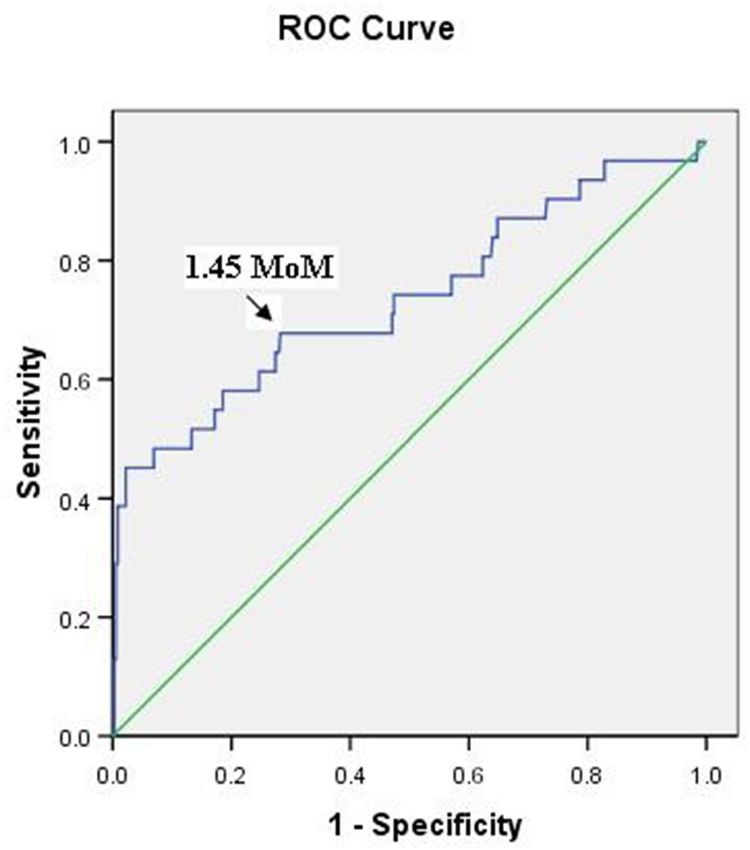
The optimal cut-off value of the serum HIF-1α levels, calculated from the receiver operating characteristic
curve (AUC = 0.735, p < 0.001), was 1.45 multiple of median (MoM), according to the gestational age at the time
of measurement (Fig. 1). The sensitivity, specificity, PPV, and NPV were 66.7%, 71.5%, 17.2%, and 96.2%, respec-
tively, when using serum levels above the cut-off value for predicting preeclampsia. To predict early-onset preec-
lampsia, the sensitivity, specificity, PPV, and NPV were 33.3%, 68.3%, 1.6%, and 98.5%, respectively (Table 3).
The 95th percentile of the mean uterine artery Doppler PI in this study, stratified into three groups accord-
ing to the gestational age at the time of measurement, were 2.77, 2.72, and 2.38 at 11–11+6, 12–12+6, and
13–13+6 weeks, respectively. Using the mean uterine artery Doppler PI above the 95th percentile as a cut-off
Scientific Reports | (2021) 11:6674 | https://doi.org/10.1038/s41598-021-86073-w 3
Vol.:(0123456789)www.nature.com/scientificreports/
Early-onset preeclampsia Late-onset preeclampsia
Controls (n = 354) Preeclampsia (n = 31) (n = 6) (n = 25) p value
699.5 (426.8, 1107.1) 1315.2 (645.9, 5128.7) < 0.001
HIF-1α (pg/ml) 646.7 (438.9, 5151.9) 0.629
1715.5 (852, 5134) < 0.001
1.72 ± 0.48 1.74 ± 0.48 0.775
UtA PI 2.21 ± 0.47 0.013
1.63 ± 0.41 0.38
Table 2. Serum HIF-1α levels and UtA Doppler PI in women with preeclampsia, early-onset preeclampsia,
and late-onset preeclampsia compared to healthy controls. Data are presented as median (IQR), mean ± SD or
n (%). HIF-1α hypoxia-inducible factor-1 alpha, UtA PI uterine artery pulsatility index.
Figure 1. Receiver-operating characteristic curve for the relationship between the serum hypoxia-inducible
factor-1α level and the diagnosis of preeclampsia (area under the curve: 0.735; 95% confidence interval:
0.625–0.844; p < 0.001).
value for predicting preeclampsia, the sensitivity, specificity, PPV, and NPV were 6.5%, 95.2%, 10.5%, and 92.1%,
respectively. To predict early-onset preeclampsia, the sensitivity, specificity, PPV, and NPV were 16.7%, 95.3%,
5.3%, and 98.6%, respectively (Table 3).
A combination of abnormal serum HIF-1α levels (higher than 1.45 MoM) and abnormal uterine artery Dop-
pler PI (above the 95th percentile) was used as a predictive value for preeclampsia and its sensitivity, specificity,
PPV, and NPV were 74.2%, 67.2%, 16.6%, and 96.8%, respectively. A combination of abnormal serum HIF-1α
levels (higher than 1.45 MoM) and abnormal uterine artery Doppler PI (above the 95th percentile) was used as
a predictive value for early-onset preeclampsia and its sensitivity, specificity, PPV, and NPV were 50%, 64.1%,
2.2%, and 98.8%, respectively (Table 3).
The relative risk of other pregnancy complications in the participants with an abnormal serum HIF-1α levels
and/or abnormal uterine artery PI compared to the controls is shown in Table 4. Preterm delivery and neonatal
respiratory distress syndrome were significantly different between preeclamptic women and non-preeclamptic
women.
Discussion
This study found that the serum HIF-1α levels with or without the uterine artery Doppler in the first trimester
was effective in predicting preeclampsia. The sensitivity of serum HIF-1α levels with or without the uterine artery
Doppler in predicting preeclampsia were 74.2% and 66.7%, respectively.
Scientific Reports | (2021) 11:6674 | https://doi.org/10.1038/s41598-021-86073-w 4
Vol:.(1234567890)www.nature.com/scientificreports/
Sensitivity (%) Specificity (%) PPV (%) NPV (%) Positive LR Negative LR
Overall preeclampsia
HIF-1α levels > 1.45 MoM 66.7 (48.6, 83.3) 71.5 (66.5, 76.1) 17.2 (13.4, 21.8) 96.2 (93.8, 97.7) 2.4 (1.8, 3.2) 0.5 (0.3, 0.8)
UtA PI > 95th percentile 6.5 (0.8, 21.4) 95.2 (92.4, 97.2) 10.5 (2.8, 32.7) 92.1 (91.4, 92.7) 1.3 (0.3, 5.6) 0.9 (0.8, 1.1)
Abnormal HIF-1α levels and/
74.2 (55.4, 88.1) 67.2 (62.1, 72.1) 16.6 (13.3, 20.4) 96.8 (94.2, 98.2) 2.3 (1.8, 2.9) 0.4 (0.2, 0.7)
or UtA PI
Early-onset preeclampsia
HIF-1α levels > 1.45 MoM 33.3 (4.3, 77.7) 68.3 (63.4, 72.9) 1.6 (0.5, 4.9) 98.5 (97.3, 99.1) 1.1 (0.3, 3.3) 0.9 (0.6, 1.7)
UtA PI > 95th percentile 16.7 (0.4, 64.1) 95.3 (92.6, 97.2) 5.3 (0.9, 26) 98.6 (98.1, 99) 3.5 (0.6, 22.2) 0.9 (0.6, 1.3)
Abnormal HIF-1α levels and/
50 (11.8, 88.2) 64.1 (59.1, 68.9) 2.2 (1, 4.7) 98.8 (97.3, 99.5) 1.4 (0.6, 3.1) 0.8 (0.4, 1.7)
or UtA PI
Late-onset preeclampsia
HIF-1α levels > 1.45 MoM 76 (54.9, 90.6) 71.4 (66.4, 76) 15.6 (12.3, 19.5) 97.7 (95.5, 98.9) 2.7 (2, 3.5) 0.3 (0.2, 0.7)
UtA PI > 95th percentile 4 (0.1, 20.4) 95 (92.2, 97) 5.3 (0.8, 28.5) 93.4 (92.9, 93.9) 0.8 (0.1, 5.8) 1 (0.9, 1.1)
Abnormal HIF-1α levels and/
80 (59.3, 93.2) 66.9 (61.8, 71.8) 14.4 (11.6, 17.7) 97.9 (95.6, 99.1) 2.4 (1.9, 3.1) 0.3 (0.1, 0.7)
or UtA PI
Table 3. Predictive value of serum HIF-1α levels and UtA Doppler for preeclampsia. HIF-1α hypoxia-
inducible factor-1 alpha, UtA PI uterine artery pulsatility index, PPV positive predictive value, NPV negative
predictive value, LR likelihood ratio, MoM multiple of median.
Relative risk 95% Confidence interval
Fetal growth restriction 1.848 0.693–4.925
Preterm delivery 1.259 1.006–1.577
Gestational diabetes 1.138 0.949–1.366
Neonatal respiratory distress syndrome 1.866 1.059–3.288
Perinatal death 1.843 0.461–7.373
Table 4. Predictive value of serum HIF-1α levels and UtA Doppler for other pregnancy complications. HIF-1α
hypoxia-inducible factor-1 alpha, UtA uterine artery.
In the present study, the serum HIF-1α levels were significantly higher in preeclamptic women compared
to non-preeclamptic women. Previous studies showed that women with preeclampsia had persistent elevated
placental HIF-1α levels that enhanced the transcription of genes that encoded soluble fms-like tyrosine kinase-1
(sFlt-1), soluble endoglin (sEng), and endothelin-1 (ET-1), all known to contribute to p reeclampsia11,16–18. Rath
19
et al.’s study found significant upregulated concentration of HIF-1α not only in the placental tissues but also in
the serum samples of preeclamptic woman collected at the time of diagnosis (mean 6.581 pg/ml in preeclamptic
women vs. 4.947 pg/ml in healthy control women, p = 0.0001). However, the discrepancy of the mean serum
HIF-1α levels between this study and that observed in Rath et al.19 might be due to the difference in the study
population and gestational age at measurement.
In this study, serum HIF-1α concentrations were higher in late-onset preeclamptic women than early-onset
preeclamptic ones. HIF-1α may be a marker of late-onset preeclampsia as HIF-1α has been described as a marker
of cardiovascular d isease24.
The incidence of overall and early-onset preeclampsia in this study was higher than expected. This may be
due to the high number of advanced maternal age in the study. Advanced maternal age is one of the risk factor
for preeclampsia25.
The mean PI of the uterine artery Doppler was significantly higher in women with early-onset preeclampsia.
Neither overall preeclampsia nor late-onset preeclampsia showed any significant differences in mean PI of the
uterine artery Doppler when compared to the controls. This observation was similar to previous s tudies26,27. In
this study, the use of uterine artery Doppler to predict preeclampsia and early-onset preeclampsia appeared to
have high specificity (95.2% and 95.3%, respectively) but low sensitivity (6.5% and 16.7%, respectively). Similarly,
Velauthar et al.’s meta-analysis28 as well as Townsend et al.’s review10 concluded that the first-trimester uterine
artery Doppler may be used as a predictor for preeclampsia because of its high specificity (92.1% and 93.4%,
respectively) even though the sensitivity was low (47.8% and 26.4%, respectively).
The results of this study revealed that the serum HIF-1α level had a good predictive value when used alone or
in combination with uterine artery Doppler to predict preeclampsia during the first trimester screening process.
The result was similar to a previous study that found a combination of Doppler of uterine artery with maternal
serum markers had good sensitivity for predicting preeclampsia during the first trimester26. On the other hand,
previous studies found that a combination of Doppler of uterine artery with maternal serum markers had poor
sensitivity and specificity in predicting preeclampsia during the first trimester. But it could only predict early-
onset preeclampsia27,29. This discrepancy may be due to the different serum markers used to detect preeclampsia.
Scientific Reports | (2021) 11:6674 | https://doi.org/10.1038/s41598-021-86073-w 5
Vol.:(0123456789)www.nature.com/scientificreports/
In addition, the sensitivity to predict early-onset preeclampsia in this study was not good when compared to
previous studies27,29. Once again, this may be due to the different serum markers used. It is also possible that the
pathogenesis between early and late-onset preeclampsia are different. Some biomarkers are good in detecting
certain type of preeclampsia.
Regarding first trimester angiogenic factors and HIF-1α in preeclampsia, there has been no study to evaluate
first trimester angiogenic factors as their expression is enhanced by increased HIF-1α. Further studies should
be conducted to evaluate first trimester angiogenic factors and HIF-1α in preeclampsia.
The strength of this study was that this is the first prospective study that used serum HIF-1α levels combined
with uterine artery Doppler in the first trimester to predict preeclampsia. Early screening of preeclampsia by
using a combination of serum HIF-1α levels and uterine artery Doppler during the first trimester (11–13+6 weeks
of gestation) allows the timing for using early low-dose aspirin prophylaxis in order to prevent preeclampsia,
which may be more effective if started prior to 16 weeks of g estation7. Moreover, these two screening tests can
be performed at the same visit and at the time of the first trimester ultrasonographic screening which will be
convenient for the patients. The limitation of this study was that there were small cases of early-onset preec-
lampsia. Additional studies with a larger sample size of early-onset preeclampsia and other models using serum
HIF-1α levels combined with uterine artery Doppler, maternal characteristic risk factors, or other biomarkers
should be conducted.
Conclusion
The serum HIF-1α levels with or without the uterine artery Doppler at 11–13+6 weeks of gestation were effective
in predicting preeclampsia.
Received: 1 December 2020; Accepted: 23 February 2021
References
1. Duley, L. The global impact of pre-eclampsia and eclampsia. Semin. Perinatol. 33, 130–137 (2009).
2. Khan, K. S., Wojdyla, D., Say, L., Gulmezoglu, A. M. & Van Look, P. F. A. WHO analysis of causes of maternal death: A systematic
review. Lancet 367, 1066–1074 (2006).
3. Creanga, A. A. et al. Pregnancy-related mortality in the United States, 2006–2010. Obstet. Gynecol. 125, 5–12 (2015).
4. Chaiworapongsa, T., Chaemsaithong, P., Yeo, L. & Romero, R. Pre-eclampsia part 1: Current understanding of its pathophysiology.
Nat. Rev. Nephrol. 10, 466–480 (2014).
5. Lam, C., Lim, K. H. & Karumanchi, S. A. Circulating angiogenic factors in the pathogenesis and prediction of preeclampsia.
Hypertension 46, 1077–1085 (2005).
6. Croke, L. Gestational hypertension and preeclampsia: A practice bulletin from ACOG. Am. Fam. Physician 100, 649–650 (2019).
7. Porter, T. F., Gyamfi-Bannerman, C., Manuck, T., Practice, C. O. & Med, S. M. F. Low-dose aspirin use during pregnancy. Obstet.
Gynecol. 132, E44–E52 (2018).
8. National Institute for Health and Care Excellence. NICE guideline [NG133]. Hypertension in pregnancy: Diagnosis and manage-
ment (2019).
9. Poon, L. C. et al. ASPRE trial: Incidence of preterm pre-eclampsia in patients fulfilling ACOG and NICE criteria according to risk
by FMF algorithm. Ultrasound Obstet. Gynecol. 51, 738–742 (2018).
10. Townsend, R. et al. Prediction of pre-eclampsia: Review of reviews. Ultrasound Obstet. Gynecol. 54, 16–27 (2019).
11. Iriyama, T. et al. Hypoxia-independent upregulation of placental hypoxia inducible factor-1 alpha gene expression contributes to
the pathogenesis of preeclampsia. Hypertension 65, 1307–1315 (2015).
12. Pringle, K. G., Kind, K. L., Sferruzzi-Perri, A. N., Thompson, J. G. & Roberts, C. T. Beyond oxygen: Complex regulation and activity
of hypoxia inducible factors in pregnancy. Hum. Reprod. Update 16, 415–431 (2010).
13. Caniggia, I. et al. Hypoxia-inducible factor-1 mediates the biological effects of oxygen on human trophoblast differentiation through
TGF beta(3). J. Clin. Investig. 105, 577–587 (2000).
14. Highet, A. R. et al. Hypoxia induced HIF-1/HIF-2 activity alters trophoblast transcriptional regulation and promotes invasion.
Eur. J. Cell Biol. 94, 589–602 (2015).
15. Caniggia, I., Winter, J., Lye, S. J. & Post, M. Oxygen and placental development during the first trimester: Implications for the
pathophysiology of pre-eclampsia. Placenta 21, S25–S30 (2000).
16. Rajakumar, A., Brandon, H. M., Daftary, A., Ness, R. & Conrad, K. P. Evidence for the functional activity of hypoxia-inducible
transcription factors overexpressed in preeclamptic placentae. Placenta 25, 763–769 (2004).
17. Nevo, O. et al. Increased expression of sFlt-1 in in vivo and in vitro models of human placental hypoxia is mediated by HIF-1. Am.
J. Physiol. Regul. Integr. Comp. Physiol. 291, R1085–R1093 (2006).
18. Ali, L. E., Salih, M. M., Elhassan, E. M., Mohmmed, A. A. & Adam, I. Placental growth factor, vascular endothelial growth factor,
and hypoxia-inducible factor-1 alpha in the placentas of women with pre-eclampsia. J. Maternal-Fetal Neonat. Med. 32, 2628–2632
(2019).
19. Rath, G., Aggarwal, R., Jawanjal, P., Tripathi, R. & Batra, A. HIF-1 alpha and placental growth factor in pregnancies complicated
with preeclampsia: A qualitative and quantitative analysis. J. Clin. Lab. Anal. 30, 75–83 (2016).
20. Akhilesh, M. et al. Hypoxia-inducible factor-1α as a predictive marker in pre-eclampsia. Biomed. Rep. 1, 257–258 (2013).
21. Galbiati, S. et al. HIF1A and MIF as potential predictive mRNA biomarkers of pre-eclampsia: A longitudinal prospective study in
high risk population. Clin. Chem. Lab. Med. 53, 1339–1347 (2015).
22. Sotiriadis, A. et al. ISUOG Practice Guidelines: Role of ultrasound in screening for and follow-up of pre-eclampsia. Ultrasound
Obstet. Gynecol. 53, 7–22 (2019).
23. Davutoglu, E. A. et al. Evaluation of maternal serum hypoxia inducible factor-1, progranulin and syndecan-1 levels in pregnancies
with early- and late-onset preeclampsia. J. Maternal-Fetal Neonat. Med. 31, 1976–1982 (2018).
24. Abe, H., Semba, H. & Takeda, N. The roles of hypoxia signaling in the pathogenesis of cardiovascular diseases. J. Atheroscler.
Thromb. 24, 884–894 (2017).
25. Luealon, P. & Phupong, V. Risk factors of preeclampsia in Thai women. J. Med. Assoc. Thai 93, 661–666 (2010).
26. Prakansamut, N. & Phupong, V. Serum SHARP1 and uterine artery Doppler for the prediction of preeclampsia. Sci. Rep. 9, 12266
(2019).
27. Aksornphusitaphong, A. & Phupong, V. Combination of serum histidine-rich glycoprotein and uterine artery Doppler to predict
preeclampsia. Hypertens. Res. 41, 275–281 (2018).
Scientific Reports | (2021) 11:6674 | https://doi.org/10.1038/s41598-021-86073-w 6
Vol:.(1234567890)www.nature.com/scientificreports/
28. Velauthar, L. et al. First-trimester uterine artery Doppler and adverse pregnancy outcome: A meta-analysis involving 55 974 women.
Ultrasound Obstet. Gynecol. 43, 500–507 (2014).
29. Odibo, A. O. et al. First-trimester placental protein 13, PAPP-A, uterine artery Doppler and maternal characteristics in the predic-
tion of pre-eclampsia. Placenta 32, 598–602 (2011).
Acknowledgements
The authors would like to thank the staff and nurses of Division of Maternal-Fetal Medicine, Department of
Obstetrics and Gynecology, Faculty of Medicine, Chulalongkorn University for their helpful suggestions and
assistance. The authors also would like to thank Miss Natnicha Houngham and Miss Walailak Thongthab for
their technical assistance.
Author contributions
V.P. contributed to the conception and design of the study. W.T. assisted by V.P., conducted the study and collected
the data. W.T. and V.P. performed the data analysis and interpretation. W.T. wrote the draft, and V.P. critically
revised the draft.
Funding
A grant for International Research Integration: Research Pyramid, Ratchadaphiseksomphot Endowment Fund,
Chulalongkorn University and Placental Related Diseases Research Unit, Chulalongkorn University (11/60).
Competing interests
The authors declare no competing interests.
Additional information
Correspondence and requests for materials should be addressed to V.P.
Reprints and permissions information is available at www.nature.com/reprints.
Publisher’s note Springer Nature remains neutral with regard to jurisdictional claims in published maps and
institutional affiliations.
Open Access This article is licensed under a Creative Commons Attribution 4.0 International
License, which permits use, sharing, adaptation, distribution and reproduction in any medium or
format, as long as you give appropriate credit to the original author(s) and the source, provide a link to the
Creative Commons licence, and indicate if changes were made. The images or other third party material in this
article are included in the article’s Creative Commons licence, unless indicated otherwise in a credit line to the
material. If material is not included in the article’s Creative Commons licence and your intended use is not
permitted by statutory regulation or exceeds the permitted use, you will need to obtain permission directly from
the copyright holder. To view a copy of this licence, visit http://creativecommons.org/licenses/by/4.0/.
© The Author(s) 2021
Scientific Reports | (2021) 11:6674 | https://doi.org/10.1038/s41598-021-86073-w 7
Vol.:(0123456789)You can also read