Value of FEops HEARTguide patient-specific computational simulations in the planning of left atrial appendage closure with the Amplatzer Amulet ...
←
→
Page content transcription
If your browser does not render page correctly, please read the page content below
Open access Protocol
Value of FEops HEARTguide patient-
Open Heart: first published as 10.1136/openhrt-2020-001326 on 6 August 2020. Downloaded from http://openheart.bmj.com/ on July 13, 2021 by guest. Protected by copyright.
specific computational simulations in
the planning of left atrial appendage
closure with the Amplatzer Amulet
closure device: rationale and design of
the PREDICT-LAA study
Philippe Garot,1 Xavier Iriart,2 Adel Aminian,3 Joelle Kefer,4 Xavier Freixa,5
Ignacio Cruz-Gonzalez,6 Sergio Berti,7 Liesbeth Rosseel,8 Reda Ibrahim,9
Kasper Korsholm,10 Jacob Odenstedt,11 Jens-Erik Nielsen-Kudsk,10
Jaqueline Saw,12 Lars Sondergaard,13 Ole De Backer 13
►► Additional material is Abstract Background
published online only. To view Background Optimal preprocedural planning is essential Percutaneous left atrial appendage (LAA)
please visit the journal online
to ensure successful device closure of the left atrial closure is being increasingly used as a treat-
(http://dx.doi.org/10.1136/
openhrt-2020-001326).
appendage (LAA). ment strategy to prevent stroke in patients
Design The PREDICT-LAA study is a prospective, with non- valvular atrial fibrillation (NVAF)
To cite: Garot P, Iriart X, international, multicentre, randomised controlled trial ( and contraindication(s) to oral anticoagulant
Aminian A, et al. Value of FEops ClinicalTrials.gov NCT04180605). Two hundred patients
therapy. In order to obtain a successful LAA
HEARTguide patient-specific eligible for LAA closure with an Amplatzer Amulet device
computational simulations in the
closure, correct LAA closure device size selec-
(Abbott, USA) will be enrolled in the study. Patients
planning of left atrial appendage will be allocated to a computational simulation arm
tion as well as optimal implantation should
closure with the Amplatzer
(experimental) or standard treatment arm (control) using be pursued.1–5
Amulet closure device: rationale Various cardiac imaging techniques are
a 1:1 randomisation. For patients randomised to the
and design of the PREDICT-LAA
computational simulation arm, preprocedural planning currently used to assess the anatomy and size
study. Open Heart
2020;7:e001326. doi:10.1136/ will be based on the analysis of cardiac computed of the LAA, ranging from two-dimensional
openhrt-2020-001326 tomography (CCT)-based patient-specific computational transoesophageal echocardiographic (TEE)
simulations (FEops HEARTguide, Ghent, Belgium) in to cardiac computed tomography (CCT),
order to predict optimal device size and position. For which allows three-dimensional (3D) evalu-
Received 28 April 2020 patients in the control arm, preprocedural planning will
Revised 26 May 2020 ation of the LAA and its surrounding struc-
be based on local practice including CCT analysis. The tures. Although more detailed preprocedural
Accepted 27 May 2020
LAA closure procedure and postprocedural antithrombotic LAA imaging by CCT helps to better under-
therapy will follow local practice in both arms. The
stand and size the patient’s LAA anatomy,3
primary endpoint of the study is incomplete LAA closure
predicting the actual ‘landing zone’ of the
and device-related thrombus as assessed at 3 months
postprocedural CCT. Secondary endpoints encompass LAA closure device still remains difficult.
procedural efficiency (number of devices used, number of The use of printed 3D-LAA models has been
repositioning, procedural time, radiation exposure, contrast reported as a method to improve the prepro-
dye), procedure-related complications within 7 days cedural planning4 5; however, this approach
postprocedure and a composite of all-cause death and is not feasible in all cases due to the logistics
© Author(s) (or their thromboembolic events at 12 months. required with 3D printing.
employer(s)) 2020. Re-use Conclusion The objective of the PREDICT-LAA study is The use of CCT- based patient- specific
permitted under CC BY-NC. No to test the hypothesis that a preprocedural planning for computational models to virtually deploy
commercial re-use. See rights LAA closure with the Amplatzer Amulet device based on
and permissions. Published
the closure device into the reconstructed
patient-specific computational simulations can result in a patient-specific LAA anatomy is a more versa-
by BMJ.
more efficient procedure, optimised procedural outcomes
For numbered affiliations see tile alternative to bench testing in 3D printed
and better clinical outcomes as compared with a standard
end of article. models and provides additional information
preprocedural planning.
Trial registration number ClinicalTrials.gov Registry
to cardiac imaging techniques only. The
Correspondence to
(NCT04180605). patient-specific computational simulation
Dr Ole De Backer; o le.
debacker@gmail.com aims to reproduce the mechanical interaction
Garot P, et al. Open Heart 2020;7:e001326. doi:10.1136/openhrt-2020-001326 1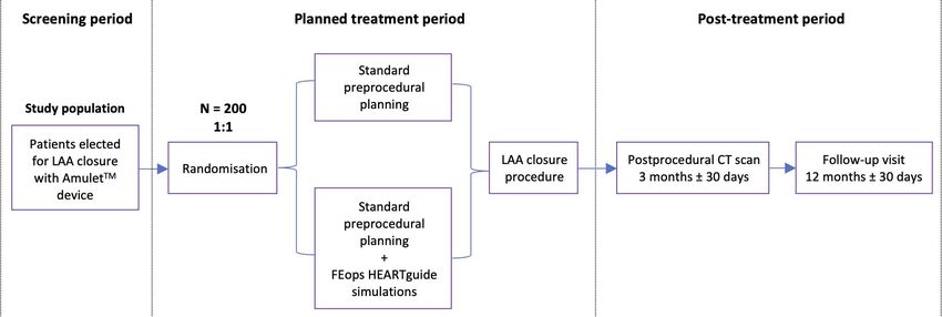
Open Heart
between device and the patient’s anatomy and provides Randomisation and treatment protocol
Open Heart: first published as 10.1136/openhrt-2020-001326 on 6 August 2020. Downloaded from http://openheart.bmj.com/ on July 13, 2021 by guest. Protected by copyright.
a deeper insight into the behaviour of the LAA closure Consenting subjects will be randomised in a 1:1 ratio to
device before the procedure. The FEops HEARTguide a standard treatment arm or computational simulation
simulation technology (FEops NV, Ghent, Belgium) has arm. Randomisation will be performed through a secure
been validated for LAA closure6 as well as for transcath- web-based REDCap application at the time of inclusion of
eter aortic valve replacement procedures.7 8 the patient in the trial. The randomisation will be strati-
The PREDICT- LAA clinical study investigates the fied by site. The study design is shown in figure 1. Patients
hypothesis that a better preprocedural planning can be randomised to the standard treatment arm will be treated
achieved by consulting FEops HEARTguide, simulating according to the site’s routine practice—as preproce-
different LAA closure device sizes and positions in a dural imaging, a CCT scan has to be performed; this can
patient-specific LAA anatomy and thereby providing the also be complemented with TEE at the discretion of the
implanter an overview of possible optimal and subop- operator. For patients randomised to the computational
timal scenarios. This better preprocedural planning may simulation arm, the planning of the LAA closure will be
result in higher rates of complete LAA closure with lower performed according to the standard practice of the site
rates of DRT as assessed on postprocedural CCT imaging. integrated with a careful review of the FEops HEART-
guide simulation results. The only prerequisite is that
the preprocedural CCT scan has to be uploaded into the
FEops HEARTguide platform in a pseudo-anonymised
Methods
fashion. The results of the computational simulation will
Study objectives
be provided to the operator within two working days,
The purpose of this trial is to study the possible added
containing a range of options in terms of device size and
value of FEops HEARTguide patient- specific compu- implant position in the selected patient (figure 2). The
tational simulations in the preprocedural planning of operator should use the computational simulations as
percutaneous LAA closure with the Amplatzer Amulet an additional preprocedural planning tool, aiming for a
device, with focus on procedural safety and efficiency as 10% to 25% compression of the Amulet lobe, complete
well as on clinical outcomes. LAA closure with full coverage of all trabeculations and
a concave-shaped disc that is not retracted into the LAA
Study population and patient selection neck (online supplementary video 1). The preproce-
Two hundred patients with NVAF and eligible for LAA dural planning should be performed integrating the
closure with an Amplatzer Amulet LAA closure device standard planning with the analysis of the patient-specific
will be enrolled at up to 12 European and Canadian sites. computational results. For all patients, the LAA closure
Only patients referred to and approved for percutaneous procedure should be performed following the routine
LAA closure—according to local practice and legisla- practice of the site with TEE, micro-TEE or intracardiac
tion—can be considered for enrolment in the trial. All echocardiography guidance, either in general or local
patients should be 18 years or older and should sign a anaesthesia. All patients should receive postprocedural
written informed consent. Key exclusion criteria are a antithrombotic medical therapy according to the partici-
reduced renal function (with glomerular filtration rate pating sites’ routine practice, which is at the discretion ofProtocol
Open Heart: first published as 10.1136/openhrt-2020-001326 on 6 August 2020. Downloaded from http://openheart.bmj.com/ on July 13, 2021 by guest. Protected by copyright.
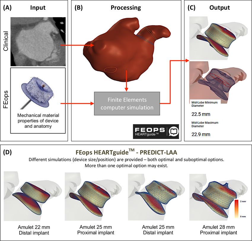
Figure 2 FEops HEARTguide workflow and simulations. (Panel A) The preprocedural CT scan of the selected patient is
uploaded on the web-based platform FEops HEARTguide and is—together with the in-house computational model of the
Amulet device with adequate material properties for device and anatomy—used as input to the workflow. (Panel B) The
received images are further processed to extract three-dimensional patient-specific anatomical reconstructions and landmarks
for the procedure. This, in combination with the device model, serves as input for the computational finite element analysis.
(Panel C) As output, several options in terms of device size and position are provided, including LAA wall apposition plots
(colour-scale indicating the distance between the device and the anatomy), deformation visualisation and measurements (at
the section indicated in red in panel C). (Panel D) The use of the simulation output in clinical practice: for one single patient,
different simulations in terms of device size and position are provided to the operator, who can gain additional insights on the
device–host interaction before the procedure.
and device position. All patients enrolled in the study endpoints evaluated in this study are listed in box 1. The
should also have a clinical follow-up visit at 12 months CCT CoreLab evaluation will be performed at Rigshospi-
after the procedure to record possible thromboembolic talet, Copenhagen, Denmark9; the readers of the CCT scan
events or all-cause mortality (table 1A). The list of medical will be blinded from the baseline patient and procedural
investigations required is shown in table 1B. data as well as from patient’s randomisation arm.
Study endpoints Sample size calculation
The primary endpoint of the PREDICT-LAA study is the The primary endpoint is expected to occur in 30% of
percentage of patients with incomplete LAA closure patients in the standard treatment arm. An equal number
(defined as any remaining contrast leakage into the LAA of patients will be enrolled in both treatment arms. A 65%
distal of the Amulet lobe) and/or a definite DRT at post- reduction of the primary endpoint requires inclusion of 174
procedural CCT imaging at 3 months after the procedure. patients in order to demonstrate superiority (power 0.8, α
Definite DRT is defined as ‘high-grade’ hypoattenuating 0.05). Taking into account an estimated loss at follow-up of
thickening at the atrial surface of the closure device— 10% to 15% of patients (due to mortality and inconclusive
as previously described by Korsholm et al.9 Secondary CCT imaging at 3 months postprocedure), the total sample
Garot P, et al. Open Heart 2020;7:e001326. doi:10.1136/openhrt-2020-001326 3Open Heart
Open Heart: first published as 10.1136/openhrt-2020-001326 on 6 August 2020. Downloaded from http://openheart.bmj.com/ on July 13, 2021 by guest. Protected by copyright.
Table 1A Schedule for patients enrolled in PREDICT-LAA study
Standard treatment arm Computational simulation arm
Preprocedural CT scan Standard Upload images to FEops HEARTguide
Preprocedural planning Standard Standard+FEops HEARTguide results
LAA closure procedure Standard Standard
Antithrombotic therapy Standard Standard
Postprocedural CT scan At 3 months±30 days At 3 months±30 days
Follow-up visit At 12 months±30 days At 12 months±30 days
Table 1B Overview of investigations required for patients enrolled in the study
Preprocedure At 3 months At 12 months
Routine medical check Yes Yes Yes
12-Lead ECG Yes Yes Yes
Transthoracic echocardiography Yes Yes No
Cardiac CT scan Yes Yes No
size needed to demonstrate superiority has been calculated (Denmark) and Institut Cardiovasculaire Paris Sud,
to be 200 patients. The primary endpoint will be analysed Massy, Paris (France). The study will be conducted at up
using Fisher’s exact test or a χ2 test, as required. Data/statis- to 12 European and Canadian sites in compliance with
tical analysis will be performed according to the ‘intention- the Declaration of Helsinki, International Conference
to-treat’ principle as a first approach and ‘as-treated’ as a on Harmonization, Good Clinical Practice Guidelines
second approach. and applicable regulatory requirements. The first patient
was enrolled in the study on 14 January 2020 and enrol-
Study organisation ment of all 200 patients is expected being completed
PREDICT- LAA is an investigator- initiated, prospec- in December 2021 with the last patient coming for the
tive, international, randomised clinical trial that will be 1 year follow-up visit in December 2022. Five patients
executed under the academic leadership of investiga- were enrolled as of the date of manuscript submission.
tors at Rigshospitalet University Hospital, Copenhagen The final study protocol and informed consent have
been reviewed and approved by the local ethics boards,
institutional review boards and corresponding health
Box 1 PREDICT-LAA study endpoints
authorities of all participating sites. Rigshospitalet takes
Primary endpoint the sponsor role in this clinical trial. The study is funded
►► Incomplete closure of the LAA with remaining contrast leakage by Abbott (Minneapolis, Minnesota, USA) and FEops
into the LAA distal of the Amulet lobe and/or presence of a defi- NV (Ghent, Belgium). The academic sponsor will have
nite device-related thrombus as assessed at postprocedural CCT at full access to the trial data and will submit the results for
3 months after LAA closure. publication in a peer-reviewed medical journal.
Secondary endpoints
►► Number of LAA closure devices used per procedure.
►► Number of LAA closure device repositionings per procedure. Conclusion
Repositioning is defined as full deployment of the Amulet lobe in the The PREDICT-LAA clinical trial is the first randomised
LAA, followed by either full or partial recapture and redeployment clinical trial studying the efficacy of the preprocedural
of the lobe. planning for percutaneous LAA closure, comparing a
►► Procedural time, radiation exposure and amount of contrast medi- standard approach with a preprocedural planning that
um used per procedure. integrates patient- specific computational simulations.
►► Procedure-related complications encompassing device embolisa- Hence, the PREDICT-LAA study will provide, as a first,
tion, pericardial effusion requiring intervention, procedure-related randomised data on the possible added value of patient-
stroke and procedure-related death within 7 days of the procedure.
specific computational simulations in the preprocedural
►► Different degrees of contrast leakage into the LAA.
planning for LAA closure.
►► Full coverage of all LAA trabeculations by the Amulet device and
a concave-shaped disc without retraction of the disc into the LAA
Author affiliations
neck as assessed at postprocedural CCT scan. 1
Department of Cardiology, Institut Cardiovasculaire Paris Sud, Massy, Île-de-
►► Composite of all-cause death and thromboembolic event (transient France, France
ischaemic attack, ischaemic stroke or systemic embolism) at 12 2
Pediatric and Congenital Cardiology, University Hospital of Bordeaux, Pessac, MS,
months after randomisation. France
3
Department of Cardiology, Centre Hospitalier Universitaire de Charleroi, Charleroi,
CCT, cardiac computed tomography; LAA, left atrial appendage.
Hainaut, Belgium
4 Garot P, et al. Open Heart 2020;7:e001326. doi:10.1136/openhrt-2020-001326Protocol
4
Division of Cardiology, Cliniques Universitaires Saint-Luc, Brussels, Belgium ORCID iD
Open Heart: first published as 10.1136/openhrt-2020-001326 on 6 August 2020. Downloaded from http://openheart.bmj.com/ on July 13, 2021 by guest. Protected by copyright.
5
Cardiovascular Institute, Hospital Clinic de Barcelona, Barcelona, Catalunya, Spain Ole De Backer http://orcid.org/0000-0002-9674-0278
6
Department of Cardiology, Hospital Clínico Universitario de Salamanca, Salamanca,
Spain
7
Cardiology Unit, Fondazione CNR Regione Toscana, Massa, Italy References
8
Department of Cardiology, University Hospital Galway, Galway, Ireland 1 Reddy VY, Sievert H, Halperin J, et al. Percutaneous left atrial
9 appendage closure vs warfarin for atrial fibrillation: a randomized
Department of Cardiology, Montreal Heart Institute, Montreal, Quebec, Canada
10 clinical trial. JAMA 2014;312:1988–98.
Department of Cardiology, Aarhus Universitetshospital Skejby, Aarhus, Denmark 2 De Backer O, Arnous S, Ihlemann N, et al. Percutaneous left atrial
11
Department of Cardiology, Sahlgrenska University Hospital, Goteborg, Sweden appendage occlusion for stroke prevention in atrial fibrillation: an
12
Department of Cardiology, Vancouver General Hospital, Vancouver, British update. Open Heart 2014;1:e000020.
Columbia, Canada 3 Korsholm K, Berti S, Iriart X, et al. Expert Recommendations on
13 Cardiac Computed Tomography for Planning Transcatheter Left Atrial
Department of Cardiology, Rigshospitalet, Copenhagen, Denmark
Appendage Occlusion. JACC Cardiovasc Interv 2020;13:277–92.
4 Otton JM, Spina R, Sulas R, et al. Left atrial appendage closure
Contributors PG and ODB designed the study and prepared the first draft of the guided by personalized 3D-printed cardiac reconstruction. JACC
manuscript. XI, AA, JK, XF, ICG, SB, LR, RI, KK, JO, JENK, JS and LS contributed to Cardiovasc Interv 2015;8:1004–6.
manuscript revision. All authors will contribute to enrolment and patient treatment 5 Ciobotaru V, Combes N, Martin CA, et al. Left atrial appendage
occlusion simulation based on three-dimensional printing:
in the study.
new insights into outcome and technique. EuroIntervention
Funding The study is funded by Abbott (Minneapolis, MN, USA) and FEops NV 2018;14:176–84.
(Ghent, Belgium). 6 Bavo AM, Wilkins BT, Garot P, et al. Validation of a computational
model aiming to optimize preprocedural planning in percutaneous
Competing interests ODB and LS have received institutional research grants from left atrial appendage closure. J Cardiovasc Comput Tomogr
Abbott and FEops NV. 2020;14:149–54.
7 de Jaegere P, Rocatello G, Prendergast BD, et al. Patient-Specific
Patient consent for publication Not required.
computer simulation for transcatheter cardiac interventions: what a
Provenance and peer review Not commissioned; externally peer reviewed. clinician needs to know. Heart 2019;105:s21–7.
8 Rocatello G, El Faquir N, De Santis G, et al. Patient-Specific
Data availability statement Data may be obtained from a third party and are not computer simulation to elucidate the role of contact pressure in the
publicly available. This is a study protocol, not containing study results/data yet. development of new conduction abnormalities after Catheter-Based
Open access This is an open access article distributed in accordance with the implantation of a self-expanding aortic valve. Circ Cardiovasc Interv
2018;11:e005344.
Creative Commons Attribution Non Commercial (CC BY-NC 4.0) license, which
9 Korsholm K, Jensen JM, Nørgaard BL, et al. Detection of device-
permits others to distribute, remix, adapt, build upon this work non-commercially, related thrombosis following left atrial appendage occlusion:
and license their derivative works on different terms, provided the original work is a comparison between cardiac computed tomography and
properly cited, appropriate credit is given, any changes made indicated, and the use transesophageal echocardiography. Circ Cardiovasc Interv
is non-commercial. See: http://creativecommons.org/licenses/by-nc/4.0/. 2019;12:e008112.
Garot P, et al. Open Heart 2020;7:e001326. doi:10.1136/openhrt-2020-001326 5You can also read