Clinical efficacy of Invisalign treatment with weekly aligner changes: Two case reports
←
→
Page content transcription
If your browser does not render page correctly, please read the page content below
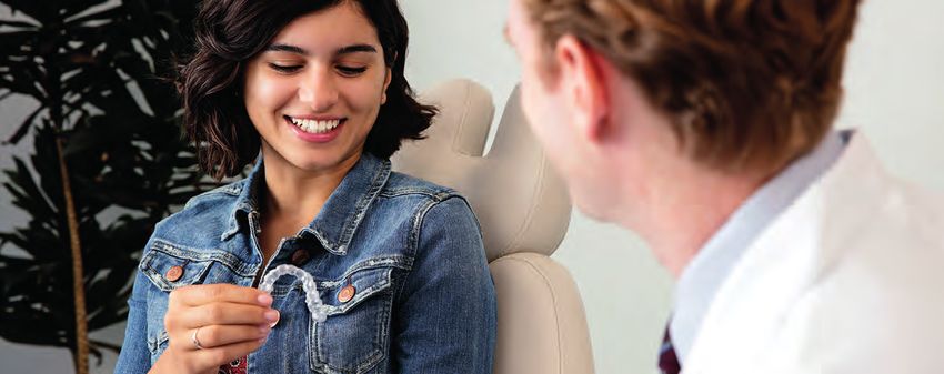
Growing your practice with Invisalign®

     Clinical efficacy of Invisalign® treatment with weekly
     aligner changes: Two case reports
     Class II and deep bite correction with the Invisalign® System and weekly aligner changes. Dr Schupp and Dr Haubrich
     Treating a teenage patient with deep bite, increased overbite and impacted first premolar with the Invisalign® System
     and weekly aligner changes. Dr Castroflorio

Summary
Background and objective: The efficacy of using Invisalign® treatment to achieve
major tooth movements and successfully treat a variety of complex malocclusions
in adults and teenagers is documented in the published literature.1–6 Align
Technology now recommends orthodontists prescribe weekly aligner changes                                                  Dr Werner Schupp
in their Invisalign® treatments; this may reduce treatment times by up to 50%                                              DDS, Ortho Spec.
compared with changes every 2 weeks.7 This recommendation is based on clinical                                          Prof. Capital Medical
analysis of more than 200 ongoing Invisalign® cases.8                                                                      University Beijing

The objective of these case reports is to describe the use of the Invisalign®
System with weekly aligner changes in an adult patient and a teenage patient; then
to show the clinical efficacy and impact of the new 1-week wear recommendation
in these cases.

Methodology: For the first case in this document, Drs Werner Schupp and                                                    Dr Julia Haubrich
Julia Haubrich provide detailed accounts of the procedures undertaken in                                                   DDS, Ortho Spec.
treating an adult patient with craniomandibular disorder (CMD) issues, Class II
division 2 relationship and deep bite, with Invisalign® aligners and weekly aligner
changes. In the second case report, Dr Castroflorio describes the treatment
details of a teenage patient presenting with deep bite, increased overjet and an
impacted first premolar, prescribed Invisalign® treatment with 1-week aligner
wear. The authors provide clinical tips on how to optimise the treatment outcomes                                               Dr Tommaso
of the presented cases and comment on the likely impact of the weekly aligner                                                   Castroflorio
changes compared with the previous recommendation of changing aligners every                                           DDS, Ortho Spec., PhD
2 weeks.

The opinions expressed in this White Paper are those of the author(s) and may not reflect those of Align Technology.                            01
The authors were paid an honorarium by Align Technology in connection with the White Paper.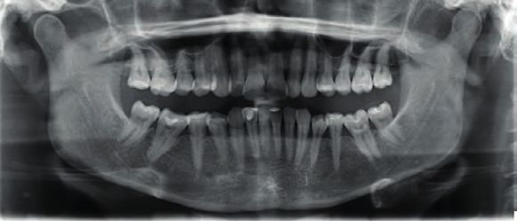
Growing your practice with Invisalign®

Results: These cases show the clinical efficacy of the Invisalign® System with weekly aligner changes in both a teenager and an adult
patient (age 37) with complex malocclusions. All treatment goals were achieved in both patients, with similar results to those that
would be expected from a 2-week wear treatment, and good tooth movement control as shown by the good aligner fit during the
whole treatment. The reduced wearing time led to highly motivated patients, which was perceived as a major benefit – especially for
teenage patients. The combination of Invisalign® aligners with auxiliaries such as buttons and elastics is shown as a fully compatible
treatment approach to accomplish very complex tooth movements, such as orthodontic traction of impacted premolars.

Conclusions: The doctors all concluded that, for these cases, weekly aligner changes provided the same results in up to half the
time that would have been anticipated for these patients had they been making aligner changes every 2 weeks. They highlighted
major benefits of weekly aligner changes, including high acceptability to patients of reduced treatment time, and a reduction in office
management costs.

References
1.	Schupp W and Haubrich J, eds. Aligner Orthodontics. 2015,                                  7.	Weekly aligner changes are recommended for all Invisalign
    Quintessenz Berlin.                                                                            treatments (with default staging protocol) for Invisalign Full,
2.	Boyd RL, Oh H, Fallah M, Vlaskalic V. An update on present                                     Invisalign Teen and Invisalign Assist products. The decision to
    and future considerations of aligners. J Calif Dent Assoc                                      prescribe weekly aligner changes is at the doctor’s discretion.
    2006;34(10):793–805.                                                                           Monitor tooth movements such as rotations, extrusions,
                                                                                                   and significant root movements; particularly blue and black
3.	Giancotti A, Mampieri G, Greco M. Correction of deep bite in
                                                                                                   movements in the Tooth Movement Assessment (TMA).
    adults using the Invisalign® system. JCO 2008;XLII(12):719–26.
                                                                                                   Depending on the patient response to treatment, particularly
4.	Boyd RL. Esthetic orthodontic treatment using the Invisalign®                                  mature adults, consider longer periods between aligner changes.
    appliance for moderate to complex malocclusions. J Dental                                      See: Align Technology introduces one-week aligner wear for
    Educ 2008;72(8):948–67.                                                                        Invisalign® Teen and Full products. Press release, 11 October
5.	Krieger E, Seiferth J, Marinello I, et al. Invisalign® treatment                               2016. Available at: http://investor.aligntech.com/releasedetail.
    in the anterior region: were the predicted tooth movements                                     cfm?ReleaseID=992964. Accessed 31 December 2016.
    achieved? J Orofac Orthop 2012;73(5):365–76.                                               8.	Align Technology, data on file.
6.	Simon M, Keilig L, Schwarze J, Jung BA, Bourauel C.
    Treatment outcome and efficacy of an aligner technique
    – regarding incisor torque, premolar derotation and molar
    distalization. BMC Oral Health 2014;14:68.

The opinions expressed in this White Paper are those of the author(s) and may not reflect those of Align Technology.                                              02
The authors were paid an honorarium by Align Technology in connection with the White Paper.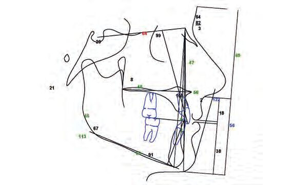
Growing your practice with Invisalign®
Case 1: Class II and deep bite correction with the
Invisalign® System and weekly aligner changes.
Dr Schupp and Dr Haubrich
Sex: Female Treatment goals
Age: 54 years To correct Class II
Chief complaint: The patient was in severe pain due to To improve deep bite
craniomandibular disorder (CMD) To resolve crowding
To end with a 0.5 mm overjet (Shimstock foil open incisor area)
The patient presented to the clinic with pain and CMD. She had To end with canine guidance without hyperbalance contacts.
previously been treated with a removable splint. Initially, the splint
was removed and, after obtaining a pain-free position, initial Treatment plan
records for Invisalign® treatment were taken. Distalization in the upper arch with Class II elastics
Torque on upper central incisors
Clinical findings
Derotation of upper laterals
CMD issues
Intrusion of lower anteriors and extrusion of lower premolars
Class II division 2 relationship to reduce the dental deep bite
Crowding and rotation in the upper and lower arch Interproximal reduction and distalization to resolve crowding.
Retruded teeth: 11, 21 with pre-existing contact points on
upper anteriors Treatment details
Protruded and rotated: 12, 22 Total treatment time
Deep bite with severe incisor contacts. – 15 months.
Number of aligners
FIGURE 1. Intraoral and extraoral images before treatment – 49 + 10
A total of 59 aligners were prescribed (49 aligners Phase 1,
–
10 aligners Phase 2), with a change of aligner every 7 days.
Attachments
– Attachments were bonded prior to scans on teeth 13, 23, 33,
34, 35, 43, 44, 45
– Vertical rectangular attachments were bonded on teeth
13 and 23, hooks for Class II elastics were also placed on
the gingival region of these teeth. In patients with hooks on
canines, we also bond attachments on these teeth to avoid
undesired rotations or angulations due to the elastic force
– In the lower arch, the patient had horizontal ellipsoid/
bevelled shaped attachments to secure anchorage on the
lower premolars and canines for intrusion and alignment of
the lower anteriors.
Interproximal reduction
– Interproximal reduction was performed during the first
FIGURE 2. Panoramic radiograph before treatment phase on the upper-right and lower anteriors, as well as on
the upper anteriors in the second phase to aid alignment.
Use of auxiliaries
Class II elastics from hooks on the upper canines to buttons
–
on the lower molars (36, 46). The patient was instructed to
wear elastics at night, and for 2–3 hours during the day for
anchorage during the distalization in the upper arch.
Retention
After two phases of Invisalign® treatment, retention was
–
ensured with a removable retainer in the upper arch for
night-time wear; in the lower arch, a fixed lingual retainer
was bonded from first premolar to first premolar.
The opinions expressed in this White Paper are those of the author(s) and may not reflect those of Align Technology. 03
The authors were paid an honorarium by Align Technology in connection with the White Paper.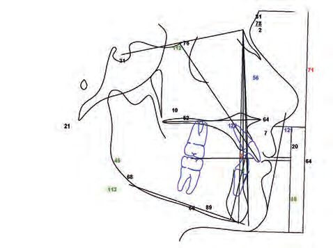
Growing your practice with Invisalign®

FIGURE 3. Intraoral images of aligner 19 in situ, showing a good fit                          FIGURE 4. Intraoral and extraoral images after treatment

Clinical tips
The aligner fitting was documented
and controlled in appointments every
7–10 weeks. Two scans were performed                             Impact of weekly aligner changes on clinical practice
                                                                 Long treatment periods with complex movements, such as
during Weeks 10 and 19 to monitor progress, distalization of upper molars, can be time consuming and may
                                                                 lead to reduced patient motivation.
and a final scan was performed at the end
of the treatment.                                                Reducing the aligner wear time to
During treatment, the aligners showed a perfect fit on all teeth 7 days instead of 14 can reduce the
except a small discrepancy on teeth 12, 22 as shown in the
intraoral pictures with aligners in situ at Stage 19 (FIGURE 3). overall treatment time by up to 50%.
Teeth 12, 22 demonstrated good aligner fit during the later
aligner numbers.
                                                                 We found that, in this patient, there was
A 22-hour wearing time is crucial in complex aligner treatments.
                                                                 a higher motivation to comply with the
                                                                 Invisalign® System.
Treatment outcome
Following treatment with 59 aligners over a 15-month period,                                  Conclusion
the planned treatment goal of the distalization of the upper arch                             The patient was suffering from CMD presenting a Class II
was achieved.                                                                                 division 2 relationship with pre-existing contacts on anteriors
                                                                                              and deep bite. Following removable splint therapy, orthodontic
The patient showed a Class I relationship with aligned arches in                              treatment with Invisalign® aligners with weekly aligner changes
the upper and lower anteriors, with physiological canine guidance                             was initiated to solve the pre-existing anterior contacts and
and incisor relationship (FIGURE 4). The patient was pain-free                                distalize the upper arch into a full Class I relationship, with
and showed no further signs of CMD.                                                           torque of the upper retruded central incisors and alignment
                                                                                              of the arches.

Comparison of this case with patients prescribed aligner
changes every 2 weeks                                                                         All treatment goals were achieved
Distalization with the Invisalign® System is highly predictable using                         and good aligner fit was documented
additional anchorage. However, due to the sequential distalization
protocol of the ClinCheck® Software, this can result in a high                                during the whole treatment. The patient
number of aligners; in this case 59. With an aligner change every
2 weeks, the overall treatment time in this case would have been                              did not report any issues with the faster
29 months, with a potential additional phase for refinement.
Changing the aligners every 7 days was successful for this
                                                                                              aligner change. No temporomandibular
patient, with no reported problems; there was a good aligner fit                              joint issues, muscle pain, headache or
during the whole treatment. In addition, a minimal second phase
of only 10 aligners was required for finishing in detail.                                     back pain were reported.

The opinions expressed in this White Paper are those of the author(s) and may not reflect those of Align Technology.                                            04
The authors were paid an honorarium by Align Technology in connection with the White Paper.
Growing your practice with Invisalign®
Case 2: Treating a teenage patient with deep bite,
increased overbite and impacted first premolar with
the Invisalign® System and weekly aligner changes.
Dr Castroflorio
Sex: Female FIGURE 5. Intraoral and extraoral images before treatment
Age: 12.9 years
Chief complaint: Missing lower-right first premolar and
spacing in the upper arch
Clinical findings
Clinical examination revealed a dental Class I malocclusion
with increased overjet due to proclination of the upper incisors.
Deep bite related to an increased Curve of Spee with extrusion
of the lower incisors. Furthermore, there was extrusion of the
upper-right premolars. Both the arches were constricted on
the frontal plane and the upper molars were rotated mesially.
A dento-dental discrepancy was also detected with a mandibular
excess of about 1 mm.
The radiographic examination showed a skeletal Class I
malocclusion in a normodivergent patient and a circular, FIGURE 6. Panoramic radiograph before treatment
well-defined unilocular radiolucent area (follicular cyst)
surrounding the crown of the mandibular right first premolar
without tooth displacement (FIGURES 5–7).
Treatment goals
T
o remove the follicular cyst and bring the lower-right first
premolar into the arch
To close the upper spacing
To correct the proclination of the upper incisors
To correct the Curve of Spee
To increase orthopaedic stability.
FIGURE 7. Pre-treatment tracing
Treatment plan
1. First Invisalign® treatment phase
An initial Invisalign® treatment phase was conducted to
expand the arches on the frontal plane and derotate the upper
first molars. When the upper molars were in their final position,
the intrusion of the upper-right premolars was completed.
When the premolars were in their final position, en-masse
retraction of the upper canines and incisors was conducted.
The lower incisors and second molars were intruded to flatten
the Curve of Spee.
The opinions expressed in this White Paper are those of the author(s) and may not reflect those of Align Technology. 05
The authors were paid an honorarium by Align Technology in connection with the White Paper.
Growing your practice with Invisalign®
FIGURE 8. Buttons and elastics in place with Invisalign® aligners vertical attachments were bonded to the upper canines and
during treatment the lower first molars
Optimised attachments were located on the premolars and
–
the lower canines
In Phase 2, conventional and optimised attachments were
–
located on the same teeth as in Phase 1 and were used for
finishing purposes.
Other features
During the first treatment phase, Precision Bite ramps
–
were used on the upper incisors to facilitate the posterior
disclusion for levelling the Curve of Spee and, thus, the
expansion of the arches and derotation of posterior teeth
Pressure areas and Power Ridge features were located on
–
the lower incisors to control the intrusion movement and
the lingual root torque of those teeth. Precision cuts were
applied to use Class II elastics (1/4” 4 to 1/2 oz.) to facilitate
the en-masse retraction of the upper incisors.
Retention
Retention was provided with Vivera® retainers.
–
Treatment outcome
2. Surgical removal of follicular cyst and traction of implanted premolar The case was finished in canine and molar Class I relationship,
After the upper premolars were intruded, the lower follicular with functional overbite and overjet. The impacted lower-right
cyst was surgically removed and, once the tooth had been first premolar was tractioned into the arch without braces or
isolated, a gold chain was bonded to the coronal aspect. archwires. The chain was anchored to the aligner with 1/4”
The chain emerged through the incision at the mid-crestal 4.5 oz. elastics. The aligners were modified with clear aligner
region and an elastic traction was fixed to the lower aligner. pliers to create hooks to anchor the elastics. The patient was
When the tooth was partially extruded and the chain was no instructed to change the elastics at least three times per day
longer useful, it was removed and a button was bonded on the in order to maintain a constant force on the impacted premolar.
buccal aspect of the lower first premolar and upper premolars. Once the occlusal third of the buccal surface was erupted,
Aligners were modified to receive buttons and a 3/16” 3 oz. buttons were placed on the buccal aspects of the lower impacted
elastic was used with a triangle geometry to complete the premolar and the upper premolars in order to use elastics to
extrusion movement (FIGURE 8). complete the guided eruption of the lower premolar (3/16”,
3 oz.). Aligners were modified using clear aligner pliers to allow
3. Second Invisalign® treatment phase the positioning of the buttons. A good final intercuspation was
achieved to guarantee orthopaedic stability. Furthermore, the
The second and final treatment phase started when the smile was improved considerably with perfect control of the
lower first premolar was fully extruded and was conducted buccolingual inclination of the upper and lower incisors, as
to complete the en-masse retraction of the upper incisors demonstrated by the cephalometric analysis (FIGURES 9–11).
and the alignment of the lower canines and incisors.
FIGURE 9. Intraoral and extraoral images after treatment
Treatment details
Total treatment time
– 54 weeks
The orthodontic traction of the impacted lower-right first
–
premolar tooth lasted for 6 months.
Number of aligners
– 32 + 18
The patient was instructed to wear the aligner for at least
–
21 hours per day
For this patient, the first two stages were prescribed for
–
longer wear time with close monitoring to assess tooth
tracking and patient compliance. From the third aligner,
a regimen of weekly aligner changes was adopted.
Attachments
– Rectangular and horizontal attachments were bonded to the
upper molars and the lower second molars. Rectangular and
The opinions expressed in this White Paper are those of the author(s) and may not reflect those of Align Technology. 06
The authors were paid an honorarium by Align Technology in connection with the White Paper.Growing your practice with Invisalign®
FIGURE 10. Panoramic radiograph after treatment
Comparison of this case with patients prescribed aligner
changes every 2 weeks
A regimen of aligner changes every 2 weeks could provide similar
results but would extend the treatment time by up to twice the
duration. Furthermore, the orthodontic traction of impacted
teeth can be extremely variable in terms of required time. This
is the main reason why, in cases where no complex movements
are required to prepare the traction, weekly aligner changes are
recommended, in order to avoid a long treatment duration and to
bring the impacted tooth into its final position.
Impact of weekly aligner changes on clinical practice
FIGURE 11. Post-treatment tracing The major benefits of weekly aligner
changes in teenage patients are the higher
acceptability of the shorter treatment time to
these patients, as well as reduced time over
which management costs for the treatment
are incurred by the practice.
Conclusions
This case was treated with 50 aligners over a period of 54 weeks.
A further 24 weeks was needed to complete the impacted lower
first premolar traction. Total treatment time was therefore
19.5 months. A longer treatment time for a regimen of aligner
changes every 2 weeks would be expected, in the region of
39 months. This estimated difference in treatment time is
important from both the patient’s and clinician’s perspective.
Clinical tips
The use of attachments on all premolars and molars is useful to
control the maxillary expansion during buccal movement, which
releases an extrusive force on those attachments. The result
is the creation of a couple of forces generating a movement,
thereby facilitating the buccolingual control of premolars and
molars. In other words, a controlled buccal movement of their
roots can be achieved. In addition, the extrusive force will improve
the final intercuspation in the premolar and molar areas, reducing
the bite-block effect described for aligner orthodontics.
This case shows how it is possible to
perform the orthosurgical traction of an
impacted lower premolar with aligners,
buttons and elastics, without braces and
wires. This was greatly appreciated by the
patient and the parents. Comfortable and
aesthetic solutions can be a fundamental
requirement for teenagers.
The opinions expressed in this White Paper are those of the author(s) and may not reflect those of Align Technology. 07
The authors were paid an honorarium by Align Technology in connection with the White Paper.Dr Werner Schupp graduated in Dentistry in 1985 from the University of Münster, Münster, Germany, and continued
his studies there as a postgraduate student of Orthodontics. Since 1990, he has been in private practice as an
orthodontic specialist in Cologne, Germany. He is certified in Invisalign® treatments and in Manual Medicine and
Osteopathy for Orthodontics. Dr Schupp is a foundation member and Past President of the German Board of
Orthodontics and Orofacial Orthopedics and board member of the German Society for Aligner Orthodontics.
He has authored several articles and two books concerning orthodontics, aligners, function and pain therapy.
He has lectured in Europe, Brazil, the USA, China, Taiwan and Japan. Dr Schupp is visiting professor at the
Capital University, Beijing, China.
Dr Julia Haubrich studied Dentistry at the University of Freiburg, Freiburg, Germany, in 2002 and was a
postgraduate student in Orthodontics from 2003–2005. She then studied as a postgraduate student in
Orthodontics at the University of Berlin, Berlin, Germany, and became a certified specialist of Orthodontics in
2007. Since then, she has been working in private practice with Dr Werner Schupp. She has authored several
publications and two books concerning orthodontics, aligners, function and pain therapy. Dr Haubrich is a
founding member and Conference President of the German Society for Aligner Orthodontics and has spoken
in both Europe and Asia. She is currently a lecturer at the Medical University of Innsbruck, Innsbruck, Austria.
Dr Tommaso Castroflorio is an orthodontist living and working in Torino, Italy. He obtained his DDS degree
and postgraduate degree in Orthodontics from the University of Torino, and a PhD in Human Anatomy from
the University of Milan, Milan, Italy. Since 2008, Dr Castroflorio has run a private practice in Torino where he
has focused his work on orthodontics and temporomandibular disorders. He started to work with aligners
in 2007. Since 2012, he has been an adjunct professor of the Postgraduate School of Orthodontics at the
University of Torino where he teaches “Orthodontics with Thermoplastic Aligners”. He is a founding member
and the Scientific Chairman of the European Aligner Society. Since 2011, he has lectured in Europe, South
America and Japan.
Author disclosures
Drs Haubrich and Schupp are both clinical speakers for Align Technology BV.
Dr Castroflorio is a member of the European Advisory Board of Align Technology and he has given lectures and contributed
to courses for Align Technology since 2011. In 2015, he received research funds from Align Technology.
Align Technology BV
Arlandaweg 161,
1043 HS Amsterdam
The Netherlands
Other (English): +31 (0)20 586 3615
www.invisalign.eu
© 2017 Align Technology (BV). All Rights Reserved.
Invisalign®, ClinCheck® and SmartTrack®, among others,
are trademarks and/or service marks of Align Technology, Inc.
or one of its subsidiaries or affiliated companies and may be
registered in the U.S. and/or other countries.
200961 Rev AYou can also read