Guide to 4-week post-operative cataract follow-up by community optometrists - LOC Online
←
→
Page content transcription
If your browser does not render page correctly, please read the page content below
Guide to 4-week post-operative cataract follow-up by community optometrists
Contents
Introduction _______________________________________________________________________ 4
Purpose of follow-up __________________________________________________________ 4
Accreditation process _________________________________________________________ 5
Clinical procedure for post-op cataract follow-up ________________ 5
Annex A-G __________________________________________________________________________ 9
A National Cataract Audit minimum dataset
(as recommended by RCOphth)
B Conditions associated with higher surgical risk
C Instilling Eye Drops – College of Optometrists
D SUN Grading scheme for Anterior Chamber Cells and Flare
E Reference guide for post-op cataract complications
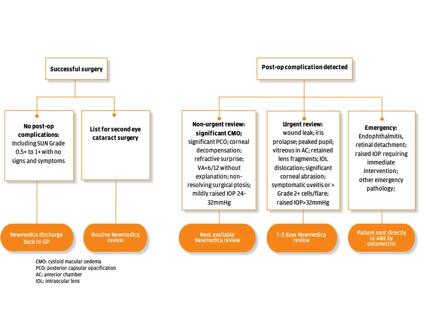
F Post-op follow-up outcome pathways
G Further informationIntroduction
After routine cataract surgery, all patients need to have a post-operative
follow-up appointment at around 4 weeks following surgery to assess the
final outcome once the eye has fully healed. It is generally acknowledged
that this period coincides with the stabilisation of the new refractive status
of the post-operative eye. Since optometrists assess both the clinical and
refractive status of patients as part of an eye examination, utilising these
skills in the community setting rather than in an ophthalmological setting
makes logical sense. By linking the delivery of these post-operative checks
with the practice which initiated the referral, a circle of continuous care is
created. This also allows suitably accredited optometrists a greater degree
of involvement in their patients’ care and enhanced use of core skills. The
post-operative follow-up is also an ideal time to consider if a second eye
operation is applicable for individual patients.
In order to maintain high standards of patient care throughout the
cataract pathway, this guide provides optometrists with best practice
standards for post-operative follow-up in the community, as mandated
by the Newmedica Medical Advisory Committee, alongside a guide to
the more common and important potential post-operative complications
which may be encountered.
Purpose of the follow-up
The role of the post-operative follow-up appointment is to determine the
visual outcome in the operated eye, to undertake a specific examination of
the eye to rule out post-operative complications, and to act on the findings
appropriately. The opportunity to re-assess the post-surgery refractive
status may also be realised through a General Ophthalmic Service
(GOS) sight test, which in turn, will herald the essential information for
National Cataract Audit dataset to be returned to the provider (see Annex
A). Suitable discussion around appropriate refractive correction and
consideration of second eye surgery would be expected to follow.
At the time of booking the follow-up, ensure that the patient is aware that
pupil dilation of the operated eye is an essential part of the assessment
and will therefore impact their ability to drive following the appointment.
4Accreditation process
Once optometry practices have satisfied the PEC/LOC criteria to be sub-
contracted for enhanced services, optometrists wishing to participate in the
service must evidence that they have achieved the following:
Safeguarding Newmedica
WOPEC
Adult & Children Cataract live
Cataract Level 1
Level 2 CET event
Contact your local Newmedica clinic for information about the next live CET
event in your area. For those who cannot attend a live event, an online lecture
is available (details on request).
Clinical procedure for post-op cataract follow-up
Note that this section specifies the post-operative focus of the follow-up and does
not stipulate all checks necessary to meet requirements of GOS 1 Sight Test.
Minimum examinations
• Interval H&S
• VA: unaided, aided and pinhole
• Accurate refraction
• IOP measure (GAT if >21mmHg)
• Slit lamp anterior segment
• Dilated BIO of posterior segment
Preparation
• Open the patient record and mentally note which eye has been operated
on, the level of pre-op Best Corrected Visual Acuity (BCVA) and whether
a good outcome was expected
• Practitioner should be aware of conditions which may have been
associated with a higher surgical risk (see Annex B)
• Perform appropriate infection control procedures
• Confirm the patient identity matches the patient record and they are
attending a post-operative cataract follow-up
5Interim history and symptoms
Ensure the following information is sought from the patient:
• Satisfaction with surgical outcome including vision
• Current ocular discomfort
• New floaters or flashes since surgery
• Pain or discomfort since surgery
• Compliant with drops since surgery
• Sensitivity to eye drops used since surgery
• Desire for second eye surgery if appropriate
• Any other particular problems with their eye(s)
Assess vision
• Unaided distance for operated eye, plus pinhole vision if worse than 6/9
• Habitual distance visual acuity of fellow eye
Accurate refraction
• Distance sphere, cylinder and axis of operated eye and fellow eye.
• Best corrected distance visual acuity of both eyes
Pupil assessment
• Confirm pupils are round, centred and are equal size in both eyes
to exclude peaked pupil
•
pupillary defect
6Intraocular pressure
• Measure and record IOP in both eyes with available tonometer
• Repeat with Goldmann tonometer if IOP>21mmHg
Dilation
• Perform the usual pre-dilation checks (see Annex C Instilling Eye
Drops by College of Optometrists) before dilating operated eye
Anterior segment examination with slit lamp
Perform a comprehensive slit-lamp assessment of the anterior eye,
including assessment of:
• Lids for ptosis or bruising
• Cornea for abrasion, striae or oedema
• Wound site for leak or dehiscence. Employ Seidel test particularly
if low IOP and shallow AC
• Anterior chamber contents for signs of inflammation, vitreous, lens
matter, nuclear fragments or foreign material. Grade cells and flare
if present (See Annex D)
• Iris for prolapse, distortion or surgical trauma
• Centration of IOL implant
• Capsule for anterior capsulophimosis (fibrosis) and posterior capsule
opacification
Posterior segment examination with slit-
lamp binocular indirect ophthalmoscopy (BIO)
Perform complete exam of the posterior segment including assessment of:
• Vitreous for pigment, blood or inflammatory cells
• Macula for cystoid macula oedema
• Optic disc to exclude optic neuropathy missed pre-operatively
• Peripheral retina for breaks, tears or detachment
7Record card
In tandem with the optometry practice-based system, record all
significant findings, including normal findings, in each of the sections on
the Post-Operative electronic form as detailed in the Post Cataract User
Guide, provided by the Primary Eyecare Services, when you signed up to
the PEC scheme. Processing the outcome of the post-operative assessment
in this way will automatically trigger payment. Ensure that the minimum
dataset for National Cataract Audit as stipulated by the RCOphth is
included (see Annex A).
Recommendations
• Indicate your recommendations to Newmedica on the Post-operative
electronic form:
i. If local referral criteria met, list second eye for cataract surgery,
indicating right or left eye
ii. Discharge if successful surgery with satisfactory visual outcome
and no second eye surgery required
• Further clinic follow-up on finding:
i. Post-operative complication(s) requiring review (for diagnosis guide
and management pathways see Annex E and F)
ii. Best corrected visual acuity is 6/12 or worse with no known cause
iii. Patient dissatisfied with refractive outcome (refractive surprise)
iv. Symptomatic cataract in the other eye which may benefit from
surgery but does not clearly meet referral criteria and hence requires
discussion with the ophthalmologist
v. Co-existing ocular pathology requiring ophthalmological review
(check local referral criteria)
In the unlikely event of a retinal tear/detachment or endophthalmitis,
the optometrist must initiate immediate emergency referral to the
local ophthalmic A&E, with notification to both Newmedica clinic and
patient GP.
8Annex A: National Cataract Audit minimum
dataset (as recommended by RCOphth)
Tests of Refractive outcome Post-operative
visual outcome complications
Sph/cyl/axis
Unaided distance vision None
of operated eye
Best corrected distance visual Sph/cyl/axis Specify complication
acuity fellow eye (see table below)
Pinhole distance visual acuity
(if VA worse than 6/9)
Ptosis Hypopyon Anterior capsulophimosis
External eye infection Endophthalmitis Posterior capsular opacity
Hypotony Hyphaema Retained soft lens matter
Raised IOP Vitreous to section Cystoid macular oedema
Corneal oedema/striae Vitreous in chamber Choroidal haemorrhage
Wound leak Iris prolapse Retinal tear
Wound dehiscence Pupil block Retinal detachment
Shallow anterior chamber IOL decentred/subluxed Globe perforation
Uveitis IOP dislocated into vitreous Other
9Annex B: Conditions associated
with higher surgical risk
High ametropia > +/-6D Pseudoexfoliation syndrome
Shallow AC Fuchs’ Endothelial Dystrophy
Small pupils Mature or posterior polar cataracts
Uveitis Medications e.g. Alpha-1 agonists and anticoagulants
Previous ocular surgery Co-existing pathology e.g. PDR, ERM, glaucoma, amblyopia
1006/03/2019 Instilling eye drops - The College of Optometrists
Annex C: Instilling Eye Drops –
Privacy notice
College of Optometrists
This site makes use of cookies. If you continue we'll assume you are happy to receive them.
Continue
Read our cookie policy
Instilling eye drops
Checking risks
A368
You must consider the cautions and contraindications for each drug you use in practice.131
A369
There is potential for interaction with some systemic drugs. For example, phenylephrine may
interact with systemically administered monoamine-oxidase inhibitors and anti-hypertensive drugs.
Making the appointment
A370
If pupils are likely to be dilated, tell patients when they make an appointment that they might not be
able to drive after the examination. Suggest that they bring sunglasses with them.
Administering drugs
A371
When you use drugs that dilate the pupil, you should consider whether to:
a check the depth of the anterior chamber, for example using the van Herick technique, for the
possibility of angle closure, and
b measure intra-ocular pressures as appropriate, for example before and/or after dilation.
A372
The NHS Diabetic Eye Screening Programme does not consider these checks necessary when
using tropicamide alone.
A373
06/03/2019 Instilling eye drops - The College of Optometrists
https://guidance.college-optometrists.org/guidance-contents/knowledge-skills-and-performance-domain/use-and-supply-of-drugs-or-medicines-in-optometric-pr… 1/3
You should check corneal integrity, if appropriate.
A374
You should ask the patient if they:
a have experienced adverse reactions to eye drops in the past
b have a history of drug-induced adverse incidents
c have any relevant medical conditions, or
d take any systemic drugs. 11
A375
You should check for possible interactions with any systemic medication the patient may be taking.Annex C: Instilling Eye Drops –
College of Optometrists... continued
06/03/2019 Instilling eye drops - The College of Optometrists
You should check corneal integrity, if appropriate.
A374
You should ask the patient if they:
a have experienced adverse reactions to eye drops in the past
b have a history of drug-induced adverse incidents
c have any relevant medical conditions, or
d take any systemic drugs.
A375
You should check for possible interactions with any systemic medication the patient may be taking.
A376
You should check:
a that you are administering the correct drug and dosage, and
b the expiry date.
A377
You should record all drugs used, including the batch number and expiry date, on the patient
record.
A378
You may keep a logbook of which drugs are used on each patient. This will help you if you need to
recall patients.
A379
You should explain to the patient:
a why you are instilling the drug
b what effects the drops might have
c how long the effects might last
d the side effects they might experience
e if you are dilating their pupils, that they might not be able to drive and must not undertake any
activity which is not advised after dilation, and for how long
f if you are using anaesthetic drops, that they should avoid wearing contact lenses for an
appropriate period of time after anaesthesia, and
g what to do if they experience an adverse reaction.
A380
You may give the patient an information sheet.132
A381
https://guidance.college-optometrists.org/guidance-contents/knowledge-skills-and-performance-domain/use-and-supply-of-drugs-or-medicines-in-optometric-pr… 2/3
12Annex C: Instilling Eye Drops –
College of Optometrists... continued
06/03/2019 Instilling eye drops - The College of Optometrists
You should instruct the patient to attend the local Accident and Emergency department if you are
not available to deal with any emergency or adverse reaction that may arise following the instillation
of the drug.
A382
You should inform the patient’s GP of any suspected adverse reaction. See also para A398.
Delegating the instillation of eye drops
A383
There is no legal restriction on who can instil eye drops to a person as the law only restricts supply
of the drops.
A384
You are responsible for the instillation and if you decide to delegate this to another member of staff
you must be on the premises whilst this is being done so you can intervene if necessary.133 You are
responsible for the management of the patient and the work of the person to whom you have
delegated the procedure. See section on Working with colleagues.
References
131 General Optical Council (2016) Standards of practice for optometrists and dispensing opticians para
7.6 [Accessed 22 Oct 2017]
132 College of Optometrists. Patient leaflets, instillation of eye drops, tear-off pads [College members only]
[Accessed 30 Oct 2017]
133 General Optical Council (2016) Standards of practice for optometrists and dispensing opticians para
9.3 [Accessed 22 Oct 2017]
© Copyright 2019 The College of Optometrists - Registered Charity no: 1060431
https://guidance.college-optometrists.org/guidance-contents/knowledge-skills-and-performance-domain/use-and-supply-of-drugs-or-medicines-in-optometric-pr… 3/3
13Annex D: SUN Grading scheme
for Anterior Chamber Cells and Flare
Grading scheme for Grading Scheme for
Anterior Chamber Cells Anterior Chamber Flare
Grade Cells in 1x1mm Grade Description
0 50
14Reference guide for post-op cataract complications
Annex
MINIMUM E: Reference guide VA: unaided, aided
for post-op IOP measure
cataract complications Dilated BIO posterior
Interval H&S SL anterior segment
EXAMINATIONS: and pinhole (GAT if >21mmHg) segment
~3% Same day referral back to
4 weeks: abnormal +/- photophobia facility
Mild red eye
~2.5% GAT or NCT Same day referral back to
21mmHg, repeat with GAT Newmedica, contact local
>4 weeks rare facility
~2% Same day referral back to
During surgery +/-corneal oedema Dynamic vitreous view, full peripheral Newmedica, contact local
fundus exam facility
SL and BIO with med-high mag, narrow Non-urgent referral
6 weeks: as and adjacent to fovea, look for granular commenced only when
self-resolving VA16
~1% Vision change: blur, diplopia, lens edge Same day referral back to
Days to years awareness Newmedica, contact local
VA drop mild–severe facility
IOL edge apparent within pupil
6wks (very rare) media (depends on pathogen). Hypopyon for cells anterior and posterior segments
may be present Hypopyon height in mm
Blurred or “frosted glass” vision Non-urgent referral.
6 weeks: rare ~0.3% membrane folds
Poor outcome with FED present Tonopen and icare 4–6 weeks post-opAnnex F: Post-op follow-up outcome pathways
17Annex G: Further Information Recommended reading Allen D & Vasavada A. Cataract and surgery for cataract: A Clinical Review BMJ 2006;333:128–32 ncbi.nlm.nih.gov/pmc/articles/PMC1502210/ Chan E, Mahroo OAR & Spalton DJ. Complications of cataract surgery. Clin Exp Optom 2010;93:6:379-389 https://onlinelibrary.wiley.com/doi/full/10.1111/j.1444-0938.2010.00516.x NICE Guidance 77 Cataract in Adults: Management nice.org.uk/guidance/ng77 (particularly Section 1.1 Patient Information & 1.2 Referral) Patient information RNIB website section on cataracts includes downloadable patient information leaflets and “What to expect during cataract surgery” video. rnib.org.uk/eye-health/eye-conditions/cataracts College of Optometrists produce free patient leaflets including Cataracts to all members. college-optometrists.org/ Homepage > Membership > Free Patient Resources
You can also read