Case Report "Orofacial Dystonia-A Silent Killer": Mandibular Fractures with Orofacial Dystonia, A Report of a Case and Review
←
→
Page content transcription
If your browser does not render page correctly, please read the page content below
Hindawi
Case Reports in Dentistry
Volume 2021, Article ID 6675961, 6 pages
https://doi.org/10.1155/2021/6675961

Case Report
“Orofacial Dystonia—A Silent Killer”: Mandibular Fractures with
Orofacial Dystonia, A Report of a Case and Review

          Anand deep Shukla , G. Srikanth, A. Chitra, Anupam Singh, and Sunil Nayak
          Department of Oral and Maxillofacial Surgery, MCODS, Manipal, India

          Correspondence should be addressed to Anand deep Shukla; drandymanipal@gmail.com

          Received 28 October 2020; Revised 14 December 2020; Accepted 17 January 2021; Published 27 January 2021

          Academic Editor: Leandro Napier de Souza

          Copyright © 2021 Anand deep Shukla et al. This is an open access article distributed under the Creative Commons Attribution
          License, which permits unrestricted use, distribution, and reproduction in any medium, provided the original work is
          properly cited.

          Mandibular parasymphysis fracture is very commonly observed especially in old age when there is resorptions of the alveolar ridges.
          In cervical dystonia, there is centrally mediated disease in which there is uncontrolled and spasmodic contraction of the facial and
          the masticatory muscles. Due to the application of this sudden and uncontrolled force, there is a tendency of the bone to
          unfavourably remodel and weaken. The case presented here is of a geriatric patient who presented to us with a fracture at the
          right parasymphysis and left dentoalveolar region of the mandible and was suffering from cervical dystonia. Management of this
          case posed a challenge in every step, and it needed a resurgery where the fracture was managed by the placement of
          reconstruction plate. Not many cases in the literature have been reported where dystonic movements have resulted in the
          fracture of the mandible.

1. Introduction                                                          because of the dystonic movements. The case presented with
                                                                         challenge at each step.
Orofacial dystonia is a neuromuscular disorder of central
origin which causes involuntary, spasmodic, and periodic                 2. Case Report
movements of the muscles of the orofacial, masticatory, and
lingual region [1].                                                      This is a case of 56 years old male, who presented to us with
    Most of the cases are idiopathic (63%), other causes                 the complaint of pain in the lower jaw since last one month
include drug induced (22.8), peripheral induced (9.3), post              ago (Figures 1 and 2). On thorough clinical and radiological
anoxic (2.55%), neurodegenerative disorder associated                    examination, the patient was diagnosed with infected right
(1.8%), and head injury associated (0.8%) [2]. The triggering            mandibular parasymphysis and left dentoalveolar fracture
factors for this basically include physical or emotional stress,         (Figures 3, 4, and 5) and was admitted under our unit for
depression, or orofacial surgical procedures [3]. There is no            the management of the same. The patient was also suffering
clear-cut outline for the diagnosis and the management of                from dystonia for the past one year. After clearances from
this condition. The options mentioned in the literature for              the concerned units, the patient was taken up for surgery,
the management of dystonia include chemodenervation [4],                 where it was decided to do a thorough debridement of the
medical management, and CNS procedures [5]. In the case                  infected area, send the bone for biopsy, and perform an open
that is presented, we came across a case of dystonia of long-            reduction and internal fixation of the fractured mandible.
term duration, which used to increase and decrease in                        During the surgery, the mandibular parasymphysis
intensity. The chronic dystonia led to the weakening of the              and the dentoalveolar fracture were approached intrao-
mandible which resulted in the fracture of the parasymphysis             rally by giving a vestibular incision and performing layered
region ultimately. There was also loosening of the teeth                 dissection, after approaching the mandibular parasymphysis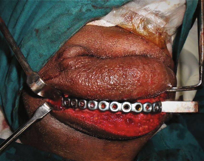
2 Case Reports in Dentistry
Figure 3: Preop CT scan showing right parasymphysis and
dentoalveolar factures.
Figure 1: Preop photograph.
Figure 4: Lingual view of the same CT scan.
Figure 2: Intraoral view presop. by the medicine department. Neurology was consulted, and
the same medication was continued. ENT consultation was
sought in view of difficulty in speech for which conservative
region, thorough debridement was done and the infected management was advised, and the patient recovered well post
chunks of bone were removed. Following this, the fractures surgery. Malignancy and osteomyelitis were ruled out by his-
were fixed using two miniplates. Following the fixation, the topathology report, and the fracture was attributed to the
incision site was thoroughly debrided and closed in layers constant occlusal trauma due to cervical dystonia. Following
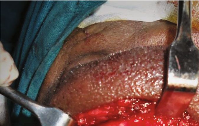
(Figures 6 and 7). this, the patient was discharged.
Following the surgery, the patient was shifted to postop Following this surgery, the patient again reported to our
ICU and then to the ward. outpatient department after three months with similar com-
On postop day one, the patient was having persistent plaints of pain in the anterior region of the mandible. On
cough in view of which blood and sputum culture was taken examination, there was slight opening in the mucosa on the
which revealed E. Faecalis sensitive to Linezolid and K. Pneu- right parasymphysis region and segmental mobility over the
monia sensitive to Gentamycin. The patient was given IV right parasymphysis region. Clinical and radiological exami-
Ampicillin, IV Gentamycin, and Tab Linezolid as advised nation confirmed hardware failure to be the cause of pain and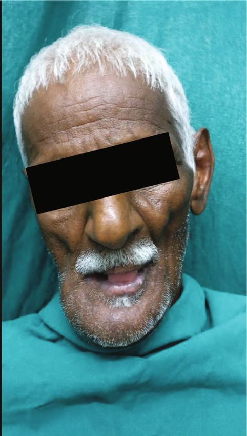
Case Reports in Dentistry 3
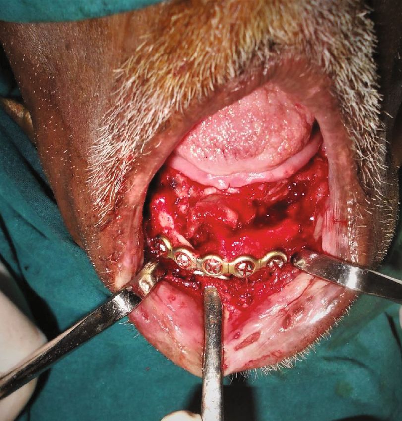
Figure 7: Plates in situ 9 (first surgery).
Figure 5: Coronal CT image.
Figure 8: CT scan after hardware failure.
chunks were curetted out, and the mandible was fixed using a
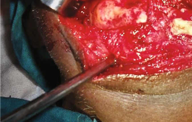
Figure 6: On-table exposure of the fracture site (first surgery).
15-hole 2.5 mm reconstruction cut plate and 8 (2 × 14 mm)
locking screws (Figures 12 and 13). Homeostasis was
achieved, and the wound was closed in layers.
mobility over the right mandibular parasymphysis region The patient recovered well post surgery and was subse-
(Figures 8 and 9). quently discharged after a week (Figure 14).
The patient was again taken up for the surgical interven-
tion where after securing adequate anesthesia, extraoral 3. Discussion
incision from the right body region of the mandible to the left
body region of the mandible was given. After layered dissec- Meige was the first person to describe orofacial dystonia in
tion, the right mandibular parasymphysis region was detail [6].This condition is also known by the name of Meige
exposed. On exposure, a dumbbell plate was found attached syndrome when it occurs in conjunction with blepharospasm
to the soft tissues with its two screws, which was subsequently [7]. This disease still poses a dilemma as far as its under-
retrieved (Figures 10 and 11). The superior 6-holed plate and standing is concerned. This condition may affect the muscles
5 screws were also retrieved. Following this, the infected bone on one side or both the sides of the face, hence causing a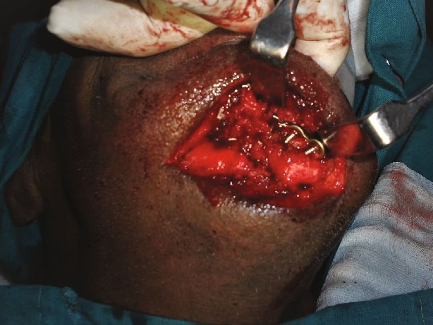
4 Case Reports in Dentistry
Figure 9: PA view of the mandible taken at the same time.
Figure 11: Exposure of the site, revealing a loose hardware.
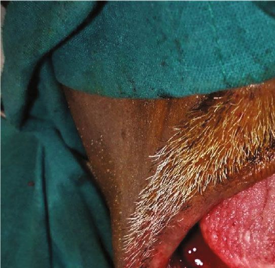
Figure 10: Extraoral markings for incision (second surgery).
different clinical presentation. All the jaw movements that Figure 12: After the removal of the hardware and necrotic bone.
include protrusion, retrusion, and side-to-side movements
may be affected. When the muscles of facial expression and
lingual musculature are affected, there may be spasmodic
contraction; there are a number of features like twitching promptly, the condition becomes very serious resulting in
of the face, abnormal lip movements, nasal contractions, intolerable pain and discomfort [11].
clenching/grinding of teeth, retractions of oral commis- Stimulating factors for this episode are stress, depression,
sures, and/or abnormal movements of tongue [8]. or any kind of activity involving the orofacial musculature.
There may be involvement of platysma also resulting in Even trauma has also been associated with the triggering of
contractions of the neck. Involvement of laryngeal muscles the episode of dystonia [12].
has also been reported resulting in difficulty in breathing, Over a period of time, the patient learns to live with the
dysphagia, dysphonia, and dysarthria [9]. condition and they tend to deal with it by engaging in more
Slowly, these uncontrolled spasms result in accumulation pleasurable activities. The contraction of facial musculature
of products of anaerobic metabolism in the muscles and thus gradually results in bruxism, clenching of the teeth, wearing
results in myalgia [10]. away and loosening of the teeth, and even dislocation of the
The pain further involves the antagonist muscles. On the condyles [13]. In a few instances, it has also resulted in the
other hand, spasms consistently lead to further damage to the fracture of the mandible due to excessive masticatory force
soft tissue envelope. If the condition is not managed generated, as was the presentation in our case.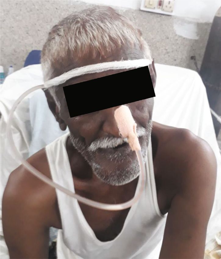
Case Reports in Dentistry                                                                                                   5

                                                                   (3) Surgical Management including Peripheral/Central
                                                                       Nervous System Procedures. This is used as the last
                                                                       retort when nothing is helping. Peripheral proce-
                                                                       dures include TMJ arthroscopy and surgery, myo-
                                                                       tomies, rhizectomies, and ramisectomies. CNS
                                                                       procedures involve deep brain stimulation or identi-
                                                                       fication and ablation of a desired nucleus in the brain.
                                                                       Thalamotomy and pallidotomy are also reported to
                                                                       provide relief in some cases [15]
                                                                    In our case, the patient was a 56-year-old male who
                                                                was suffering from dystonia for the past 1.5 years. The
                                                                patient came from a very low socioeconomic background
                                                                and was not undergoing any treatment for cervical dysto-
                                                                nia. The patient showed signs of twisting of the head and
                                                                chin towards the side which was involuntary. He had dif-
                                                                ficulty in coordinating the movements of his jaw muscles.
                                                                He was partially edentulous and was not using any dental
                                                                prosthesis. When the patient presented to us, his chief
                                                                complaint was the pain and tenderness over the right
                                                                mandibular parasymphysis region; on examination and
                                                                radiological examination, a diagnosis of fracture of the
                                                                right mandibular parasymphysis region was made. There
                                                                was no history of any trauma on that region. In all likeli-
                   Figure 13: Plates in situ.                   hood, the fracture had occurred some time back since
                                                                there were obvious signs of infection evident. Since there
                                                                was no history of trauma which was confirmed by the his-
                                                                tory taken from the patient as well as the relatives accom-
                                                                panying the patient, the most obvious cause of the fracture
                                                                can be the occlusal forces due to dystonia resulting in the
                                                                fracture of the mandibular parasymphysis. The patient also
                                                                showed signs of bruxism which was confirmed by the
                                                                presence of occlusal facets on the remainder of teeth.
                                                                There were only two lower molar present which may have
                                                                caused improper distribution of occlusal forces resulting in
                                                                the fracture of the mandibular region. The patient and the
                                                                patient’s party gave the history that the dystonia that the
                                                                patient was suffering from was idiopathic. He was also
                                                                partially edentulous with loss of few anterior teeth and
                                                                did not used a denture. Due to these factors, the occlusal
                                                                load on the anterior mandible would have been increased
                                                                multiple times leading to the eventual weakening of the
                                                                region and leading to fracture of the anterior mandible.
      Figure 14: Postop photograph after second surgery.        The primary surgery was aimed at thorough debridement
                                                                of the infected bone, curettage, biopsy to know if it was
                                                                a pathological fracture, and fixation of the mandible. The
                                                                fixation that was done using two miniplates which were
    Currently, there is no standard cure for dystonia,
                                                                load sharing.
although various treatments have been documented. Treat-
                                                                    The biopsy report ruled out any pathological involvement
ment options for dystonia are as follows.
                                                                in the form of malignancy or osteomyelitis. When the patient
                                                                reported to us again, he had a hardware failure most probably
   (1) Medical Management. This includes administration
                                                                due to dystonia and excessive masticatory forces. He was then
       of pharmacological agents like anticholinergics, anti-
                                                                taken up for surgery again, and the loosened hardware were
       convulsants, antiparkinsonians, benzodiazepines,
                                                                removed, followed by fixation with a reconstruction plate,
       carbamezipine, lithium, and gabapentin, which are
                                                                which is a load-bearing plate. The use of a heavy-load-
       very effective in this condition [14]
                                                                bearing plate gives additional strength to the mandible and
   (2) Chemodenervation. It implies injections of botuli-       hence bears the excessive forces of mastication in dystonia
       num neurotoxin in the form of Botulinum toxin A          patients. The patient recovered well after the surgery and
       and B in focal dystonia                                  had shown good improvement on his follow-up visits.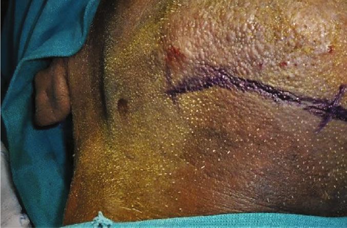
6                                                                                                            Case Reports in Dentistry

4. Conclusion                                                           [12] A. E. Lang, “Psychogenic dystonia: a review of 18 cases,” The
                                                                             Canadian Journal of Neurological Sciences, vol. 22, no. 2,
Orofacial dystonia is a very distressing condition which                     pp. 136–143, 1995.
causes a lot of problems to the person concerned. Some gen-             [13] P. J. Blanchet, O. Abdillahi, C. Beauvais, P. H. Rompre, and
eral recommendations to be followed in these patients are as                 G. J. Lavigne, “Prevalence of spontaneous oral dyskinesia in
follows:                                                                     the elderly: a reappraisal,” Movement Disorders, vol. 19,
                                                                             no. 8, pp. 892–896, 2004.
    (1) Periodic dental visits to look for health of the teeth          [14] J. Jankovic, S. Leder, D. Warner, and K. Schwartz, “Cervical
        and the surrounding structures                                       dystonia: clinical findings and associated movement disor-
                                                                             ders,” Neurology, vol. 41, no. 7, pp. 1088–1091, 1991.
    (2) Periodic X-rays to be taken of the maxillofacial
        region, to rule out facial fractures or pathology               [15] I. S. Cooper, “20-year follow-up study of the neurosurgical
                                                                             treatment of dystonia musculorum deformans,” Advances in
        caused due to dystonia
                                                                             Neurology, vol. 14, pp. 423–452, 1976.
    (3) Good maintenance of oral hygiene
    (4) Proper administration of the medications for orofa-
        cial dystonia
    If all these measures are undertaken and a periodic
check-up, any problem arising can be diagnosed early and
patients suffering can be reduced considerably.

Conflicts of Interest
The authors declare that they have no conflicts of interest.

References
 [1] E. S. Tolosa, “Clinical features of Meige’s disease (idiopathic
     orofacial dystonia). A report of 17 cases,” Archives of Neurol-
     ogy, vol. 38, no. 3, pp. 147–151, 1981.
 [2] E. K. Tan and J. Jankovic, “Botulinum toxin A in patients with
     oromandibular dystonia: Long-term follow-up,” Neurology,
     vol. 53, no. 9, pp. 2102–2107, 1999.
 [3] B. L. Scott, “Evaluation and treatment of dystonia,” Southern
     Medical Journal, vol. 93, no. 8, pp. 746–751, 2000.
 [4] R. Tintner and J. Jankovic, Botulinum toxin type A in the man-
     agement of oromandibular dystonia and bruxism. Scientific
     and therapeutic aspects of Botulinum toxin, PA: Lippincott
     Williams & Wilkins, Philadelphia, 2002.
 [5] K. H. Lee, “Oromandibular dystonia,” Oral Surgery, Oral Med-
     icine, Oral Pathology, Oral Radiology, and Endodontology,
     vol. 104, no. 4, pp. 491–496, 2007.
 [6] E. S. Tolosa, “Clinical features of Meige’s disease (idiopathic
     orofacial Dystonia),” Archives of Neurology, vol. 38, no. 3,
     pp. 147–151, 1981.
 [7] E. Tolosa and M. J. Marti, “Blepharospasm-oromandibular
     dystonia syndrome (Meige’s syndrome): clinical aspects,”
     Advances in Neurology, vol. 49, pp. 73–84, 1988.
 [8] J. Jankovic, “Etiology and differential diagnosis of blepharo-
     spasm and oromandibular dystonia,” Advances in Neurology,
     vol. 49, pp. 103–116, 1988.
 [9] E. K. Tan and J. Jankovic, “Botulinum toxin As in patients with
     oromandibular dystonia: long-term follow-up,” Neurology,
     vol. 53, no. 9, pp. 2102–2107, 1999.
[10] M. Hallett, “Is dystonia a sensory disorder?,” Annals of Neurol-
     ogy, vol. 38, no. 2, pp. 139-140, 1995.
[11] E. K. Tan, L. L. Chan, and M. C. Wong, “Levodopa-induced
     oromandibular dystonia in progressive supranuclear palsy,”
     Clinical Neurology and Neurosurgery, vol. 105, no. 2,
     pp. 132–134, 2003.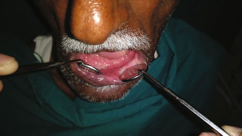
You can also read