Neuroanatomy of Two Species of Genus Myliobatis (Chondrichthyes: Myliobatoidea)
←
→
Page content transcription
If your browser does not render page correctly, please read the page content below
Int. J. Morphol.,
38(2):499-504, 2020.

                         Neuroanatomy of Two Species of Genus
                         Myliobatis (Chondrichthyes: Myliobatoidea)

          Neuroanatomía de Dos Especies del Género Myliobatis (Chondrichthyes: Myliobatoidea)

                         Héctor Marcos Montes-Domínguez1,3; Luis Amado AyalaPérez2; Manuel Arnoldo
                                Castillo Rivera4; Mónica González Isáis3 & Víctor Hugo Reynoso5

MONTES-DOMÍNGUEZ, H. M.; AYALA-PÉREZ, L. A.; CASTILLO-RIVERA, M. A.; GONZÁLEZ-ISÁIS, M. & REYNOSO,
V. H. Neuroanatomy of two species of genus Myliobatis (Chondrichthyes: Myliobatoidea). Int. J. Morphol., 38(2):499-504, 2020.

         SUMMARY: Several studies on the elasmobranchs neuroanatomy have shown that their brain is more complex than previously
thought, and had significant intra and interspecific variations. The objective of this work was conducting a comparative encephalic
neuroanatomy study of two species of genus Myliobatis. In total, 16 organisms of genera Myliobatis californica and Myliobatis longirostris,
collected in the coasts of Kino Bay, Sonora, Mexico, were used. In Myliobatis, the brain has a long telencephalon and the posterior
central nucleus is poorly developed. Their cerebellum is asymmetric, has several sulci, most of which are transversally oriented, with
four lobes (anterior, medium and two posterior), a condition which has not been reported for any other species. It was observed that,
despite the morphology of M. californica and M. longirostris is similar, there are some significant differences. Both species have moderate
foliation, but M. californica has more sulci. In the diencephalon of M. californica, it was observed that the lobes of the infundibulum are
oval-shaped and separated, while in M. longirostris, such lobes are rounded and near the medium line. It has to be highlighted that
Myliobatis belongs to the most derived batoid group; nevertheless, its brain is considerably less complex, as compared to what has been
reported for the most derived milyobatoids species.

        KEY WORDS: Elasmobranchii; Myliobatis; Gross brain; Telencephalon; Cerebellum.

INTRODUCTION

       Batoids or rays, are a specialized group, derived                 has also been shown that some structures have differences
from neoselachii sharks, and distribute in marine,                       during the development of organisms, including the optic
estuaries, rivers and freshwater systems. They include                   lobes, the mesencephalon (Lisney & Collin, 2006; Lisney
over 630 species in four orders, 17 families and 83 gene-                et al., 2007, 2017), and the cerebellum foliation level
ra, which is over one-half of all living elasmobranchii                  (Ari). However, only a few works have considered
known (Nelson et al., 2016). The order Myliobatiformes                   species of family Myliobatidae. Northcutt (1978) and
makes up 35 % of all batoid species (Compagno, 1990),                    Smeets, published a diagram of M. californica in dorsal
and includes the family Myliobatidae, which, according                   view. All other neuroanatomy works have focused on
to White & Last (2016) is represented by two genera                      different batoid groups (Northcutt, 1977, 1978, 1989;
(Aetomylaeus and Myliobatis), and 18 species. Both ge-                   Smeets; Hoffman), including Urobatis jamaicensis
nera are characterized by having a short, rounded rostral                (Walker & Sherman, 2001), Diplobatis ommata (Mon-
lobe, and rhombic disc (Nelson et al.; White & Last).                    tes Domínguez et al., 2014), potamotrygonids (Fontanelle
                                                                         & Carvalho, 2016), Gymnura micrura (Kobelkowsky,
       Works conducted on the batoids neuroanatomy                       2017), and mobulids (Ari). The objective of this work
have revealed a significant variation among different                    was conducting a comparative anatomy study of the
groups, particularly in the telencephalon and cerebellum                 external morphology of the encephalon in two species
structures (Smeets, 1998; Hoffman, 1999; Ari, 2011). It                  of the genus Myliobatis.

1
  Doctorado en Ciencias Biológicas y de la Salud, Universidad Autónoma Metropolitana, Unidad Xochimilco, México.
2
  Universidad Autónoma Metropolitana, Unidad Xochimilco, México.
3
  Facultad de Estudios Superiores Iztacala, UNAM, México.
4
  Universidad Autónoma Metropolitana, Unidad Iztapalapa, México.
5
  Instituto de Biología, Universidad Nacional Autónoma de México, México.

                                                                                                                                         499
MONTES-DOMÍNGUEZ, H. M.; AYALA-PÉREZ, L. A.; CASTILLO-RIVERA, M. A.; GONZÁLEZ-ISÁIS, M. & REYNOSO, V. H. Neuroanatomy of two species of genus Myliobatis
                                             (Chondrichthyes: Myliobatoidea). Int. J. Morphol., 38(2):499-504, 2020.

MATERIAL AND METHOD                                                             RESULTS

         Ten specimens of Myliobatis californica (Gill, 1865),                         The brain of both Myliobatis species is quite similar.
and six of Myliobatis longirostris (Applegate & Fitch, 1964),                   Therefore, a general description is presented, and the analysis
all sexually mature, were used. Animals were collected in                       will focus on the differences.
the coast of Kino Bay, Son., Mexico. In the field, the brain
was exposed and fixed with 4 % saline formaline. In the lab,                            The Myliobatis’ brain is relatively long and is inside
each cranium was processed with the conventional brain                          the neurocranium cavity, which can be totally or partially
extraction technique. In the dissection, the dorsal, frontal,                   occupied by it, depending on the organism size. Eyes are
lateral, and ventral regions of the brain were exposed, which                   relatively large, and are in front of the telencephalon (Fig.
were later described. Subsequently, the cerebellum, the pos-                    1a, c).
terior choroid plexus and the auricles upper lip were remo-
ved, to observe structures underneath. Terminology used was                     Telencephalon. The anterior region of the telencephalon is
the same used by Northcutt (1977, 1978, 1989), Smeets and                       wider than the posterior one, and the posterior central nucleus
Fontanelle & Carvalho.                                                          is covered by the cerebellum. Olfactory tracts arise from the
                                                                                                           anterolateral part of the
                                                                                                           telencephalon,         and      runs
                                                                                                           anterolaterally to the ventral
                                                                                                           region, as can be observed in late-
                                                                                                           ral view (Fig. 2). The tracts length
                                                                                                           increases with the organism size.
                                                                                                           Tracts are considerably thicker
                                                                                                           than the optic nerve (II), they are
                                                                                                           long and widen anteriorly, where
                                                                                                           are continued by the olfactory
                                                                                                           bulbs. The medial and lateral
                                                                                                           surface of the bulbs reaches the
                                                                                                           ventral region of the olfactory
                                                                                                           epithelium, as can be observed in
                                                                                                           lateral view (Fig. 2).

                                                                                                                 The telencephalon is relatively
                                                                                                                 long. The anterior edge has three
                                                                                                                 commissures, one central and two
                                                                                                                 laterals. In the latter, the lateral
                                                                                                                 pallium is quite developed, as can
                                                                                                                 be observed in lateral view (Fig.
                                                                                                                 2). When the cerebellum is remo-
                                                                                                                 ved, it can be observed that the
                                                                                                                 central     nucleus       of    the
                                                                                                                 telencephalon is split in one ante-
                                                                                                                 rior and one posterior regions (Fig.
                                                                                                                 1 c, f). The anterior division is
                                                                                                                 larger than the posterior one, and
                                                                                                                 has a fosseta on the medial line.
Fig. 1. Dorsal view of the encephalon: a) and b) Myliobatis californica, b) and c) Myliobatis
longirostris. In c) and d) dorsal view when the cerebellum is removed. ACN, anterior central                     Diencephalon. In the cephalic
nucleus; A, anterior lobe of the cerebellum; DON, dorsal octavolateralis nucleus; F, fossa; FV,                  region, the diencephalon is limited
fourth ventricle; IA, inferior auricle; L, lateral pallium; LP, lateral peduncles; M, medium
                                                                                                                 by the optic nerve (II). It extends
lobe of de cerebellum; MLF, medial longitudinal fasciculus; MO, medulla oblongata; MON,
medial octavolateral nucleus; P, posterior lobe of the cerebellum; PCN, posterior central nucleus;
                                                                                                                 ventrally in caudal position to the
OB, olfactory bulbs; OE, olfactory epithelium; OT, olfactory tracts; OpL, optic lobes; UA,                       telencephalon, and is relatively
upper auricle. Cranial nerves: II, optic; IV, trochlear; X, vagus. Scale = 10 mm.                                small. In ventral view, the inferior

500
MONTES-DOMÍNGUEZ, H. M.; AYALA-PÉREZ, L. A.; CASTILLO-RIVERA, M. A.; GONZÁLEZ-ISÁIS, M. & REYNOSO, V. H. Neuroanatomy of two species of genus Myliobatis
(Chondrichthyes: Myliobatoidea). Int. J. Morphol., 38(2):499-504, 2020.
lobes of the infundibulum can be observed clearly defined,
on both sides of the hypophysis. In M. californica, the infe-
rior lobes are elongated and separated, while in M.
longirostris they are rounded and located near the medium
line (Fig. 4). The hypophysis is an elongated, three-lobed
structure. The saccus vasculosus pair is located on both sides
of the hypophysis, towards the posterior part of the
infundibulum lobes.
Mesencephalon. The mesencephalon is partially covered
by the cerebellum, and is wider than long. In the dorsal
region, the mesencephalon is formed by the optic tectum;
and in the ventral region by the optic tegmentum. The tectum
forms the bigeminated lobes, which are separated by a sulcus.
In M. longirostris these lobes are rounded, and in M.
californica are slightly oval (Fig. 1). In ventral view, a part
of the optic tegmentum is visible, and on both sides of the
posterior part of the hypophysis, a pair of protuberances,
poorly developed can be observed, which are less evident in
Myliobatis longirostris (Fig. 4).
Cerebellum. The cerebellum is formed by a long cerebellar
body and the cerebellar auricles. The cerebellar body is on
the fourth ventricle, extending rostrally on the mesencephalic
tectum, and caudally on the inferior lip (auricle) of the
cerebellum.
Fig. 2. Lateral view of the encephalon: a) Myliobatis californica,
b) Myliobatis longirostris. A, anterior lobe of the cerebellum; IL,
inferior lobes of the infundibulum; L, lateral pallium; M, medium The cerebellar body is not symmetrical and is formed
lobe of the cerebellum; MO, medulla oblongata; OB, olfactory by four lobes, one anterior, one medium and two posterior –
bulbs; OE, olfactory epithelium; OT, olfactory tracts; OpL, optic one in dorsal and one in ventral position (Figs. 1a, c). The
lobes; P, posterior lobes of the cerebellum; T, telencephalon; UA, anterior lobe has the higher number of sulci, and is slightly
upper auricles. Cranial nerves: II optic; III, oculomotor; IV, smaller than the dorsal posterior lobe. The medium lobe is
trochlear. Scale = 10 mm. relatively small and can be found on the left or right side.
Most sulci in the cerebellum are transversal, but a few
longitudinal ca be observed, which are shallower (Fig. 3).
In the basal part of the body, on both sides of the
fourth ventricle, the lateral peduncles can be found, linking
the cerebellum with the median brain. These are rostrally
limited by the posterior part of the mesencephalic tectum,
and caudally by the upper lip. In the median region, on both
sides of the lateral peduncles, a paired, rounded structure
projects ventrally. This structure is the stem, which occupies
the anterior part of the fourth ventricle (Figs. 3c, f).
Fig. 3. Cerebellum of Myliobatis californica: a) and b) dorsal view, c)
ventral view. Myliobatis longirostris: d) and e) dorsal view, f) ventral
view. A, anterior lobe of the cerebellum; LP, lateral peduncle; M,
medium lobe of the cerebellum; P, posterior lobe of the cerebellum; S,
steam. Scale = 10 mm.
501MONTES-DOMÍNGUEZ, H. M.; AYALA-PÉREZ, L. A.; CASTILLO-RIVERA, M. A.; GONZÁLEZ-ISÁIS, M. & REYNOSO, V. H. Neuroanatomy of two species of genus Myliobatis
(Chondrichthyes: Myliobatoidea). Int. J. Morphol., 38(2):499-504, 2020.
The cerebellar auricles are clearly visible when the the posterior leaf. The inferior auricles are in a posterior
cerebellum is removed (Figs. 1b, d). On each side, they position, as related to lateral peduncles, at level of nerve
are formed by two leaves – one rostromedial superior and VIII; and the superior auricles are in an anterior position,
one caudolateral inferior. The first one is in anterior relative to the octavolateralis area. The anterior auricles
position, and the second in posterior position. In these are ornamented, i. e., their profile is circumvoluted. The
species, in lateral view (Fig. 2), it can be observed that the posterior auricles have a medial joint, forming an “infe-
anterior leaf is located in an inferior position, relative to rior lip” which creates a bridge in the fourth ventricle. These
auricles are smooth and thin, and are transversally arranged.
Medulla oblongata. The medulla oblongata is in the pos-
terior region of the brain. In dorsal view, it is almost totally
covered by the cerebellum. When the cerebellum and the
posterior choroid plexus are removed, the relatively long,
fourth ventricle can be observed. At its base, the medial
longitudinal fascicle arises, which has a sulcus on its
midline. The lateral peduncles of the cerebellum are at both
sides of the ventricle, in the anterior region. On both sides,
towards the ventricle posterior region, the developed dor-
sal octavolateralis nucleus can be observed. In lateral
position relative to this nucleus, is the medial
octavolateralis nucleus. In ventral view, the medulla
oblongata has a sulcus in its middle region (Fig. 4).
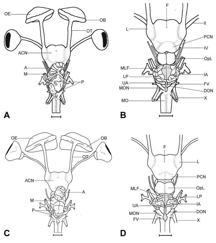
Cranial nerves: The olfactory nerve (I) is inside the
olfactory bulbs. The olfactory peduncles have been
reported by several authors as the cranial pair I; however,
they are formed by the secondary olfactory fibers, as
reported by Smeets. The optic nerve (II), arises from the
ventral part of the diencephalon, and is the boundary
between the telencephalon and the diencephalon. On other
vertebrate groups, the optic chiasma indicates the crossing
over of optic nerves after leaving the cerebellum; in
elasmobranchs, however, where this crossing over is
absent, the term is used to indicate the origin of cranial
pair II. (Fig. 5)
The oculomotor nerve (III) originates in the ven-
tral part of the mesencephalon, in front of the
protuberances; and the trochlear nerve (IV), arises from
the dorsal region of the mesencephalon, in the
posterolateral part of optic lobes. Both nerves run
anteriorly. The abducens nerve (VI) is an extremely thin
structure. In M. californica the origin is in the ventral region
of the medulla oblongata, at the level of nerve VIII, and
runs anteriorly towards the origin of nerve V, to continue
its trajectory on the ventral part of it. In M. longirostris,
the abducens nerve follows a similar trajectory, but its
origin is slightly posterior to the origin of nerve VIII.
Fig. 4. Ventral view of the encephalon: a) Myliobatis californica,
b) Myliobatis longirostris. H, hypophysis; IL, inferior lobes of the
Trigeminal (V), facial (VII) and acoustic (VIII)
infundibulum; MO, medulla oblongata; OB, olfactory bulbs; OE,
olfactory epithelium; OT, olfactory tracts; OTg, optic tegmentum; nerves, originate in the lateral wall of the medulla
PRO, protuberance; SV, saccus vasculosus. Cranial nerves: II, optic. oblongata, and have and independent origin although they
Scale = 10 mm. are very close. Pair VII is split in two main branches. After
502MONTES-DOMÍNGUEZ, H. M.; AYALA-PÉREZ, L. A.; CASTILLO-RIVERA, M. A.; GONZÁLEZ-ISÁIS, M. & REYNOSO, V. H. Neuroanatomy of two species of genus Myliobatis
(Chondrichthyes: Myliobatoidea). Int. J. Morphol., 38(2):499-504, 2020.
grows, as has been reported by
Smeets, for other species. In
Myliobatis, these structures have
strongly migrated towards the
ventral region, contrary to what
has been reported for Diplobatis
ommata (Montes Domínguez et
al.), Urobatis jamaicensis (Walker
& Sherman), and other
potamotrygonids (Fontanelle &
Carvalho), where the peduncles
run either anteriorly or slightly
bent towards the ventral region in
the anterior part.
In different elasmobranch
species, it has been described that
the cerebellum is formed by three
lobes, and the anterior lobe is the
largest (Northcutt, 1989;
Puzdrowski & Leonard, 1992;
Walker & Sherman; Fontanelle &
Carvalho). However, in the two
Myliobatis species in this study,
four lobes were observed: one an-
terior, one medium and two pos-
terior – the anterior being slightly
smaller than the posterior one. In
most derived species, such as
Mobula and Manta, the cerebellum
Fig. 5. Cranial nerves. Myliobatis longirostris: a) Dorsal view of the brain when the
cerebellum is removed, b) lateral view c) ventral view. Myliobatis californica d) ventral has a number of lobes (Ari). It has
view. II, optic; III, oculomotor; IV, trochlear; V, trigeminal; VI, abducens; VII, facial; to be noted that neither the number
VIII, acoustic; IX, glossopharyngeal; X, vagus. Scale = 10 mm. of lobes nor their size have been
reported for other species.
leaving the medulla oblongata, the glossopharyngeal nerve
(IX) runs anteriorly, but after leaving the cranium, it changes Another relevant
direction and runs to the posterior region. The vague nerve characteristic in the Myliobatis brain is the medium lobe.
(X), originates in the lateral wall of the medulla oblongata, This structure, besides being significantly smaller, as
has several divisions and runs caudally. compared to species above, can be on the right or the left
side (Fig. 3). Variations in the orientation of the cerebellar
lobes have been reported only for Dasyatis sabina
DISCUSSION (Puzdrowski & Leonard).
Cranial pairs VII and VIII, have an independent
In species of genus Myliobatis, the brain had origin, as reported by Montes Domínguez et al. for
distinctive characteristics as related to the neuroanatomy Diplobatis ommata, and oppose to what was reported by
reported for other batoid genera. In M. californica and M. Walker & Sherman for Urobatis jamaicensis. These authors
longirostris, the telencephalon is long and well developed. state that nerves VII and VIII have a common origin. It should
However, the central posterior nucleus is poorly developed, be noted that in dorsal view, it is observed that nerve VIII is
unlike most derived batoid species such as Mobula and on nerve VII and apparently, these have a common origin;
Manta, where the posterior central nucleus is well but it actually originates at a point slightly posterior, right
developed (Ari), and considered to be a derived character. below nerve VII. This arrangement could lead to the wrong
The olfactory tracts increase their length as the organism conclusion that they have a common origin.
503MONTES-DOMÍNGUEZ, H. M.; AYALA-PÉREZ, L. A.; CASTILLO-RIVERA, M. A.; GONZÁLEZ-ISÁIS, M. & REYNOSO, V. H. Neuroanatomy of two species of genus Myliobatis
(Chondrichthyes: Myliobatoidea). Int. J. Morphol., 38(2):499-504, 2020.
Based on the above characteristics, it can be stated that Compagno, L. J. V. Alternative life-history styles of cartilaginous fishes in
the brains of M. californica and M. longirostris are quite si- time and space. Environ. Biol. Fish., 28:33-75, 1990.
Fontanelle, J. P. & Carvalho, M. R. Systematic implications of brain
milar, with the following significant differences: 1) in M. morphology in Potamotrygonidae (Chondrichthyes: Myliobatiformes).
longirostris, the peduncles migration towards the ventral region J. Morphol., 277(2):252-63, 2016.
is stronger than in M. californica. 2) In M. californica, the Hoffman, M. H. Nervous System. In: Hamlet, W. C. (Ed.). Sharks, Skates,
cerebellum has a larger number of sulci. 3) The infundibulum and Rays: The Biology of Elasmobranch Fishes. Baltimore, Johns
Hopkins University Press, 1999.
lobes are more separated in M. californica. 4) In M. californica, Kobelkowsky, A. Comparative anatomy of the neurocranium and
nerve VI arises at the level of nerve VIII, while in M. encephalon of the butterfly ray, Gymnura micrura (Batoidea:
longirostris it originates posteriorly to nerve VIII. Gymnuridae). Int. J. Morphol., 35(2):644-50, 2017.
Lisney, T. J. & Collin, S. P. Brain morphology in large pelagic fishes: a
comparison between sharks and teleosts. J. Fish Biol., 68(2):532-54,
Myliobatis, together with Aetomylaeus form the sister 2006.
group of more derived myliobatoids (Naylor et al., 2016). Lisney, T. J.; Bennett, M. B. & Collin, S. P. Volumetric analysis of sensory
However, and despite its brain having characteristics exclusive brain areas indicates ontogenetic shifts in the relative importance of
to this genus, it is relatively simple in contrast to what has sensory systems in elasmobranchs. Raffles Bull. Zool., 14:7-15, 2007.
Lisney, T. J.; Yopak, K. E.; Camilieri-Asch, V. & Collin, S. P. Ontogenetic
been reported for more derived myliobatoids, such as Mobula shifts in brain organization in the bluespotted stingray Neotrygon kuhlii
and Manta. (Chondrichthyes: Dasyatidae). Brain Behav. Evol., 89(2):68-83, 2017.
Montes Domínguez, H. M.; López Bárcenas, R. & González Isáis, M.
Morphological study of the brain and cranial nerves of Diplobatis
ommata (Elasmobranchii: Narcinidae). Int. J. Morphol., 32(4):1152-5,
MONTES-DOMÍNGUEZ, H. M.; AYALA-PÉREZ, L. A.; 2014.
CASTILLO-RIVERA, M. A.; GONZÁLEZ-ISÁIS, M. & Naylor, G. J. P.; Yang, L.; Corrigan, S. & de Carvalho, M. R. Phylogeny
REYNOSO, V. H. Neuroanatomía de dos especies del género and Classification of rays. In: Last, P. R.; White, W. T.; de Carvalho,
Myliobatis (Chondrichthyes: Myliobatoidea). Int. J. Morphol., M. R.; Séret, B.; Stehmann, M. F. W. & Naylor, G. J. P. (Eds.). Rays of
38(2):499-504, 2020. the World. Sidney, Csiro Publishing, 2016.
Nelson, J. S.; Grande, T. C. & Wilson, M. V. H. Fishes of the World. 5th ed.
RESUMEN: Diversos estudios sobre la neuroanatomía de los Hoboken, John Wiley & Sons, 2016.
elasmobranquios han demostrado que el cerebro es más complejo de Northcutt, R. G. Brain Organization in the Cartilaginous Fishes. In:
lo que se pensaba y presenta considerables variaciones tanto intra como Hodgson, E. S. & Mathewson R. F. (Eds.). Sensory Biology of Sharks,
Skates, and Rays. Arlington, Office of Naval Research, 1978.
interespecíficas. El objetivo de este trabajo fue realizar un estudio de
Northcutt, R. G. Brain variation and phylogenetic trends in elasmobranch
neuroanatomía comparada del encéfalo de dos especies del género
fishes. J. Exp. Zool. Suppl., 2:83-100, 1989.
Myliobatis. Se utilizaron un total de 16 organismos de Myliobatis Northcutt, R. G. Elasmobranch central nervous system organization and
californica y Myliobatis longirostris, los cuales fueron colectados en its possible evolutionary significance. Am. Zool., 17(2):411-29, 1977.
las costas de Bahía Kino, Son., México. El cerebro de Myliobatis tiene Puzdrowski, R. L. & Leonard, R. B. Variations in cerebellar morphology
un telencéfalo largo, el núcleo central posterior está poco desarrolla- of the Atlantic stingray, Dasyatis sabina. Neurosci. Lett., 135(2):196-
do; el cerebelo es asimétrico, presenta surcos que en su mayoría están 200, 1992.
orientados transversalmente, con cuatro lóbulos (anterior, medio y dos Smeets, W. J. A. J. Cartilaginous Fishes. In: Nieuwenhuys, R. & Roberts,
posteriores), condición que no ha sido reportada para otra especie. Se B. L. (Eds.). The Central Nervous System of Vertebrates. Berlin,
observó que, aunque M. californica y M. longirostris presentan una Springer-Verlag, 1998.
morfología similar existen ciertas diferencias. En ambas especies pre- Walker, K. B. & Sherman, L. R. Gross brain morphology in the yellow
sentan una foliación moderada; sin embargo, en M. californica se ob- stingray, Urobatis jamaicensis. Florida Sci., 64(4):246-9, 2001.
White, W. T. & Last, P. R. Eagle Rays. Family Myliobatidae. In: Last, P.
servan más surcos. En el diencéfalo de M. californica se observa que
R.; White, W. T.; de Carvalho, M. R.; Séret, B.; Stehmann, M. F. W. &
los lóbulos del infundíbulo son ovalados y están separados, mientras
Naylor, G. J. P. (Eds.). Rays of the World. Sidney, Csiro Publishing,
que en M. longirostris son redondeados y se encuentran próximos a la 2016.
línea media. Es importante señalar que, pese a que Myliobatis pertene-
ce al grupo de batoideos más derivado, su cerebro es considerable-
mente menos complejo de lo que se ha reportado para las especies de
Corresponding autor:
miliobatoideos más derivadas.
Héctor Marcos Montes Domínguez
FES Iztacala, UNAM
PALABRAS CLAVE: Elasmobranchii; Myliobatis; Cere-
Av. de los Barrios N° 1
bro; Telencéfalo; Cerebelo.
Los Reyes Iztacala
Tlalnepantla de Baz
MÉXICO
REFERENCES
Email: goritec@unam.mx
Ari, C. Encephalization and brain organization of Mobulid rays
(Myliobatiformes, Elasmobranchii) with ecological perspectives. Open Received : 21-09-2019
Anat. J., 3:1-13, 2011. Accepted : 17-10-2019
504You can also read