Revision of the muscular anatomy of the paired fins of the living coelacanth Latimeria chalumnae (Sarcopterygii: Actinistia)
←
→
Page content transcription
If your browser does not render page correctly, please read the page content below
Biological Journal of the Linnean Society, 2021, 133, 949–989. With 9 figures.

Revision of the muscular anatomy of the paired
fins of the living coelacanth Latimeria chalumnae
(Sarcopterygii: Actinistia)
ALESSIA HUBY1,+, , ROHAN MANSUIT2,3,*,+, , MARC HERBIN3 and ANTHONY HERREL3,

                                                                                                                                     Downloaded from https://academic.oup.com/biolinnean/article/133/4/949/6261035 by guest on 17 September 2021
1
  Laboratory of Functional and Evolutionary Morphology, FOCUS Research Unit, Department of Biology,
Ecology and Evolution, University of Liège, 4000 Liège, Belgium
2
  UMR 7207 Centre de Recherche en Paléontologie, Paris, Département Orgines & Evolution, Muséum
national d’Histoire naturelle – Sorbonne Université – CNRS, 8 rue Buffon, CP38, 75005 Paris, France
3
  UMR 7179 Mécanismes Adaptatifs et Evolution, Département Adaptations du Vivant, Muséum national
d’Histoire naturelle – Sorbonne Université – CNRS, 57 rue Cuvier, CP55, 75005 Paris, France

Received 18 November 2020; revised 8 March 2021; accepted for publication 8 March 2021

As a sarcopterygian fish, the extant coelacanth Latimeria has muscular paired fins, different in their skeletal and
muscular anatomy from the paired fins of actinopterygians. Although the muscular anatomy of the pectoral and pelvic
fins of Latimeria has been described by several studies, a detailed functional description of the muscles and their
architecture has never been performed. Our detailed functional description of the muscles of the paired fins shows
a more complex organization than previously described. The pectoral and pelvic fins have a different organization
of their muscular anatomy, and the pelvic fin shows a more plesiomorphic configuration of the muscles since most
of them are poly-articular and run from the pelvic girdle to the fin rays, an organization typical of actinopterygians.
We found that the pectoral fins are stronger than the pelvic fins which is likely to be associated with the greater
contribution of the pectoral fins to locomotion and manoeuvring. Finally, the study of the joint mobility of the paired
fins showed that the pectoral fins show greater mobility than the pelvic fins. The reduced mobility of the pelvic fin is
possibly a consequence of the morphology of the mesomeres and the large pre-axial radials.

ADDITIONAL KEYWORDS: anatomical cross-section area – mobility – muscles – pectoral fin – pelvic fin
– sarcopterygians.

                    INTRODUCTION                                      The pectoral and pelvic fins of actinopterygian fishes
                                                                      are internally supported by bony spines (fin rays or
Most living vertebrates are bony fishes (Osteichthyes)
                                                                      lepidotrichia) that are directly attached to the body at
whose evolutionary success is in part due to the
                                                                      the level of the pectoral and pelvic girdles by several
morphological diversification of the paired appendages
                                                                      basal skeletal elements (radials) connected to three
allowing their invasion of novel environments
                                                                      basal cartilages (polybasal articulation). By contrast,
(Drucker & Lauder, 2002; Kardong, 2018). Based on
                                                                      the lepidotrichia of the paired fins of sarcopterygian
the anatomical organization of pectoral and pelvic
                                                                      fishes are connected to an endoskeleton composed of
appendages, osteichthyan fishes are divided into
                                                                      several adjacent elements (mesomeres) forming an
two groups: the ray-finned fishes (Actinopterygii)
                                                                      axis (metapterygial axis) that is joined to the body by a
grouping the vast majority of extant bony fishes and
                                                                      single endoskeletal element (monobasal articulation),
the sarcopterygians including lobe-finned fishes
                                                                      the first mesomere (Johanson et al., 2007; Zhu & Yu,
(extant coelacanths, lungfishes) and tetrapods (Diogo
                                                                      2009; Kardong, 2018). In addition to endoskeletal
& Abdala, 2007; Amemiya et al., 2013; Nelson et al.,
                                                                      differences, fin movements in actinopterygians are
2016; Amaral & Schneider, 2018; Kardong, 2018).
                                                                      mainly controlled by relatively small abductor and
                                                                      adductor muscles located within the body, whereas
*Corresponding author. E-mail: rohan.mansuit@mnhn.fr                  these muscles are larger and located largely outside
+
 Joint first authors                                                  the body within the lobed fins in sarcopterygians

© 2021 The Linnean Society of London, Biological Journal of the Linnean Society, 2021, 133, 949–989                            949
This is an Open Access article distributed under the terms of the Creative Commons Attribution-NonCommercial License
(http://creativecommons.org/licenses/by-nc/4.0/), which permits non-commercial re-use, distribution, and reproduction in any
medium, provided the original work is properly cited. For commercial re-use, please contact journals.permissions@oup.com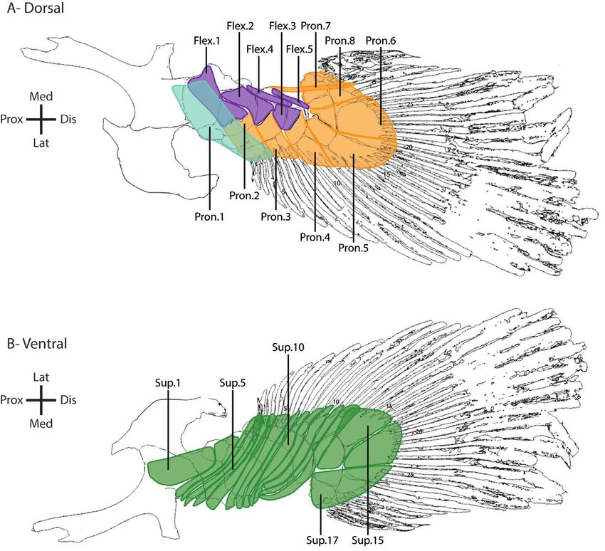
950    A. HUBY ET AL.

(Millot & Anthony, 1958; Wilhelm et al., 2015; Diogo             living models to infer the basal tetrapod condition
et al., 2016; Miyake et al., 2016; Kardong, 2018).               in terms of anatomy, development and genetics
   Among sarcopterygians, coelacanths (Actinistia)               (Ahlberg, 1992; Coates, 1994; Diogo & Abdala, 2007;
incl u de t wo exta n t s pe c i e s, i . e. , th e A f ri can   Diogo et al., 2016; Miyake et al., 2016; Amaral &
coelacanth Latimeria chalumnae (Smith, 1939) and                 Schneider, 2018). Despite several studies describing
the Indonesian coelacanth Latimeria menadoensis                  the muscular anatomy of the pectoral and pelvic fins
(Erdmann et al., 1998). However, fossil forms were               in coelacanths, little is known about the architecture
extremely diverse from the Early Devonian to the                 of the musculoskeletal system of these paired
Late Cretaceous and varied in shape, size and ecology            appendages. However, an understanding of the
(Agassiz, 1839; Erdmann et al., 1998; Forey, 1998;               muscle architecture is essential to understand the
Nulens et al., 2011). Living coelacanths are large-              function and role of the different appendages during

                                                                                                                                  Downloaded from https://academic.oup.com/biolinnean/article/133/4/949/6261035 by guest on 17 September 2021
bodied marine fishes, that measure up to 2 m in length           locomotion and the changes that occurred during the
and may weigh up to 105 kg (Nulens et al., 2011).                water-to-land transition.
They are predatory fishes that live around deep-water               The aim of the present study is to provide a detailed
caves and rugged terrain at depths between 110 and               functional description of the muscular anatomy
400 m (Fricke et al., 1987; Fricke & Hissmann, 1992;             (i.e., muscle arrangement, muscle mass, anatomical
Hissmann et al., 2006). The anatomy of the pectoral              cross-sectional area) of the paired fins of the living
and pelvic fins of the African coelacanth L. chalumnae           coelacanth L. chalumnae. Moreover, we provide data
was extensively studied by Millot & Anthony (1958)               on the mobility of the endoskeletal axis (e.g., joint
and subsequently revised by many authors (Ahlberg,               mobility) and compare it between the pectoral and
1992; Diogo et al., 2016; Miyake et al., 2016). More             pelvic fins. This will provide a better understanding
recently, Miyake and co-authors (2016) studied the               of the functional role of the paired fins in the unique
arrangement and function of the pectoral fin muscles.            swimming mode of the extant coelacanth in addition
   Even though coelacanths are bottom dwelling, the              to provide base-line data for future modelling studies.
hypothesis that these animals walk on substrate,
similar to tetrapods, has been refuted by numerous
observations in their natural environment (Fricke et al.,                     MATERIAL AND METHODS
1987, 1991; Fricke & Hissmann, 1992; Hissmann et al.,
2006). Coelacanths most often swim in a slow manner                                 Specimens studied
while maintaining a stiff and inflexible body. Propulsion        Two specimens of the extant African coelacanth
is generated by the lobed, paired and unpaired fins              L. chalumnae were used for dissections: CCC 14 and
(Fricke & Hissmann, 1992). The caudal fin is used                CCC 27 (Nulens et al., 2011). The specimen CCC 14
mainly during accelerations. During slow continuous              is an adult male specimen of 134 cm in total length
locomotion the second dorsal fin and anal fin as well            (TL) and weighs 39 kg. It was captured in the region of
as both pectoral fins typically show large amplitude             Dzahadjou, Hambou, off the coast of Grande Comore
movements whereas the amplitude of pelvic fins is                Island in 1956. The specimen CCC 27 is an adult male
lower (Fricke & Hissmann, 1992). Moreover, the paired            specimen of 132 cm TL and weighs 38 kg, captured
fins are highly mobile and display atypical movements            off the coast of Grande Comore Island in 1961. Both
including figure of eight motions of the pectoral fin            specimens are preserved in a 6–7% formaldehyde
during forward swimming, or elliptical motions of the            solution and stored in the collections of the Museum
pelvic fins (Décamps et al., 2017). Fricke & Hissmann            national d’Histoire naturelle (MNHN) in Paris, France,
(1992) hypothesized that the muscular part of the pelvic         under the collection numbers MNHN-ZA-AC-2012-11
fin is shorter than that of the pectoral fin and that the        and MNHN-ZA-AC-2012-21, respectively. The isolated
mobility of the pelvic fin is limited compared to the            pectoral fin of a third specimen was used for the
pectoral fin; however, this remains to be tested.                study of the joint mobility of the fins, specimen CCC
   Although the paired fins of the coelacanth are a              19 (MNHN-ZA-AC-2012-15; Nulens et al., 2011). This
derived condition in sarcopterygian fish, they are               specimen is a male of 140 cm TL and weighs 35 kg,
structurally comparable to the limbs of tetrapods                captured in 1959 off the coast of Grande Comore Island.
and appear to develop in a similar way (Shubin &                 It was dissected and the pectoral fin is preserved in a
Alberch, 1986; Ahlberg, 1992; Diogo et al., 2016).               6–7% formaldehyde solution.
Compared to lungfish fins, the paired fins of
coelacanths have been hypothesized to better reflect
the ancestral sarcopterygian condition (Coates                                          Dissections
et al., 2002; Friedman et al., 2007). Consequently,              The two specimens of L. chalumnae were immersed
coelacanths have been considered as one of the best              in water for 1 week before anatomical dissections in

                            © 2021 The Linnean Society of London, Biological Journal of the Linnean Society, 2021, 133, 949–989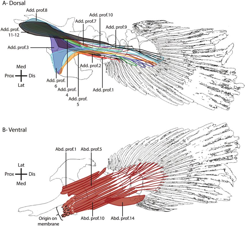
PAIRED FINS MUSCLES OF THE COELACANTH                     951

order to remove the formaldehyde. The left pectoral              movements (see below for definitions used here). For
fin of specimen CCC 14 and the left pelvic fin of                example, we introduced one needle on mesomere 1 and
specimen CCC 27 were dissected. The preservation of              the other on mesomere 2 to determine the mobility of
the specimens provided an exceptional preservation of            the joint between mesomere 1 and mesomere 2 and
the muscular tissues with no noticeable deterioration.           we photographed each maximum position. For each
The preservation of the specimens showed a slight                movement, five measures were taken, after returning
retraction of the muscle tissues, allowing us to easily          the joint to the resting position of the fin. The resting
identify the muscle bundles during the dissections               position of each fin is defined in the Results. The angle
(Supporting Information, Fig. S1). For each fin,                 formed by the needles was then determined using
the origin and insertion sites of the muscle bundles             the software Fiji (v.ImageJ 1.52p, Java 1.8.0_172),
were noted and muscle bundles were photographed,                 and the mean maximal angle was calculated for each

                                                                                                                              Downloaded from https://academic.oup.com/biolinnean/article/133/4/949/6261035 by guest on 17 September 2021
removed with care and classified into functional                 movement.
groups. Photographs were taken in situ at each stage
of the dissection (Supporting Information, Fig. S2).
After removal muscle bundles were directly placed in                                 Data availability
a 70% aqueous solution of ethanol. After the complete            The data underlying the study are available in the
dissection of each fin, the length of all muscle bundles         main tables and in the Supporting Information.
was measured using a ruler (± 1 mm), blotted dry and
weighed using an analytical balance (Mettler AE100, ±
0.00001g) or an electronical balance (Ohaus Scout Pro,
                                                                                          RESULTS
± 0.01g). The total length of each muscle bundle was
defined as the maximal distance between the origin               The first anatomical description of the paired fins of
and insertion of the muscle bundle (from the most                L. chalumnae was done by Millot & Anthony (1958).
proximal origin of the muscular part to the most distal          The nomenclature used in the present study follows
insertion, excluding tendons or aponeuroses).                    that of Diogo et al. (2016). The nomenclature used
                                                                 by Miyake et al. (2016) for the pectoral fin of the
                                                                 coelacanth is similar to that used by Diogo et al. (2016).
                Muscle architecture                              The correspondence between the nomenclature used
The mass and length of the muscles were used to                  by Millot & Anthony (1958), Diogo et al. (2016) and the
quantify the anatomical cross-section area (ACSA)                present work is shown in Tables 1 to 6. As previously
of each muscle bundle as an estimator of its force-              described, both the pectoral and pelvic fin musculature
generating capacity (Loeb & Gans, 1986). The ACSA                is organized in three different layers: the superficial
is based on the bundle mass (m), a standard muscular             layer just beneath the scales, the middle layer, and
density (ρ) and the muscle bundle length (L) using the           the deep layer that overlaps the endoskeleton of the
following equation:                                              fin (Millot & Anthony, 1958; Diogo et al., 2016; Miyake
                                                           et al., 2016).
       ACSA cm2 = m [g] / ρ g.cm−3 ∗ L [cm]
Since the value of the muscular density of L. chalumnae,
and more generally of lobe-finned fishes, remains
unknown, we here used the values reported for fish                    Osteological anatomy of the paired fins
(1.06 g.cm-3) (Dabrowski, 1978).                                 We provide here a short description of the osteology of
                                                                 the pectoral and pelvic fins of L. chalumnae (Fig. 1).
                                                                 A more extensive description is available in the
                     Joint mobility                              literature (see Millot & Anthony, 1958; Mansuit et al.,
To compare the mobility of the different articulations of        2020a, b).
the pectoral and pelvic fins, we measured the mobility of           The pectoral girdle has an arc shape and is formed
each joint along the metapterygial axis after complete           by four flattened dermal bones (the anocleithrum,
dissection but with the ligaments intact. We also had            cleithrum, extracleithrum and clavicle) and a
the opportunity to measure the joint mobility of the             massive endoskeletal scapulocoracoid element
already dissected pectoral fin of CCC 19. To do so, we           (Fig. 1A). The pelvic girdle is formed by a single
introduced two needles parallel to one another in the            endoskeletal bone containing a flat and curved
two bony elements involved in the joint (Moon, 1999).            lateral process, a short and triangular medial
Then, the elements were moved maximally without                  process, a rod-like cartilaginous anterior process
damaging ligaments or joint capsules to estimate the             and a small postero-superior process. The anterior
degree of freedom of the joint for adduction/abduction,          part of the pelvic girdle presents a highly ossified
protraction/retraction and pronation/supination                  surface and internal trabecular system (Fig. 1B).

© 2021 The Linnean Society of London, Biological Journal of the Linnean Society, 2021, 133, 949–989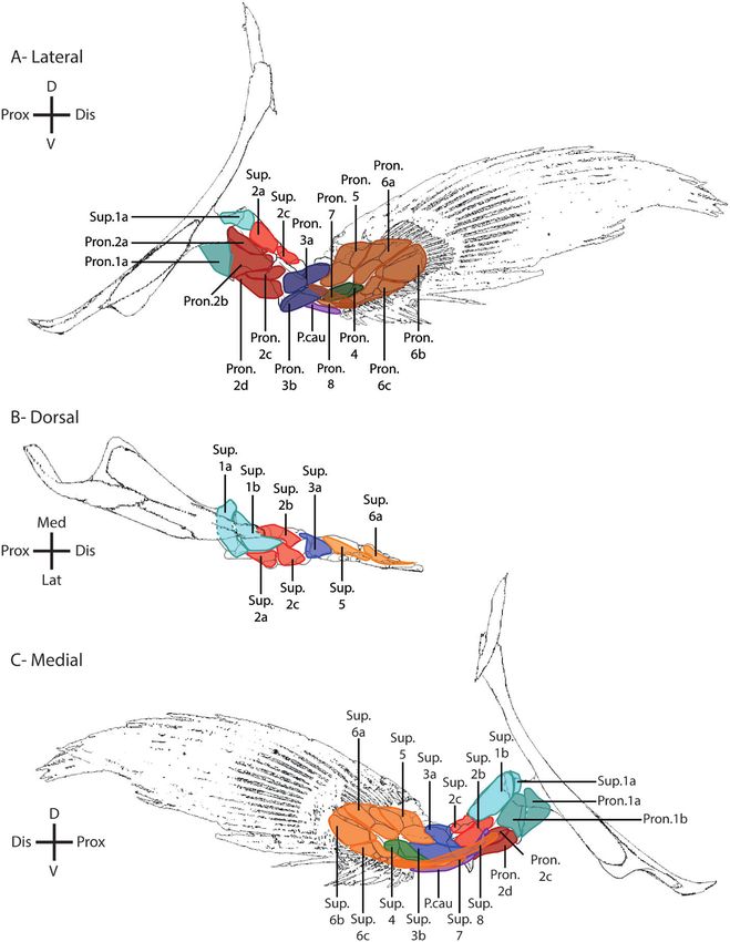
952     A. HUBY ET AL.

                                                                                                                                      Downloaded from https://academic.oup.com/biolinnean/article/133/4/949/6261035 by guest on 17 September 2021

Figure 1. Comparison of the skeletal anatomy of the pectoral (A) and pelvic (B) fin of L. chalumnae. The shaded parts
correspond to the pectoral (A) and pelvic (B) girdle. The dark grey part (B) corresponds to the dense ossification of the
pelvic girdle associated with a trabecular system. ano.: anocleithrum; ant.pro.: anterior process; cla.: clavicle; cle.: cleithrum;
dis.rad.: distal radial; ecl.: extracleithrum; lat.pro.: lateral process; lig.: “ligament-ball”; med.pro.: medial process; mes.:
mesomere; pos.sup.pro.: postero-superior process; po.rad.: post-axial radial; pr.rad.: pre-axial radial; scc.: scapulocoracoid.

The left and right medial processes are linked to each             mesomeres associated with pre-axial radial elements
other by a “ligament ball”.                                        and post-axial radial elements. The most proximal pre-
  The pectoral and pelvic fins have a similar                      axial radial elements are small and globular in shape,
organization, with the metapterygial axis formed by four           whereas the most distal pre-axial radials (pr.rad.3–4)

                            © 2021 The Linnean Society of London, Biological Journal of the Linnean Society, 2021, 133, 949–989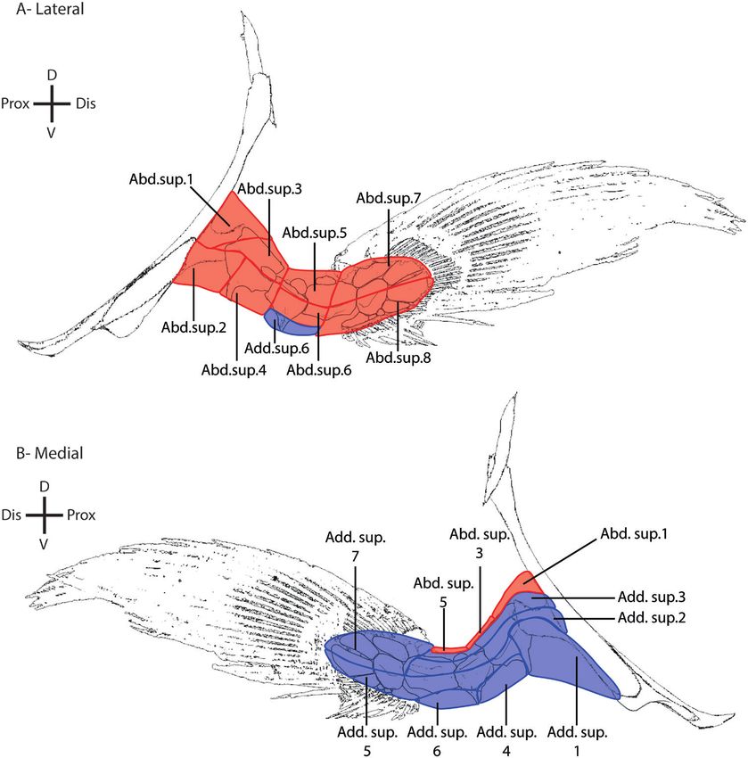
PAIRED FINS MUSCLES OF THE COELACANTH                      953

are elongated, trapezoidal in shape, and associated with         1. Protraction: the lateral side of the fin has a forward
dermal fin rays in both fins. The post-axial elements                movement.
are formed by the post-axial radials and the distal              2. Retraction: the medial side of the fin has a backward
radial (elongate and trapezoidal in shape), and are                  movement.
associated with dermal fin rays. The pelvic fin presents         3. A bduction: the pre-axial edge of the fin has an
a supernumerary pre-axial radial, called pre-axial                   upward movement.
radial 0, associated with mesomere 1 and the pelvic              4. A dduction: the pre-axial edge of the fin has a
girdle (Millot & Anthony, 1958; Mansuit et al., 2020b).              downward movement.
                                                                 5. Pronation: the lateral side of the fin has a downward
                                                                     pivoting movement around the axis of the fin.
              Pectoral muscle anatomy                            6. Supination: the lateral side of the fin has an upward

                                                                                                                               Downloaded from https://academic.oup.com/biolinnean/article/133/4/949/6261035 by guest on 17 September 2021
Eighty-six muscle bundles, organized into 13                         pivoting movement around the axis of the fin.
functional groups, were identified in the pectoral fin
of L. chalumnae. Similarly to Mansuit et al. (2020a),
the resting position of the pectoral fin is considered as
                                                                                     Superficial layer
the fin positioned along the body with its leading edge
oriented dorsally (Fig. 2). In this position, the muscles                                 (Table 1)
on the lateral side are abductor muscles protracting             The superficial layer of the pectoral fin is formed by two
the fin and those on the medial side are the adductor            muscle masses: the abductor superficialis muscles on the
muscles retracting the fin. Following this position,             lateral side and the adductor superficialis muscles on
movements of the pectoral fin are defined as follows:            the medial side of the pectoral fin (Fig. 3).

Figure 2. Pectoral and pelvic fins in their resting position in lateral (A) and ventral (B) views. The red arrows correspond
to the abduction movement of the fins, the blue arrows to the protraction movement of the fins and the green arrows to the
pronation movement of the fins, when fins are in their resting positions. The antagonist movements are not represented
here.

© 2021 The Linnean Society of London, Biological Journal of the Linnean Society, 2021, 133, 949–989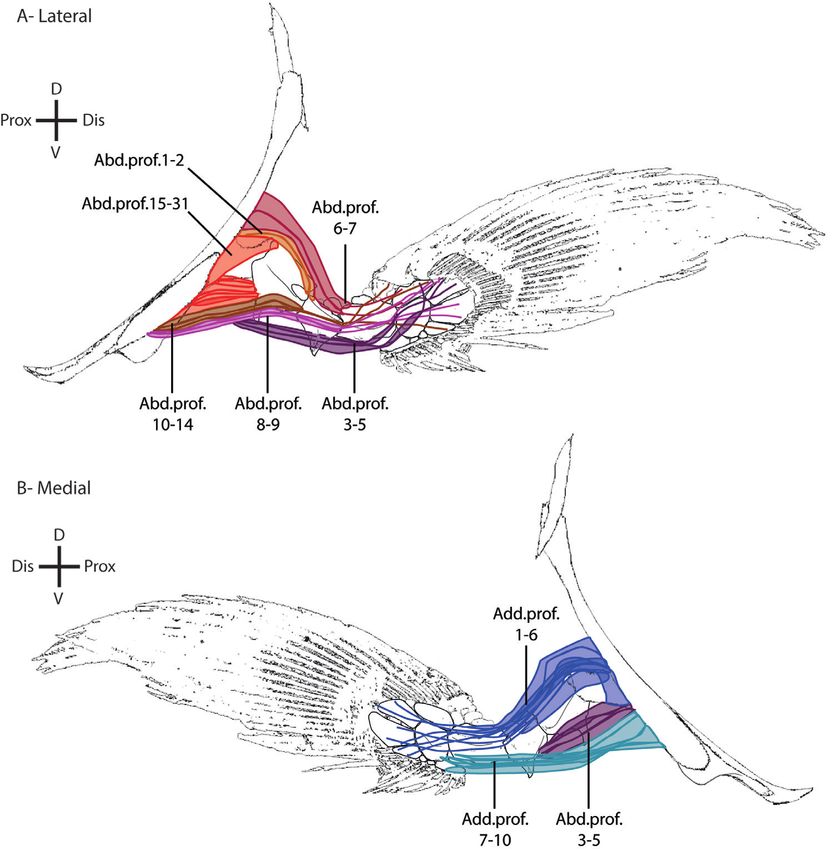
Table 1. Muscles of the superficial layer of the pectoral fin of the coelacanth L. chalumnae
954
Fin side Muscle Diogo et al. Millot & Origin(s) Insertion(s) Articulation Function Mass (g) Bundle ACSA
(2016) Anthony (1958) mode length (cm) (cm2)
Lateral Abductor Abductor “Adducteur Cleithrum: inner Mesomere 1 Mono-articular Abduction and 9.000 4.25 1.998
superficialis 1 superficialis superficiel” surface protraction
Abductor Extracleithrum: Mesomere 1 Mono-articular Adduction and 3.653 5.00 0.689
superficialis 2 posterior portion protraction
A. HUBY ET AL.
Abductor Mesomere 1: Mesomere 2 Mono-articular Abduction and 4.042 2.25 1.695
superficialis 3 distal part of and protraction
dorsal edge; pre-axial
scapulocoracoid: radial 1
glenoid process
Abductor Mesomere 1: lateral Mesomere 2 Mono-articular Adduction and 3.980 3.50 1.073
superficialis 4 edge protraction
Abductor Mesomere 2: distal Mesomere 3 Mono-articular Abduction and 2.899 2.00 1.367
superficialis 5 portion of the protraction
dorso-medial edge
Abductor Mesomere 2: Mesomere 3 Mono-articular Protraction 2.423 2.75 0.831
superficialis 6 dorso-lateral edge
Abductor Mesomere 3: lateral Fin rays 1–12 Poly-articular Protraction 1.786 3.00 0.562
superficialis 7 ridge
Abductor Mesomere 3: lateral Fin rays Poly-articular Protraction 2.732 4.50 0.573
superficialis 8 ridge 13–33
Medial Adductor Adductor “Adducteur Scapulocoracoid Mesomere 1 Mono-articular Adduction and 14.000 8.00 1.651
superficialis 1 superficialis superficiel” retraction
Adductor Cleithrum: anterior Mesomeres Poly-articular Retraction 7.000 10.00 0.660
superficialis 2 edge 1–3
Adductor Cleithrum: anterior Mesomeres Poly-articular Retraction 2.612 10.00 0.246
superficialis 3 edge 1–3
Adductor Mesomere 1: ventral Mesomere 2 Mono-articular Adduction and 2.838 4.50 0.595
superficialis 4 ridge retraction
Adductor Mesomere 1: ventral Mesomere Poly-articular Retraction 2.477 10.50 0.223
superficialis 5 ridge 3 + fin rays
22–33
Adductor Mesomere 2: dorso- Mesomere 3 Mono-articular Adduction and 2.238 3.50 0.603
superficialis 6 lateral edge retraction
Adductor Mesomere 3: distal Fin rays 1–21 Poly-articular Retraction 2.265 2.50 0.855
superficialis 7 part of dorsal edge
© 2021 The Linnean Society of London, Biological Journal of the Linnean Society, 2021, 133, 949–989
Downloaded from https://academic.oup.com/biolinnean/article/133/4/949/6261035 by guest on 17 September 2021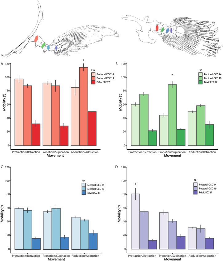
PAIRED FINS MUSCLES OF THE COELACANTH 955
Downloaded from https://academic.oup.com/biolinnean/article/133/4/949/6261035 by guest on 17 September 2021
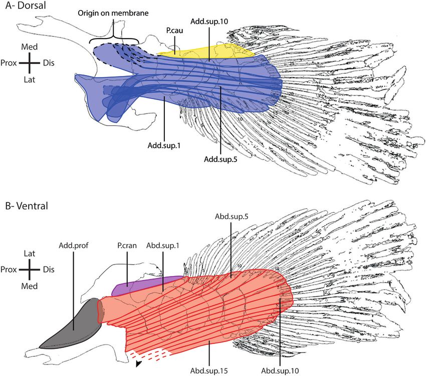
Figure 3. Superficial muscle layer of the pectoral fin of the coelacanth L. chalumnae in lateral (A) and medial (B) views. Blue:
adductor superficialis; red: abductor superficialis. Abd.sup.: abductor superficialis; Add.sup.: adductor superficialis; D:
dorsal; Dis: distal; Prox: proximal; V: ventral.
Abductor superficialis superficialis 7–8) are poly-articular, meaning they
The abductor superficialis muscle is formed by eight cross more than one joint.
different muscle bundles having different origins and
insertions (Table 1; Fig. 3A). Six bundles (abductor Abductor superficialis 1: Originates on the lateral
superficialis 1–6) are mono-articular, meaning they side of the fin, on the latero-posterior edge of the
cross only one joint, and two other bundles (abductor cleithrum dorsally to the extracleithrum. It inserts
© 2021 The Linnean Society of London, Biological Journal of the Linnean Society, 2021, 133, 949–989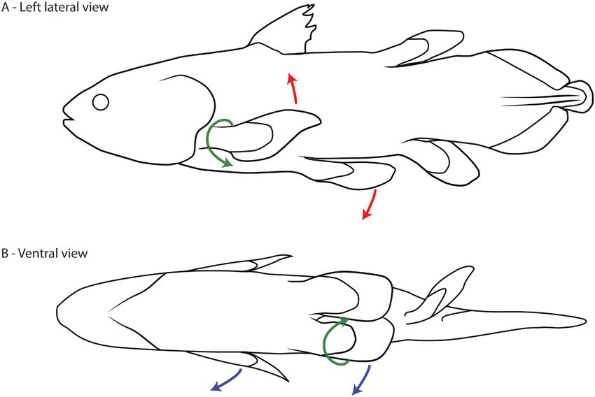
956    A. HUBY ET AL.

on the proximal portion of the dorsal ridge and                  (adductor superficialis 1, 4 and 6) are mono-articular
the dorso-lateral facet of mesomere 1. This muscle               and four bundles (adductor superficialis 2, 3, 5 and
bundle permits the abduction and protraction of                  7) are poly-articular.
mesomere 1.
                                                                 Adductor superficialis 1: Originates on the
Abductor superficialis 2: Originates on the lateral              ventro-medial edge of the cleithrum, the ventral
edge of the extracleithrum. It inserts on the proximal           side of the scapulocoracoid and the ventral edge
portion of the ventro-lateral aspect of mesomere                 of the extracleithrum. It inserts on the proximal
1. It permits the adduction and protraction of                   portion of the ventro-medial aspect of mesomere 1
mesomere 1.                                                      and on the large ventral ridge of this mesomere.
                                                                 It permits the adduction and retraction of

                                                                                                                                  Downloaded from https://academic.oup.com/biolinnean/article/133/4/949/6261035 by guest on 17 September 2021
Abductor superficialis 3: Originates on the dorso-               mesomere 1.
lateral aspect of mesomere 1 and on its dorsal ridge,
both on lateral and medial side of the fin. It inserts           Adductor superficialis 2–3: Originate from the
on the proximal portion of the dorsal part and dorso-            anterior edge of the cleithrum, and insert on the distal
medial ridge of mesomere 2. It permits the abduction             portion of the medial aspect of mesomere 3. They are
and protraction of mesomere 2.                                   also attached to mesomeres 1 and 2 with strong fibrous
                                                                 connective tissue. Adductor superficialis 3 is dorsal to
Abductor superficialis 4: Originates on the ventro-              adductor superficialis 2. They permit the retraction of
lateral aspect of mesomere 1. It inserts on the proximal         mesomeres 1 to 3.
portion of the ventral part of mesomere 2. It permits
the adduction and protraction of mesomere 2.                     Adductor superficialis 4: Originates on the distal
                                                                 portion of the ventral ridge of mesomere 1, on its
Abductor superficialis 5: Originates on the dorsal               medial side. It inserts on the proximal portion of the
part of mesomere 2, on its dorso-lateral and dorso-              ventro-medial ridge of mesomere 2. It permits the
medial edge. It inserts on the dorso-lateral aspect of           adduction and retraction of mesomere 2.
mesomere 3 and on its dorsal ridge, both on lateral and
medial sides of the fin. It permits the abduction and            Adductor superficialis 5: Originates on the proximal
protraction of mesomere 3.                                       portion of the ventro-medial aspect of mesomere 1,
                                                                 just dorsal to the origin of the adductor superficialis
Abductor superficialis 6: Originates on the proximal             4. It inserts on the medial side of mesomere 3 and at
part of lateral aspect of mesomere 2. It inserts on              the base of the fin rays 22 to 33 via an aponeurosis. It
the proximal portion of the ventro-lateral aspect                permits the retraction of mesomere 3 and fin rays 22
of mesomere 3. It permits the protraction of                     to 33.
mesomere 3.
                                                                 Adductor superficialis 6: Originates on the medial
Abductor superficialis 7: Originates on the distal               and lateral sides of the fin, on the distal portion of the
portion of the dorso-lateral aspect of mesomere                  ventro-medial ridge of mesomere 2. It inserts on the
3 and inserts at the base of fin rays 1 to 12 via an             ventral side of mesomere 3. It permits the adduction
aponeurosis at the lateral side of the fin. It permits           and retraction of mesomere 3.
the protraction of the distal part of the fin and fin
rays 1 to 12.                                                    Adductor superficialis 7: Originates on the distal
                                                                 portion of mesomere 3. It inserts at the base of fin rays
Abductor superficialis 8: Originates on the distal portion       1 to 21 via an aponeurosis at the medial side of the fin.
of the ventro-lateral aspect of mesomere 3 and inserts at        It permits the retraction of the distal part of the fin
the base of fin rays 13 to 33 via an insertion aponeurosis       and fin rays 1 to 21.
at the lateral side of the fin. It permits the protraction of
the distal part of the fin and fin rays 13 to 33.                                       Middle layer
                                                                                          (Table 2)
Adductor superficialis                                           The middle layer is also formed by two muscle masses:
The adductor superficialis muscle mass is formed                 the abductor profundus on the lateral side and the
by seven different muscle bundles having different               adductor profundus on the medial side of the pectoral
origins and insertions (Table 1; Fig. 3B). Three bundles         fin (Fig. 4).

                            © 2021 The Linnean Society of London, Biological Journal of the Linnean Society, 2021, 133, 949–989
Table 2. Muscles of the middle layer of the pectoral fin of the coelacanth L. chalumnae
Fin side Muscle Diogo et al. Millot & Origin(s) Insertion(s) Articulation Function Mass (g) Bundle ACSA
(2016) Anthony mode lenght (cm) (cm2)
(1958)
Lateral Abductor Abductor “Adducteur Cleithrum: inner Mesomere 1: distal Mono-articular Abduction and 2.327 4.30 0.511
profundus 1 profundus profond” surface portion of the protraction
dorso-lateral facet
Lateral Abductor Abd. prof. “Adducteur Cleithrum: inner Mesomere 1: distal Mono-articular Abduction and 1.545 4.00 0.364
profundus 2 profond” surface portion of the protraction
dorso-lateral facet
Lateral Abductor Abd. prof. “Adducteur Scapulocoracoid: Mesomeres 2–3 Poly-articular Adduction and 1.568 14.00 0.106
profundus 3 profond” proximal portion of along the ventral protraction
internal posterior edge (lateral face)
surface
Lateral Abductor Abd. prof. “Adducteur Scapulocoracoid: Mesomere 3 along Poly-articular Adduction and 7.000 18.00 0.367
profundus 4 profond” proximal portion of the ventral edge protraction
internal posterior (lateral face)
surface
Lateral Abductor Abd. prof. “Adducteur Scapulocoracoid: Mesomeres 1–3 Poly-articular Adduction and 1.495 10.00 0.141
profundus 5 profond” proximal portion of along the ventral protraction
internal posterior edge (lateral face)
surface
Lateral Abductor Abd. prof. “Adducteur Scapulocoracoid: Mesomeres 1–3 Poly-articular Abduction and 0.327 11.00 0.028
profundus 6 profond” proximal portion of (lateral face) protraction
internal posterior
surface
Lateral Abductor Abd. prof. “Adducteur Scapulocoracoid: Mesomeres 1–3 Poly-articular Abduction and 1.482 11.00 0.127
profundus 7 profond” proximal portion of (lateral face) protraction
internal posterior
surface
Lateral Abductor Abd. prof. “Adducteur Scapulocoracoid: Mesomeres 1–2 Mono-articular Adduction and 1.642 10.50 0.148
profundus 8 profond” proximal portion of along the ventral protraction
internal posterior edge (lateral face)
© 2021 The Linnean Society of London, Biological Journal of the Linnean Society, 2021, 133, 949–989
surface
Lateral Abductor Abd. prof. “Adducteur Scapulocoracoid: Mesomeres 1–2 Poly-articular Adduction and 1.032 8.00 0.122
profundus 9 profond” proximal portion of along the ventral protraction
internal posterior edge (lateral face)
surface
Lateral Abductor Abd. prof. “Adducteur Scapulocoracoid: Mesomere 1 along Poly-articular Adduction and 0.219 6.00 0.034
profundus 10 profond” proximal portion of the ventral edge protraction
internal posterior
PAIRED FINS MUSCLES OF THE COELACANTH
surface
957
Downloaded from https://academic.oup.com/biolinnean/article/133/4/949/6261035 by guest on 17 September 2021Table 2. Continued
958
Fin side Muscle Diogo et al. Millot & Origin(s) Insertion(s) Articulation Function Mass (g) Bundle ACSA
(2016) Anthony mode lenght (cm) (cm2)
(1958)
Lateral Abductor Abd. prof. “Adducteur Scapulocoracoid: Mesomere 1 along Mono-articular Adduction and 0.267 3.00 0.084
profundus 11 profond” proximal portion of the ventral edge protraction
internal posterior
surface
A. HUBY ET AL.
Lateral Abductor Abd. prof. “Adducteur Scapulocoracoid: Mesomere 1 along Mono-articular Adduction and 0.605 6.00 0.095
profundus 12 profond” proximal portion of the ventral edge protraction
internal posterior
surface
Lateral Abductor Abd. prof. “Adducteur Scapulocoracoid: Mesomere 1 along Mono-articular Adduction and 0.414 4.50 0.087
profundus 13 profond” proximal portion of the ventral edge protraction
internal posterior
surface
Lateral Abductor Abd. prof. “Adducteur Scapulocoracoid: Mesomere 1 along Mono-articular Adduction and 0.451 4.20 0.101
profundus 14 profond” proximal portion of the ventral edge protraction
internal posterior
surface
Lateral Abductor Abd. prof. “Adducteur Scapulocoracoid: Mesomere 1: prox- Mono-articular Adduction and 0.247 4.50 0.052
profundus 15 profond” proximal portion of imal edge (lateral protraction
internal posterior face)
surface
Lateral Abductor Abd. prof. “Adducteur Scapulocoracoid: Mesomere 1: Mono-articular Adduction and 0.027 4.00 0.006
profundus 16 profond” proximal portion of proximal edge protraction
internal posterior (lateral face)
surface
Lateral Abductor Abd. prof. “Adducteur Scapulocoracoid: Mesomere 1: Mono-articular Adduction and 0.140 4.20 0.031
profundus 17 profond” proximal portion of proximal edge protraction
internal posterior (lateral face)
surface
Lateral Abductor Abd. prof. “Adducteur Scapulocoracoid: Mesomere 1: Mono-articular Adduction and 0.101 4.00 0.024
profundus 18 profond” proximal portion of proximal edge protraction
internal posterior (lateral face)
surface
Lateral Abductor Abd. prof. “Adducteur Scapulocoracoid: Mesomere 1: Mono-articular Adduction and 0.014 3.70 0.003
profundus 19 profond” proximal portion of proximal edge protraction
internal posterior (lateral face)
surface
Lateral Abductor Abd. prof. “Adducteur Scapulocoracoid: Mesomere 1: Mono-articular Adduction and 0.383 4.00 0.090
profundus 20 profond” proximal portion of proximal edge protraction
internal posterior (lateral face)
surface
© 2021 The Linnean Society of London, Biological Journal of the Linnean Society, 2021, 133, 949–989
Downloaded from https://academic.oup.com/biolinnean/article/133/4/949/6261035 by guest on 17 September 2021Table 2. Continued
Fin side Muscle Diogo et al. Millot & Origin(s) Insertion(s) Articulation Function Mass (g) Bundle ACSA
(2016) Anthony mode lenght (cm) (cm2)
(1958)
Lateral Abductor Abd. prof. “Adducteur Scapulocoracoid: Mesomere 1: Mono-articular Adduction and 0.689 4.00 0.163
profundus 21 profond” proximal portion of proximal edge protraction
internal posterior (lateral face)
surface
Lateral Abductor Abd. prof. “Adducteur Scapulocoracoid: Mesomere 1: Mono-articular Adduction and 0.046 4.00 0.011
profundus 22 profond” proximal portion of proximal edge protraction
internal posterior (lateral face)
surface
Lateral Abductor Abd. prof. “Adducteur Scapulocoracoid: Mesomere 1: Mono-articular Adduction and 0.254 3.60 0.066
profundus 23 profond” proximal portion of proximal edge protraction
internal posterior (lateral face)
surface
Lateral Abductor Abd. prof. “Adducteur Scapulocoracoid: Mesomere 1: Mono-articular Adduction and 0.035 3.10 0.011
profundus 24 profond” proximal portion of proximal edge protraction
internal posterior (lateral face)
surface
Lateral Abductor Abd. prof. “Adducteur Scapulocoracoid: Mesomere 1: Mono-articular Adduction and 0.129 3.70 0.033
profundus 25 profond” proximal portion of proximal edge protraction
internal posterior (lateral face)
surface
Lateral Abductor Abd. prof. “Adducteur Scapulocoracoid: Mesomere 1: Mono-articular Adduction and 0.247 3.80 0.061
profundus 26 profond” proximal portion of proximal edge protraction
internal posterior (lateral face)
surface
Lateral Abductor Abd. prof. “Adducteur Scapulocoracoid: Mesomere 1: Mono-articular Adduction and 0.089 3.10 0.027
profundus 27 profond” proximal portion of proximal edge protraction
internal posterior (lateral face)
surface
Lateral Abductor Abd. prof. “Adducteur Scapulocoracoid: Mesomere 1: Mono-articular Adduction and 0.194 4.00 0.046
profundus 28 profond” proximal portion of proximal edge protraction
© 2021 The Linnean Society of London, Biological Journal of the Linnean Society, 2021, 133, 949–989
internal posterior (lateral face)
surface
Lateral Abductor Abd. prof. “Adducteur Scapulocoracoid: Mesomere 1: Mono-articular Adduction and 0.226 4.50 0.047
profundus 29 profond” proximal portion of proximal edge protraction
internal posterior (lateral face)
surface
Lateral Abductor Abd. prof. “Adducteur Scapulocoracoid: Mesomere 1: Mono-articular Adduction and 2.886 3.00 0.907
PAIRED FINS MUSCLES OF THE COELACANTH
profundus 30 profond” proximal portion of proximal edge protraction
internal posterior (lateral face)
surface
959
Downloaded from https://academic.oup.com/biolinnean/article/133/4/949/6261035 by guest on 17 September 2021Table 2. Continued
960
Fin side Muscle Diogo et al. Millot & Origin(s) Insertion(s) Articulation Function Mass (g) Bundle ACSA
(2016) Anthony mode lenght (cm) (cm2)
(1958)
Lateral Abductor Abd. prof. “Adducteur Scapulocoracoid: Mesomere 1: prox- Mono-articular Adduction and 1.173 3.25 0.340
profundus 31 profond” proximal portion of imal edge (lateral protraction
internal posterior face)
surface
A. HUBY ET AL.
Medial Adductor Adductor “Abducteur Cleithrum: medio- Mesomeres 1–3 Poly-articular Abduction and 0.869 8.30 0.099
profondus 1 profundus profond” caudal edge of the along the dorsal retraction
superior portion edge (medial face)
Medial Adductor Add. prof. “Abducteur Cleithrum: medio- Mesomeres 1–3 Poly-articular Abduction and 2.323 10.50 0.209
profondus 2 profond” caudal edge of the along the dorsal retraction
superior portion edge (medial face)
Medial Adductor Add. prof. “Abducteur Cleithrum: medio- Mesomeres 1–3 Poly-articular Abduction and 3.253 12.60 0.244
profondus 3 profond” caudal edge of the along the dorsal retraction
superior portion edge (medial face)
Medial Adductor Add. prof. “Abducteur Cleithrum: medio- Mesomeres 1–3 Poly-articular Abduction and 1.798 8.00 0.212
profondus 4 profond” caudal edge of the along the dorsal retraction
superior portion edge (medial face)
Medial Adductor Add. prof. “Abducteur Cleithrum: medio- Mesomeres 1–2 Poly-articular Abduction and 0.694 5.50 0.119
profondus 5 profond” caudal edge of the along the dorsal retraction
superior portion edge (medial face)
Medial Adductor Add. prof. “Abducteur Cleithrum: medio- Mesomeres 1–2 Poly-articular Abduction and 0.134 3.50 0.036
profondus 6 profond” caudal edge of the along the dorsal retraction
superior portion edge (medial face)
Medial Adductor Add. prof. “Abducteur Scapulocoracoid: Mesomeres 2–3 Poly-articular Adduction and 1.948 10.50 0.175
profondus 7 profond” proximal portion of along the ventral retraction
the ventral surface edge (medial face)
Medial Adductor Add. prof. “Abducteur Scapulocoracoid: Mesomeres 2–3 Poly-articular Adduction and 2.460 12.50 0.186
profondus 8 profond” proximal portion of along the ventral retraction
the ventral surface edge (medial face)
Medial Adductor Add. prof. “Abducteur Scapulocoracoid: Mesomeres 2–3 Poly-articular Adduction and 2.028 13.00 0.147
profondus 9 profond” proximal portion of along the ventral retraction
the ventral surface edge (medial face)
Medial Adductor Add. prof. “Abducteur Scapulocoracoid: Mesomeres 2–3 Poly-articular Adduction and 1.228 14.50 0.080
profondus 10 profond” proximal portion of along the ventral retraction
the ventral surface edge (medial face)
© 2021 The Linnean Society of London, Biological Journal of the Linnean Society, 2021, 133, 949–989
Downloaded from https://academic.oup.com/biolinnean/article/133/4/949/6261035 by guest on 17 September 2021PAIRED FINS MUSCLES OF THE COELACANTH 961
Downloaded from https://academic.oup.com/biolinnean/article/133/4/949/6261035 by guest on 17 September 2021
Figure 4. Middle muscle layer of the pectoral fin of the coelacanth L. chalumnae in lateral (A) and medial (B) views. Abd.
prof.: abductor profundus; Add.prof.: adductor profundus; D: dorsal; Dis: distal; Prox: proximal; V: ventral.
Abductor profundus They insert on the distal part of the dorso-lateral
The abductor profundus muscle is formed by 31 aspect of mesomere 1. They permit the abduction and
different muscle bundles (Fig. 4A). Among them, 24 protraction of mesomere 1.
bundles are mono-articular (abductor profundi 1–2, 8,
11–31) and seven are poly-articular (abductor profundi Abductor profundi 3–5: Originate on the ventral side
3–7 and 9–10). of the scapulocoracoid. They follow the ventral edge of
mesomeres 1 to 3 and insert on the lateral side of the
Abductor profundi 1–2: Originate on the medio-caudal fin via a long tendon. They permit the adduction and
edge of the superior part of the cleithrum (Table 2). protraction of the fin.
© 2021 The Linnean Society of London, Biological Journal of the Linnean Society, 2021, 133, 949–989962 A. HUBY ET AL.
Abductor profundi 6–7: Originate on the ventral side The deep layer is composed of eight antagonistic
of the scapulocoracoid. They follow the dorsal edge of pairs of pronator and supinator muscles that cover
mesomeres 1 to 3 and insert on the lateral side of the the entire endoskeletal axis of the pectoral fin and the
fin via a long tendon. They permit the abduction and proximal portion of dermal fin rays, and a post-axial
protraction of the fin. muscle (pterygialis caudalis) (Fig. 5). In the deep layer,
most of the muscles are mono-articular.
Abductor profundi 8–9: Originate on the ventral
side of the scapulocoracoid, near the edge of the
extracleithrum. They follow the ventral edge of
Pronator muscles
mesomeres 1 and 2 and insert on the lateral side of the The pronator muscle group is formed by eight
fin via a long tendon. They permit the adduction and pronator muscles at the lateral side of the pectoral
Downloaded from https://academic.oup.com/biolinnean/article/133/4/949/6261035 by guest on 17 September 2021
protraction of mesomeres 1 and 2. fin that run obliquely across the ventral margin of
the fin (Fig. 5A). Pronator 1b is the only pronator
Abductor profundi 10–14: Originate on the ventral bundle that originates and inserts on the medial
side of the scapulocoracoid, near the edge of the side of the fin.
extracleithrum on the lateral side of the girdle. They The pronator 1–4 muscles are mono-articular. Their
insert on the lateral side of mesomere 1 via a long tendon numbers correspond to the mesomere they insert
and follow the ventral edge of this mesomere. They upon (or the previous pre-axial radial), and they
permit the adduction and protraction of mesomere 1. originate from the previous element (scapulocoracoid
or mesomere). They permit the pronation of the
Abductor profundi 15–31: Originate on the ventral associated element and of the fin.
side of the fin, on the articular process of the Pronators 5–8 insert on the proximal portion of the
scapulocoracoid. They insert on the proximal edge of fin rays, and originate on mesomere 3 (pron. 5 and
mesomere 1 via a short tendon, on the lateral side of 7), mesomere 4 (pron. 6) and mesomere 2 (pron. 8).
the fin. They permit the adduction and protraction of They permit the lateral flexion of the fin rays and the
mesomere 1. pronation (pron. 5, 6a) and supination of the fin (pron.
6b, 6c, 7, 8).
Adductor profundus
Supinator muscles
The adductor profundus muscle is formed by ten poly-
articular muscle bundles that run under the surface of The supinator muscle group is formed by eight
the abductor superficialis muscle (Fig. 4B). supinator muscles on the medial side of the pectoral
fin (Fig. 5B–C). The organization of these muscles
Adductor profundi 1–6: Originate from the medio- is quite similar to that of the pronator muscles.
caudal edge of the superior portion of the cleithrum Supinators 1a, 2a and 2c insert on the lateral side
(Table 2). They follow the dorsal edge of mesomeres 1 of the fin.
to 3 and insert via a long tendon to the medial side of Supinators 1–4 are mono-articular muscles. The
the fin. They permit the abduction and retraction of number assigned to the muscles corresponds to the
the fin. mesomere they insert upon (or the previous pre-axial
radial). They originate from the previous element
Adductor profundi 7–10: Originate on the ventral (scapulocoracoid or mesomere). These muscles permit
side of the scapulocoracoid (Table 2). They follow the the supination of the associated elements and of
ventral edge of mesomeres 2 and 3, and insert via a the fin.
long tendon on the medial side of the fin. They permit Supinators 5–8 insert on the proximal portion of the
the adduction and retraction of the fin. fin rays at the medial side of the fin, and originate on
mesomere 3 (sup. 5), mesomere 4 (sup. 6), mesomere 2
(sup. 7) and mesomere 1 (sup. 8). They are symmetrical
Deep layer to pronators 5–8 and permit their antagonistic
movement (Table 3).
(Table 3)
Here, we refer to pronator and supinator muscles as
the muscles of the deep layer that insert respectively Pterygialis caudalis
on the lateral and medial sides of the pectoral fin (see The pterygialis caudalis follows the post-axial edge
Diogo et al., 2016; Miyake et al., 2016; Fig. 5). However, of the pectoral fin. It originates on the medial side of
the exact role of each muscle can be different from mesomere 1 and inserts at the base of fin ray 33. It
what the name suggests, as indicated in Table 3. permits the adduction of the fin.
© 2021 The Linnean Society of London, Biological Journal of the Linnean Society, 2021, 133, 949–989Table 3. Muscles of the deep layer of the pectoral fin of the coelacanth L. chalumnae
Fin side Muscle Diogo et al. Millot & Origin(s) Insertion(s) Articulation Function Mass (g) Bundle ACSA
(2016) Anthony (1958) mode length (cm) (cm2)
Lateral Pronator 1a Supinator 1 “Supinateur 1” Scapulocoracoid: Mesomere 1: Mono-articular Pronation 6.000 5.00 1.132
glenoid process proximal portion
(medial side) of the ventral
ridge (lateral side)
Pronator 1b Supinator 1 “Supinateur 1” Scapulocoracoid: Mesomere 1: Mono-articular Pronation 5.411 3.50 1.458
proximal part of the ventro-medial face
inferior portion of
the glenoid process
(medial side)
Pronator 2a Supinator 1 “Supinateur 1” Mesomere 1: proximal Pre-axial radial 1 Mono-articular Pronation 0.312 3.00 0.098
portion of ventral (lateral side)
edge (lateral side)
Pronator 2b Supinator 1 “Supinateur 1” Mesomere 1: ventral Pre-axial radial 1 Mono-articular Pronation 2.739 2.65 0.975
edge and ventral ridge (lateral side)
(lateral side)
Pronator 2c Supinator 2 “Supinateur 2” Mesomere 1: distal Mesomere 2: Mono-articular Pronation 3.018 3.00 0.949
portion of ventral proximal portion
edge (medial side) of the dorsal edge
Pronator 2d Supinator 2 “Supinateur 2” Mesomere 1: distal Mesomere 2: Mono-articular Pronation 2.286 6.00 0.359
portion of ventral proximal portion of
edge (medial side) the ventro-lateral
ridge
Pronator 3a Supinator 2 “Supinateur 2” Mesomere 2: proximal Pre-axial radial 2; Mono-articular Pronation 2.971 3.25 0.862
portion of the mesomere 3:
ventro-lateral ridge proximal portion
of the dorsal edge
(lateral face)
Pronator 3b Supinator 3 “Supinateur 3” Mesomere 2: distal part Mesomere 3: ventral Mono-articular Pronation 2.200 3.50 0.593
of the ventro-lateral side of the lateral
ridge ridge
© 2021 The Linnean Society of London, Biological Journal of the Linnean Society, 2021, 133, 949–989
Pronator 4 Supinator 3 “Supinateur 3” Mesomere 3: ventral Mesomere 4: Mono-articular Pronation 0.718 2.50 0.271
ridge proximal portion
of lateral ridge
Pronator 5 Supinator 3 “Supinateur 3” Mesomere 3: ventral Proximal portion of Mono-articular Lateral flexion 3.454 4.50 0.724
ridge (lateral side) fin rays 1–6 of fin rays and
pronation
Pronator 6a Supinator 4 “Supinateur 4” Mesomere 4: lateral Proximal portion of Mono-articular Lateral flexion 2.568 3.50 0.692
edge fin rays 6–12 of fin rays and
PAIRED FINS MUSCLES OF THE COELACANTH
pronation
963
Downloaded from https://academic.oup.com/biolinnean/article/133/4/949/6261035 by guest on 17 September 2021Table 3. Continued
964
Fin side Muscle Diogo et al. Millot & Origin(s) Insertion(s) Articulation Function Mass (g) Bundle ACSA
(2016) Anthony (1958) mode length (cm) (cm2)
Pronator 6b Supinator 4 “Supinateur 4” Mesomere 4: distal Proximal portion of Mono-articular Lateral flexion of 1.356 2.75 0.465
portion of lateral edge fin rays 13–21 fin rays and
supination
Pronator 6c Supinator 4 “Supinateur 4” Mesomere 4: lateral Proximal portion of Mono-articular Lateral flexion of 1.957 1.75 1.055
edge fin rays 21–33 fin rays and
A. HUBY ET AL.
supination
Pronator 7 Pterygialis “Supinateur 5” Mesomere 3: ventral Proximal portion of Poly-articular Lateral flexion of 0.164 3.80 0.041
caudalis edge fin rays 28–33 fin rays and
supination
Pronator 8 Pterygialis “Supinateur 5” Mesomere 2: distal Proximal portion of Poly-articular Lateral flexion of 0.978 5.50 0.168
caudalis portion of the fin rays 28–33 fin rays and
dorso-lateral edge supination
Medial Supinator 1a Pronator 1 “Pronateur 1” Scapulocoracoid: Mesomere 1: prox- Mono-articular Supination 1.880 1.50 1.182
superior portion of the imal portion of the
glenoid process ventral ridge (lat-
eral side)
Supinator 1b Pronator 1 “Pronateur 1” Scapulocoracoid: Mesomere 1: distal Mono-articular Supination 5.140 3.50 1.385
superior portion of the portion of
glenoid process dorso-medial face
Supinator 2a Pronator 1 “Pronateur 2” Mesomere 1: proximal Pre-axial radial 1 Mono-articular Supination 1.244 1.50 0.782
portion of the dorsal (lateral side)
ridge (lateral side)
Supinator 2b Pronator 2 “Pronateur 2” Mesomere 1: distal Mesomere 2: Mono-articular Supination 2.689 3.00 0.846
portion of the medial dorso-medial edge
edge
Supinator 2c Pronator 2 “Pronateur 2” Mesomere 1: distal Pre-axial radial 2: Poly-articular Supination 0.591 2.50 0.223
portion of the dorsal proximal portion
ridge (lateral side)
Supinator 3a Pronator 2 “Pronateur 3” Mesomere 2: distal Pre-axial radial 2; Mono-articular Supination 1.875 2.00 0.884
portion of the dorso- proximal element
medial ridge of pre-axial
accessory element
Supinator 3b Pronator 3 “Pronateur 3” Mesomere 2: distal Mesomere 3: distal Mono-articular Supination 1.699 4.00 0.401
portion of portion of the
ventro-medial ridge medial ridge
Supinator 4 Pronator 3 “Pronateur 3” Mesomere 3: distal Mesomere 4: prox- Mono-articular Supination 0.663 2.75 0.227
portion of the ventral imal portion of
ridge (medial side) the ventral edge
(medial side)
© 2021 The Linnean Society of London, Biological Journal of the Linnean Society, 2021, 133, 949–989
Downloaded from https://academic.oup.com/biolinnean/article/133/4/949/6261035 by guest on 17 September 2021Table 3. Continued
Fin side Muscle Diogo et al. Millot & Origin(s) Insertion(s) Articulation Function Mass (g) Bundle ACSA
(2016) Anthony (1958) mode length (cm) (cm2)
Supinator 5 Pronator 3 “Pronateur 3” Mesomere 3: distal Proximal portion of Mono-articular Medial flexion of 1.667 2.25 0.699
portion of the dorsal fin rays 1–7 fin rays and
ridge (medial side) supination
Supinator 6a Pronator 4 “Pronateur 4” Mesomere 4: dorsal edge Proximal portion of Mono-articular Medial flexion of 0.873 2.75 0.299
(medial side) fin rays 7–12 fin rays and su-
pination
Supinator 6b Pronator 4 “Pronateur 4” Mesomere 4: distal Proximal portion of Mono-articular Medial flexion 0.924 2.75 0.317
portion of the ventral fin rays 13–23 of fin rays and
edge (medial side) pronation
Supinator 6c Pronator 4 “Pronateur 4” Mesomere 4: ventral Proximal portion of Mono-articular Medial flexion 0.749 1.75 0.404
edge (medial side) fin rays 23–33 of fin rays and
pronation
Supinator 7 Pterygialis “Pronateur 5” Mesomere 2: distal Proximal portion of Poly-articular Medial flexion 0.364 4.50 0.076
caudalis portion of medial edge fin rays 28–33 of fin rays and
pronation
Supinator 8 Pterygialis “Pronateur 5” Mesomere 1: Proximal portion of Poly-articular Medial flexion 0.700 6.50 0.102
caudalis ventro-lateral edge fin rays 28–33 of fin rays and
pronation
Pterygialis Pterygialis Undescribed Mesomere 1: distal Proximal portion of Poly-articular Adduction 0.691 8.00 0.081
caudalis caudalis portion of the medial fin ray 33
edge
© 2021 The Linnean Society of London, Biological Journal of the Linnean Society, 2021, 133, 949–989
PAIRED FINS MUSCLES OF THE COELACANTH
965
Downloaded from https://academic.oup.com/biolinnean/article/133/4/949/6261035 by guest on 17 September 2021966 A. HUBY ET AL.
Downloaded from https://academic.oup.com/biolinnean/article/133/4/949/6261035 by guest on 17 September 2021
Figure 5. Deep muscle layer of the pectoral fin of the coelacanth L. chalumnae in lateral (A), dorsal (B) and medial (C)
views. Different colors represent different muscles. P.cau: pterygialis caudalis; Pron.: pronator; Sup.: supinator; D: dorsal;
Dis: distal; Lat: lateral; Med: medial; Prox: proximal; V: ventral.
© 2021 The Linnean Society of London, Biological Journal of the Linnean Society, 2021, 133, 949–989PAIRED FINS MUSCLES OF THE COELACANTH 967
Pelvic musculature anatomy caudalis on the post-axial side of the fin (Fig. 6). All
Eighty-three muscle bundles, organized into 12 the muscles of the superficial layer are poly-articular.
functional groups, were identified for the pelvic fin of
L. chalumnae. Moreover, most of these muscle bundles Adductor superficialis pelvicus
were separated in several sub-bundles (Supporting
The adductor superficialis pelvicus is composed of ten
Information, Table S1). The resting position of the
dorsal muscle bundles which are oriented proximally
pelvic fin was considered as the fin positioned along
to distally and numbered from the pre-axial to post-
the ventral side of the body, with the leading edge
axial side of the pelvic fin (Fig. 6A). These muscle
directed laterally. Abductor muscles are on the ventral
bundles originate on: (i) the lateral process of the
side of the fin, and adductor muscles are located on
pelvic girdle (add. sup. pelvicus 1–7), onto both the
Downloaded from https://academic.oup.com/biolinnean/article/133/4/949/6261035 by guest on 17 September 2021
the dorsal side of the fin. Compared to the pectoral fin,
dorsal and the ventral side; and (ii) on the fascia that
most muscles of the pelvic fin are poly-articular and
separates the pelvic fin muscles from the abdominal
originate from the pelvic girdle and insert at the base
muscles (add. sup. pelvicus 7–10; Table 4). They insert
of the fin rays. For the pelvic fin in its resting position,
at the base of fin rays 1 to 27 on the dorsal side of
movements are defined as follows:
the fin via short aponeuroses. Due to their origin
1. P rotraction: the pre-axial edge of the fin has a onto the curved concave lateral process of the pelvic
forward movement. girdle, these muscle bundles cross each other (Fig. 6A).
2. R etraction: the pre-axial edge of the fin has a Add. sup. pelvicus 8 and 9 also insert respectively
backward movement. on the dorsal ridge of mesomere 3 and mesomere 2
3. A bduction: the ventral side of the fin has a by a strong fibrous connective tissue band. Add. sup.
downward movement. pelvicus 1 also inserts on the pre-axial radials 1 and
4. Adduction: the dorsal side of the fin has an upward 2. Add. sup. pelvicus 2–6 have a gutter-like shape
movement. and surround the adductor profundus pelvicus of the
5. Pronation: the ventral side of the fin has a medial middle layer as follows. Add. sup. pelvicus 2 surrounds
pivoting movement around the axis of the fin. adductor profundus pelvicus 1,2, add. sup. pelvicus 3
6. Supination: the ventral side of the fin has a lateral surrounds add. prof. pelvicus 3,4, add. sup. pelvicus 4
pivoting movement around the axis of the fin. surrounds add. prof. pelvicus 5,6,7,9, add. sup. pelvicus
5 surrounds add. prof. pelvicus 8,10 and add. sup.
Similar to the pectoral fin, the pelvic fin is not
pelvicus 6 surrounds add. prof. pelvicus 11,12. All add.
maintained in this reference position during
sup. pelvicus bundles permit the adduction of the fin
swimming. The movements of the fin defined here thus
rays. Add. sup. pelvicus 1 also permits the protraction
refer to the resting position of the fin (Fig. 2). Since
of the fin.
the pectoral and pelvic fins are oriented in different
planes, the movements of the two fins are defined
differently. Thus, the protraction movement of the Pterygialis caudalis
pectoral fin corresponds to an abduction movement
The pterygialis caudalis originates from the ventral
of the pelvic fin, and an abduction movement of the
side of the postero-superior process of the pelvic
pectoral fin corresponds to a protraction movement of
girdle and inserts at the base of pelvic fin rays 28–30
the pelvic fin.
(Fig. 6A). Based on its origin and insertion, the role of
the pterygialis caudalis is to adduct the pelvic fin.
Superficial layer
(Table 4) Abductor superficialis pelvicus
Since the muscle organization is simpler compare The abductor superficialis pelvicus is composed of
to that of the pectoral fin, we provide here a general 15 ventral muscle bundles. As in the dorsal muscles,
description of the origin and insertion of the muscles these muscles are numbered from pre-axial to post-
of the superficial layer. The detailed origins and axial, and are oriented from proximal to distal (Fig.
insertions of each muscle in the superficial layer are 6B). They originate: (i) anteriorly on the lateral
provided in Table 4. process of the pelvic girdle, on the adductor profundus
The superficial layer of the pelvic fin is formed by four pelvicus, via an aponeurosis; (ii) on the ventral side
muscle masses: the adductor superficialis pelvicus on of the pelvic girdle; (iii) on the medial process of the
the dorsal side and the abductor superficialis pelvicus pelvic girdle; and (iv) on the aponeurosis between the
on the ventral side of the fin, as well as the pterygialis two pelvic fins from ventral to dorsal (Table 4). Abd.
cranialis on the pre-axial side and the pterygialis sup. 15 has the most dorsal origin on this aponeurosis.
© 2021 The Linnean Society of London, Biological Journal of the Linnean Society, 2021, 133, 949–989You can also read