Restricted compensatory movement on activation pattern of gluteal muscles during unilateral weight-bearing exercise
←
→
Page content transcription
If your browser does not render page correctly, please read the page content below
Original Article

The influences of restricted compensatory
movement on activation pattern of gluteal muscles
during unilateral weight-bearing exercise

SOO-YONG KIM1, MIN-HYEOK KANG2 1
1Department of      Marine Sports, Pukyong National University, Busan, Republic of Korea
2Department of Physical Therapy, College of Health Sciences, Catholic University of Pusan, Busan, Republic
of Korea

                                                     ABSTRACT

We investigated changes in lateral pelvic tilt and activity of the gluteal muscles during one-leg standing with
and without pressure biofeedback. Seventeen participants performed one-leg standing tests with pressure
biofeedback (the threshold was set to the minimum change in pressure) and without biofeedback (standard
one-leg standing). The lateral pelvic tilt was significantly lower in one-leg standing with pressure biofeedback
than in standard one-leg standing (p < .05). In addition, gluteus medius and gluteus maximus activity was
significantly greater during one-leg standing with pressure biofeedback than during standard one-leg standing
(p < .05). Based on our results, restriction of compensatory movement can be used to increase activity of
gluteal muscles when performing unilateral weight-bearing exercise such as one-leg standing.
Keywords: Biofeedback; Buttocks; Exercise; Posture.

    Cite this article as:
    Kim, S-Y., & Kang, M-H. (2020). The influences of restricted compensatory movement on activation
       pattern of gluteal muscles during unilateral weight-bearing exercise. Journal of Human Sport and
       Exercise, in press. doi:https://doi.org/10.14198/jhse.2021.163.11

1
    Corresponding author. Department of Physical Therapy, College of Health Sciences, Catholic University of Pusan, 57
      Oryundae-ro, Geumjeong-gu, Busan 46252, Republic of Korea. http://orcid.org/0000-0002-1530-7254
      E-mail: kmhyuk01@gmail.com
      Submitted for publication November 19, 2019
      Accepted for publication January 27, 2020
      Published in press April 24, 2020
      JOURNAL OF HUMAN SPORT & EXERCISE ISSN 1988-5202
      © Faculty of Education. University of Alicante
      doi:10.14198/jhse.2021.163.11
                                                                            VOLUME -- | ISSUE - | 2020 |        1
Kim et al. / Restricted compensatory movement changes muscle activation pattern   JOURNAL OF HUMAN SPORT & EXERCISE

INTRODUCTION

The gluteus medius (Gmed) is wide and fan shaped (Pfirrmann et al., 2001), and its primary role is as a hip
abductor. It accounts for 60% of the total cross-sectional area of the hip abductor muscles (Clark and Haynor,
1987). The gluteus maximus (Gmax) has the largest cross-sectional area of the hip muscles (Reiman et al.,
2012) and acts as a hip abductor as well as a powerful hip extensor and external rotator (Nakagawa et al.,
2008; Reiman et al., 2012; Wilson et al., 2006). These two muscles support weight-bearing activities by
assisting in load transfer and maintaining the alignment of the lower extremities (Lee, 1996). Weakness of
the Gmed and Gmax is linked to lower extremity injuries and lower back pain (LBP) (Akuthota and Nadler,
2004; Brindle et al., 2003). Gmed and Gmax strengthening exercises are used to treat and prevent lower
extremity injuries and back pain, and weight-bearing exercise is recommended because it requires greater
muscle activity than non-weight-bearing exercise (Macadam et al., 2015).

One-leg standing (OLS) is a weight-bearing exercise that is used to strengthen hip abductor muscles (Krause
et al., 2009). The hip abductors generate a force equal to twice the body’s weight to maintain the correct
pelvic and torso posture during OLS (Neumann, 1989), and they have the muscle length to produce the
greatest torque in the OLS condition (Neumann et al., 1988). However, pelvic deviation may occur to the
support side due to the shift of the body’s centre of gravity during OLS, and this leads to a contralateral pelvic
depression, which may cause weakening of the hip abductors (Fujita et al., 2017; Grimaldi, 2011). Previous
studies have shown that ipsilateral deviation of the pelvis during OLS decreases Gmed muscle activity (Fujita
et al., 2017; Prior et al., 2014). Therefore, to increase activity of the hip abductor muscles during OLS, there
is a need to minimize pelvic deviation.

Pressure biofeedback is a way to check the change in pressure at the contact area in real time, which is
useful for identifying deviations in body alignment during exercise. Thus, since the deviation of the trunk or
pelvis can be checked in real time during OLS, pressure biofeedback can be used to improve lumbopelvic
alignment and correct muscle activity (Cynn et al., 2006; Noh et al., 2014; Oh et al., 2007; Yu et al., 2018).
Cynn et al. (2006) reported a decreased pelvic lateral tilt angle and increased Gmed activity during side-lying
hip abduction with pressure biofeedback compared to without. A similar study demonstrated that the pelvic
anterior tilt angle decreased and hip extensor muscle activity increased during prone hip extension with the
addition of pressure biofeedback (Oh et al., 2007). It has also been demonstrated that the application of
pressure biofeedback during active straight leg raise (ASLR) tests increased abdominal activity while
decreasing pelvic rotation (Noh et al., 2014), and Yu et al. (2018) observed that infraspinatus activity
increased and posterior deltoid activity decreased when the pressure was maintained at 2 mmHg using
pressure biofeedback during prone shoulder external rotation.

Previous studies have shown that the pressure biofeedback method is useful to correct movement patterns
and increase muscle activity during therapeutic exercise (Cynn et al., 2006; Noh et al., 2014; Oh et al., 2007;
Yu et al., 2018). However, no studies have investigated the effects of pressure biofeedback on gluteal muscle
activity and pelvic lateral tilt during OLS. In addition, the pressure biofeedback unit used in previous studies
has a scale with a narrow range, which makes it difficult to identify accurately during exercise.

The purpose of this study was to investigate differences in the activities of the Gmed and Gmax during the
OLS test with and without pressure biofeedback. As a secondary measure, we recorded the difference in
lateral pelvic tilt during OLS with and without pressure biofeedback.

     2      | 2020 | ISSUE - | VOLUME --                                                    © 2020 University of Alicante
Kim et al. / Restricted compensatory movement changes muscle activation pattern JOURNAL OF HUMAN SPORT & EXERCISE
MATERIALS AND METHODS
Participants
Seventeen healthy male subjects (age 31.26 ± 2.53 years, height 176 ± 2.66 cm, weight 73.89 ± 3.85 kg)
participated in this study. The inclusion criteria included no history of low back pain or lower extremity pain
and the ability to maintain OLS for more than 5 s. Subjects were excluded if they could not maintain the
correct posture during OLS or if the exercise generated pain in the lower back or lower extremity. Prior to the
study, all participants read and signed an informed consent form approved by the Pukyong National
University Institutional Review Board.
G*power software was used to determine the necessary sample size (ver. 3.1.2; Franz Faul, University of
Kiel, Kiel, Germany). The power analysis was conducted based on a previous study (Fujita et al., 2017) and
indicated that at least 14 participants would be required to achieve a power of 0.80 with a large effect size of
0.8 at an α level of 0.05. Owing to the possibility of withdrawals and data loss, we recruited 17 subjects.
Procedure
Before starting the experiment, all participants completed a questionnaire related to demographic
parameters. The examiners then measured lateral pelvic tilt and activity of the Gmed and Gmax during two
types of OLS. Two examiners were involved in this study. While performing the OLS test, examiner 1
measured lateral pelvic tilt and examiner 2 measured muscle activity.
A wireless TeleMyo Direct Transmission System (Noraxon Inc., Scottsdale, AZ, USA) and Myo-Research
Master Edition 1.06 XP software were used to measure electromyographic (EMG) activity of the Gmed and
Gmax in the dominant leg. The dominant leg was defined as the kicking leg (Krause et al., 2009). Before
placing the two bipolar electrodes, the skin was shaved and cleaned using a disposable alcohol swab to
reduce skin impedance. The surface electrode pairs (AG/AGCl) had a pre-gelled diameter of 15 mm and
were attached 2 cm apart. The EMG electrode for the Gmed was placed on the proximal side, one-third of
the distance between the iliac crest and greater trochanter; the EMG electrode for the Gmax was positioned
halfway between the trochanter and sacral vertebrae in the middle of the muscle at an oblique angle at the
level of the trochanter. The EMG electrodes were placed parallel to the direction of the muscle fibres in the
middle of the muscle belly. The raw EMG signals were sampled at 1,000 Hz with a band pass filter at 20–
450 Hz and notch filter at 60 Hz. All EMG data were processed to root mean square values with a 50 ms
window.
Normalization of the EMG data was performed using a maximal voluntary isometric contraction (MVIC). The
MVIC of the Gmed muscle was measured in the side-lying position. When the subjects performed hip
abduction with maximal effort, the examiner applied manual resistance to the ankle (Kang et al., 2019). The
MVIC of the Gmax muscle was measured in the prone position with 90° of knee flexion (Willcox and Burden,
2013). The subjects were asked to extend the hip with maximal effort while the examiner provided manual
resistance to the lower part of the posterior thigh. The MVIC of each muscle was performed for 5 s and 10
min of rest time was provided between each trial. The middle 3 s of EMG data from three trials were used to
calculate a mean value, which was used as the MVIC. The mean percentage MVIC (% MVIC) of three OLS
trials was used for data analysis.
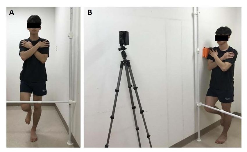
Lateral pelvic tilt was measured to analyse pelvic alignment during OLS. A marker was attached to each
anterior superior iliac spine (ASIS) and a digital camera was placed 2.5 m from the subject according to the
subject’s ASIS height. A photographic image was taken during OLS, and the lateral pelvic tilt was measured
VOLUME -- | ISSUE - | 2020 | 3Kim et al. / Restricted compensatory movement changes muscle activation pattern JOURNAL OF HUMAN SPORT & EXERCISE
as the angle between a horizontal line and a line connecting the ASISs (Prior et al., 2014; Figure 1). All
photographic images were analysed using ImageJ (ver. 1.44) (Edmondston et al., 2013). The average of
three measurements was used for data analysis.
Figure 1. Measurement of lateral pelvic fit.
All OLS tests were performed on the dominant side. Prior to the experiment, all subjects practiced OLS for
10 min under the supervision of an examiner. Subjects randomly performed standard OLS and OLS with
pressure biofeedback (OLSPB).
OLS was performed with reference to the study of Edmondston et al. (2013). The subjects were asked to
stand barefoot with their feet together and their arms across their chest. The subjects were instructed to flex
the hip 30° on the non-support side. A target bar was used to confirm that the participants performed 30° of
hip flexion during the trials (Figure 2A).
OLSPB was performed in the same way as standard OLS except for the addition of pressure biofeedback
(Figure 2B). The 4D-MT pressure sensor (ReLive, Seoul, Korea) was used to check the pressure changes in
real time during OLS. This equipment consists of a cuff device that senses pressure. The pressure data were
sent to an Android tablet PC, and the pressure changes were displayed on the screen in real time. Prior to
performing OLSPB, a pressure sensor was placed between the lateral shoulder on the non-support side and
the wall and calibration was performed. The threshold was set to the minimum change in pressure during
OLSPB. The change in pressure on the tablet screen was expressed as a bar; subjects were asked to avoid
4 | 2020 | ISSUE - | VOLUME -- © 2020 University of AlicanteKim et al. / Restricted compensatory movement changes muscle activation pattern JOURNAL OF HUMAN SPORT & EXERCISE
raising the bar, if possible, during the OLSPB. If an abnormal posture such as increased thoracic kyphosis,
lumbar lordosis or kyphosis, trunk rotation, or knee flexion of the support leg appeared, data collection was
discontinued. OLS was maintained for 5 s and rest time was provided between trials, along with a 20 min
rest between OLS conditions to reduce fatigue and learning effects.
Figure 2. Standard one-leg standing (A) and one-leg standing with pressure biofeedback (B).
Statistical analysis
The Kolmogorov-Smirnov test was used to verify the normality of the outcome measures. All variable data
are expressed as the mean ± standard deviation. The paired t-test was used to compare EMG data of the
Gmed and Gmax and lateral pelvic tilt. The level of statistical significance was set at p < .05. All variable data
were analysed using SPSS ver. 20.0 (IBM Corp., Armonk, NY, USA).
RESULTS
Table 1. Changes in activity of the gluteal muscles and lateral pelvic tilt during one-leg standing with and
without pressure biofeedback.
Standard OLS OLSPB P 95% CI
Gluteus medius (%MVIC) 19.96 ± 9.20 23.69 ± 10.19 .015* -6.65 – -0.81
Gluteus maximus (%MVIC) 4.55 ± 2.16 6.81 ± 4.72 .034* -4.33 – 0.19
Lateral pelvic tilt (°) 3.63 ± 1.59 1.04 ± 0.54 < .001* 1.88 – 3.30
CI, confidence interval; MVIC, maximal voluntary isometric contraction; OLS, one leg standing; OLSPB, one leg standing with
pressure biofeedback. *p < .05.
The Kolmogorov-Smirnov test showed normality in all outcome variables (p > .05). The muscle activity and
lateral pelvic tilt data from during OLS are shown in Table 1. The activity of the Gmed and Gmax was
VOLUME -- | ISSUE - | 2020 | 5Kim et al. / Restricted compensatory movement changes muscle activation pattern JOURNAL OF HUMAN SPORT & EXERCISE
significantly greater during OLSPB compared with standard OLS (p < .05). A significant reduction in lateral
pelvic tilt was observed during OLSPB compared to standard OLS (p < .05).
DISCUSSION
The purpose of our study was to examine the effect of pressure biofeedback on pelvic alignment and gluteal
muscle activity during OLS in healthy men. Our findings show that the lateral pelvic tilt was decreased and
activity of the Gmed and Gmax was increased during OLSPB compared to OLS.
Compared to standard OLS, the lateral pelvic tilt angle was 71% during OLSPB. This may be explained by
the subjects controlling their trunk deviation by checking the pressure changes in real time. If trunk deviation
on the non-supported side occurs, a pelvic deviation occurs on the supported side, which is related to lateral
pelvic tilt (Fujita et al., 2017; Grimaldi, 2011). An increase in pressure during OLS means that the deviation
of the trunk increased on the unsupported side. These results are consistent with previous findings (Cynn et
al., 2006; Noh et al., 2014; Oh et al., 2007; Yu et al., 2018). Noh et al. (2014) reported that the pelvic rotation
angle was decreased during ASLR tests with pressure biofeedback compared to without biofeedback in
patients with LBP. Cynn et al. (2006) demonstrated that side-lying hip abduction with pressure biofeedback
resulted in significantly lower lateral pelvic tilt than without pressure biofeedback, while Oh et al. (2007)
showed that the pelvic anterior tilt was significantly reduced during prone hip extension with pressure
biofeedback. Based on our results and those of previous studies, we conclude that pressure biofeedback is
a useful method to correct pelvic alignment during OLS.
The activity of the Gmed and Gmax was 18% and 50% greater during OLSPB than during standard OLS,
respectively. The Gmax and Gmed act as hip abductors (Clark and Haynor, 1987; Wilson et al., 2006), which
provide pelvic stability on the frontal plane during OLS (Hurwitz et al., 2003). If lateral pelvic tilt increases
during OLS, this indicates a weakening of the hip abductors (Fujita et al., 2017). In contrast, when lateral
pelvic tilt decreases, the internal moment arm decreases (Henderson et al., 2011), and the hip abductor
requires greater activation to maintain this position (Kang et al., 2019). Our findings confirm that the lateral
pelvic tilt was decreased during OLSPB. For this reason, the activity of the Gmed and Gmax seems to
increase during OLSPB. In addition, both muscles contribute to weight bearing by controlling load transfer
through the hip joint (Lee, 1996). Because pelvic alignment was better maintained under the OLSPB condition
than standard OLS, the activity of both muscles seems to be greater. Previous studies have reported
significant increases in the activity of the Gmed and Gmax during weight-bearing exercises, which supports
our findings (Macadam et al., 2015; Reiman et al., 2012). Some previous research is consistent with our
results (Cynn et al., 2006; Noh et al., 2014; Oh et al., 2007; Yu et al., 2018). Yu et al. (2018) found that
infraspinatus activity increased and posterior deltoid activity decreased when the pressure was maintained
at 2 mmHg during prone shoulder external rotation with pressure biofeedback in healthy men, and other
researchers showed that dual pressure biofeedback during ASLR significantly increased abdominal muscle
activity in patients with LBP (Noh et al., 2014). In other studies, the activities of the Gmed and hip extensor
muscles were greater during hip abduction and hip extension using pressure biofeedback, respectively (Cynn
et al., 2006; Oh et al., 2007). Therefore, OLSPB can be suggested as a useful exercise to increase the activity
of the Gmed and Gmax.
In clinical or sports settings, strengthening exercises for the Gmed and Gmax are commonly used for LBP
and to prevent lower limb injury. While OLS is used during various movements, compensatory movements
such as pelvic lateral tilt can occur. Thus, in this study, the control of trunk deviation using pressure
biofeedback during OLS resulted in increased activity of the gluteal muscles and decreased lateral pelvic tilt.
6 | 2020 | ISSUE - | VOLUME -- © 2020 University of AlicanteKim et al. / Restricted compensatory movement changes muscle activation pattern JOURNAL OF HUMAN SPORT & EXERCISE
The implication of these results is that pressure biofeedback during OLS is effective for increasing the activity
of the gluteal muscles and for correcting pelvic alignment.
Our study has some limitations. First, we only investigated healthy men. Future studies should determine the
effect of pressure biofeedback during OLS in patients with LBP or lower extremity injury. Second, we only
evaluated pelvic movements in the frontal plane. Additional studies should include measurements of pelvic
movement in the sagittal and horizontal planes as well as in the frontal plane. Third, since our study only
examined the short-term effects of pressure biofeedback, no long-term effects are known.
CONCLUSION
We investigated muscle activity in gluteal muscles and lateral pelvic tilt during OLS with and without pressure
biofeedback. Our findings demonstrated that OLSPB significantly increased Gmed and Gmax, together with
decreasing lateral pelvic tilt than standard OLS. These results may help clinicians to design effective exercise
programs to increase gluteal muscles and correct pelvic alignment.
AUTHOR CONTRIBUTIONS
1) Conception and design, or acquisition, or analysis and interpretation of data: Kim SY, Kang MH.
2) Drafting the article or revising it critically for important intellectual content: Kim SY, Kang MH.
3) Final approval of the version to be published: Kim SY, Kang MH.
SUPPORTING AGENCIES
This work was supported by the National Research Foundation of Korea (NRF) grant funded by the Korea
government (MSIT) (No. 2018R1C1B5085529).
DISCLOSURE STATEMENT
No potential conflict of interest relevant to this article was reported.
REFERENCES
Akuthota, V., & Nadler, S. F. (2004). Core strengthening. Arch Phys Med Rehabil, 85, S86-92.
Brindle, T. J., Mattacola, C., & McCrory, J. (2003). Electromyographic changes in the gluteus medius
during stair ascent and descent in subjects with anterior knee pain. Knee Surg Sports Traumatol
Arthrosc, 11(4), 244-251. https://doi.org/10.1007/s00167-003-0353-z
Clark, J. M., & Haynor, D. R. (1987). Anatomy of the abductor muscles of the hip as studied by computed
tomography. J Bone Joint Surg Am, 69(7), 1021–1031. https://doi.org/10.2106/00004623-
198769070-00010
Cynn, H. S., Oh, J. S., Kwon, O. Y., & Yi, C. H. (2006). Effects of lumbar stabilization using a pressure
biofeedback unit on muscle activity and lateral pelvic tilt during hip abduction in sidelying. Arch Phys
Med Rehabil, 87(11), 1454-1458. https://doi.org/10.1016/j.apmr.2006.08.327
Edmondston, S., Leo, Y., Trant, B., Vatna, R., Kendell, M., & Smith, A. (2013). Symmetry of trunk and
femoro-pelvic movement response to single leg loading test in asymptomatic females. Man Ther,
18(3), 231-236. https://doi.org/10.1016/j.math.2012.10.010
VOLUME -- | ISSUE - | 2020 | 7Kim et al. / Restricted compensatory movement changes muscle activation pattern JOURNAL OF HUMAN SPORT & EXERCISE
Fujita, K., Kabata, T., Kajino, Y., Iwai, S., Kuroda, K., Hasegawa, K., Fujiwara, K., & Tsuchiya, H. (2017).
Quantitative analysis of the Trendelenburg test and invention of a modified method. J Orthop Sci,
22(1), 81-88. https://doi.org/10.1016/j.jos.2016.09.007
Grimaldi, A. (2011). Assessing lateral stability of the hip and pelvis. Manu Ther, 16(1), 26-32.
Henderson, E. R., Marulanda, G. A., Cheong, D., Temple, H. T., & Letson, G, D. (2011). Hip abductor
moment arm–a mathematical analysis for proximal femoral replacement. J Orthop Surg Res, 6, 6.
https://doi.org/10.1186/1749-799x-6-6
Huritz, D. E., Foucher, K. C., & Andriacchi, T. P. (2003). A new parametric approach for modeling hip
forces during gait. J Biomech, 36(1), 113-119. https://doi.org/10.1016/s0021-9290(02)00328-7
Kang, M. H., Kim, S. Y., Yu, I. Y., & Oh, J. S. (2019). Effects of real-time visual biofeedback of pelvic
movement on electromyographic activity of hip muscles and lateral pelvic tilt during unilateral weight-
bearing and side-lying hip abduction exercises. J Electromyogr Kinesiol, 48, 31-36.
https://doi.org/10.1016/j.jelekin.2019.06.003
Krause, D. A., Jacobs, R. S., Pilger, K. E., Sather, B. R., Sibunka, S. P., & Hollman, J. H. (2009).
Electromyographic analysis of the gluteus medius in five weight-bearing exercise. J Strength Cond
Res, 23(9), 2689-2694. https://doi.org/10.1519/jsc.0b013e3181bbe861
Lee, D. (1996). Instability of the sacroiliac joint and the consequences to gait. J Manual Manip Ther, 4(1),
22-29.
Macadam, P., Cronin, J., & Contreras, B. (2015). An examination of the gluteal muscle acitivity
associated with dynamic hip abduction and hip external rotation exercise: A systematic review. Int J
Sports Phys Ther, 10(5), 573-591.
Nakagawa, T. H., Muniz, T. B., Baldon Rde, M., Dias Maciel, C., de Menezes Reiff, R. B., & Serrão, F.
V. (2008). The effect of additional strengthening of hip abductor and lateral rotator muscles in
patellofemoral pain syndrome: A randomized controlled pilot study. Clin Rehab, 22(12), 1051-1060.
https://doi.org/10.1177/0269215508095357
Neumann, D. A. (1989). Biomechanical analysis of selected principles of hip joint protection. Arthritis
Care Res, 2(4), 146-155. https://doi.org/10.1002/anr.1790020409
Noh, K. H., Kim, J. W., Kim, G. M., Ha, S. M., & Oh, J. S. (2014). The influence of dual pressure
biofeedback units on pelvic rotation and abdominal muscle activity during the active straight leg raise
in women with chronic lower back pain. J Phys Ther Sci, 26(5), 717-719.
https://doi.org/10.1589/jpts.26.717
Oh, J. S., Cynn, H. S., Won, J. H., Kwon, O. Y., & Yi, C. H. (2007). Effects of performing an abdominal
drawing-in maneuver during prone hip extension exercises on hip and back extensor muscle activity
and amount of anterior pelvic tilt. J Orthop Sports Phys Ther, 37(6), 320-324.
https://doi.org/10.2519/jospt.2007.2435
Pfirrmann, C. W., Chung, C. B., Theumann, N. H., Trudell, D. J., & Resnick, D. (2001). Greater trochanter
of the hip: Attachment of the abductor mechanism and a complex of three bursae—MR imaging and
MR bursography in cadavers and MR imaging in asymptomatic volunteers. Radiology, 221(2), 469–
477. https://doi.org/10.1148/radiol.2211001634
Prior, S., Mitchell, T., Whiteley, R., O'Sullivan, P., Williams, B. K., Racinais, S., & Farooq, A. (2014). The
influence of changes in trunk and pelvic posture during single leg standing on hip and thigh muscle
activation in a pain free population. Sports Sci Med Rehabil, 6(1), 13. https://doi.org/10.1186/2052-
1847-6-13
Reiman, M. P., Bolgla, L. A., & Loudon, J. K. (2012). A literature review of studies evaluating gluteus
maximus and gluteus medius activation during rehabilitation exercises. Physiother Theory Pract,
28(4), 257-268. https://doi.org/10.3109/09593985.2011.604981
8 | 2020 | ISSUE - | VOLUME -- © 2020 University of AlicanteKim et al. / Restricted compensatory movement changes muscle activation pattern JOURNAL OF HUMAN SPORT & EXERCISE
Wilson, J. D., Ireland, M. L., & Davis, I. (2006). Core strength and lower extremity alignment during single
leg squats. Med Sci Sports Exerc, 38(5), 945-952.
https://doi.org/10.1249/01.mss.0000218140.05074.fa
Willcox, E. L., & Burden, A. M. (2013). The influence of varying hip angle and pelvis position on muscle
recruitment patterns of the hip abductor muscles during the clam exercise. J Orthop Sports Phys
Ther, 43(5), 325-331. https://doi.org/10.2519/jospt.2013.4004
Yu, I. Y., Choo, Y. K., Kim, M. H., & Oh, J. S. (2018). The effects of pressure biofeedback training on
infraspinatus muscle activity and muscle thickness. J Electromyogr Kinesiol, 39, 81-88.
https://doi.org/10.1016/j.jelekin.2018.01.007
This work is licensed under a Attribution-NonCommercial-NoDerivatives 4.0 International (CC BY-NC-ND 4.0).
VOLUME -- | ISSUE - | 2020 | 9You can also read