Small intestinal submucosal lipoma: a rare cause of secondary intussusception in a child
←
→
Page content transcription
If your browser does not render page correctly, please read the page content below
Cheikhrouhou et al. Annals of Pediatric Surgery             (2021) 17:47                                                 Annals of Pediatric Surgery
https://doi.org/10.1186/s43159-021-00114-7

 CASE REPORTS                                                                                                                                        Open Access

Small intestinal submucosal lipoma: a rare
cause of secondary intussusception in a
child
Taycir Cheikhrouhou1,2* , Mahdi Ben Dhaw1,2, Mohamed Zouari1,2, Hayet Zitouni1,2, Rim Kallel2,3,
Naourez Gouiaa2,3, Tahya Sellami Boudawara2,3 and Riadh Mhiri1,2

  Abstract
  Background: Intestinal lipomas are benign, non-epithelial, intestinal tumors with an exceptionally rare localization
  at the ileum. Lipomas in the small intestine occur mainly in elderly patients and seldom occur in childhood. They
  are frequently asymptomatic, possibly due to their slow growth. These tumors may act as a lead point of
  intussusception.
  Case presentation: We report a rare case of double compounded ileo-ileal intussusception due to a submucosal
  intestinal lipoma in an 8-year-old female. To our knowledge, this is only the seventh pediatric case to be reported
  in the medical literature.
  Conclusions: Small intestinal submucosal lipoma should be considered in case of intussusception in pediatric
  patients. Surgical resection seems sufficient in case of symptomatic intestinal lipoma with low morbidity.
  Keywords: Lipoma, Small bowel, Intussusception, Child, Case report

Background                                                                              Case presentation
Lipomas can develop in virtually all organs. They are be-                               An 8-year-old girl was admitted to our hospital with ab-
nign soft tissue tumors derived from mature adipocytes.                                 dominal pain and vomiting that had started 2 days be-
Lipomas of the gastrointestinal tract (GIT) are rare [1].                               fore admission. During questioning, the patient
Most GIT lipomas are asymptomatic [2] and < 50% of                                      mentioned having an intermittent colicky pain in her
adult patients who have intestinal lipomas become                                       right lower abdomen over the previous month that had
symptomatic, usually because of intussusception, ob-                                    not responded to analgesia or spasmolytic. There were
struction, or hemorrhage [3]. Pediatric intestinal lip-                                 no episodes of bilious vomiting, bloody stools, or fever.
omas, although rare, have previously been reported as                                     On admission, the patient was hemodynamically stable
the leading point of intussusception [4].                                               with a blood pressure of 110/70 mmHg. Her
  We describe a pediatric case of intussusception sec-                                  temperature was 37.2 °C. Clinical examination revealed
ondary to a GIT lipoma and present a review of the lit-                                 tenderness at the right iliac fossa without abdominal dis-
erature on small bowel intussusception due to                                           tension or signs of peritoneal irritation. Rectal examin-
gastrointestinal lipomas.                                                               ation was normal. A complete blood cell count and
                                                                                        laboratory tests were normal. Abdominal X-ray revealed
                                                                                        no specific bowel gas pattern, but gaseous distension of
                                                                                        several small bowel loops (Fig. 1).
* Correspondence: cheikhrouhoutaycir@gmail.com                                            Abdominal ultrasound revealed the typical target sign
1
 Department of Pediatric Surgery, Hedi Chaker Hospital, 3029 Sfax, Tunisia              in the right lower abdomen suggestive of ileocolic intus-
2
 University of Medicine of Sfax, University of Sfax, Sfax, Tunisia
Full list of author information is available at the end of the article                  susception, extending for approximately 9.6 cm. In

                                        © The Author(s). 2021 Open Access This article is licensed under a Creative Commons Attribution 4.0 International License,
                                        which permits use, sharing, adaptation, distribution and reproduction in any medium or format, as long as you give
                                        appropriate credit to the original author(s) and the source, provide a link to the Creative Commons licence, and indicate if
                                        changes were made. The images or other third party material in this article are included in the article's Creative Commons
                                        licence, unless indicated otherwise in a credit line to the material. If material is not included in the article's Creative Commons
                                        licence and your intended use is not permitted by statutory regulation or exceeds the permitted use, you will need to obtain
                                        permission directly from the copyright holder. To view a copy of this licence, visit http://creativecommons.org/licenses/by/4.0/.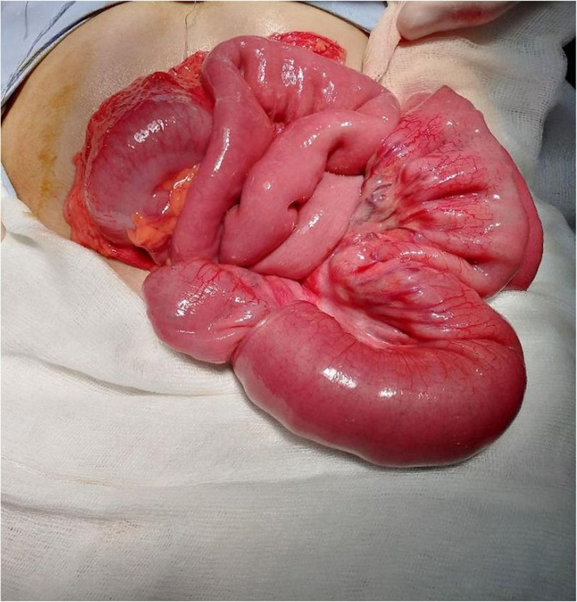
Cheikhrouhou et al. Annals of Pediatric Surgery    (2021) 17:47                                                             Page 2 of 5

                                                                   Fig. 3 Pre-operative view of compounded ileo-ileal intussusception

                                                                     Resection and end-to-end anastomosis of the ileal seg-
                                                                  ment containing the mass were performed with a 5-cm
                                                                  safety margin on each side. Histopathological examin-
 Fig. 1 Abdominal X-ray showing no specific gas pattern
                                                                  ation confirmed the diagnosis of a submucosal intestinal
                                                                  lipoma (Fig. 5). The postoperative course was uneventful
                                                                  and the patient was discharged after 1 week.
transverse section, images showed a mass composed of
multiple concentric circles (triple circle mass) (Fig. 2).        Discussion
   The patient underwent emergency exploratory laparot-           Intussusception is a common pediatric surgical problem
omy, which revealed a long intussuscepted ileal tract             in infants and toddlers, with the highest incidence be-
(Fig. 3). Manual reduction was performed and a com-               tween 3 and 12 months. It is defined as the telescoping
pound ileo-ileo-ileal intussusception was found. The lead         of one segment of the intestine into a distal segment.
point of the intussusception was a 3-cm hemispherical             Most intussusceptions are idiopathic with a good prog-
mass located 100 cm away from the ileocecal junction              nosis. Secondary intussusceptions are caused by patho-
and originating from the antimesenteric border of the             logic lead points (PLPs) (such as Meckel’s diverticulum,
ileum (Fig. 4).

 Fig. 2 Abdominal ultrasound revealed an intussusception with
 typical target sign (arrow)                                       Fig. 4 Pre-operative view of submucosal lipoma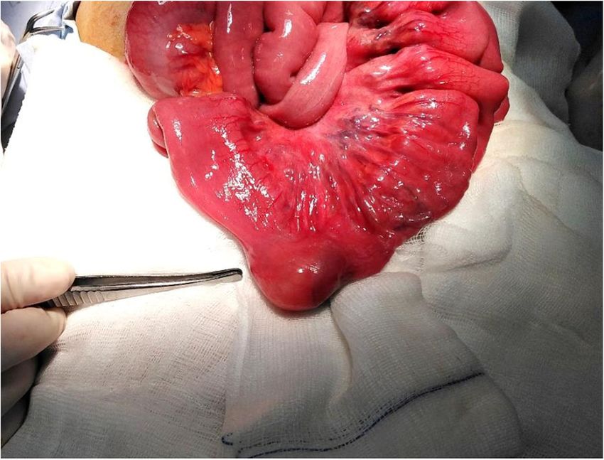
Cheikhrouhou et al. Annals of Pediatric Surgery (2021) 17:47 Page 3 of 5
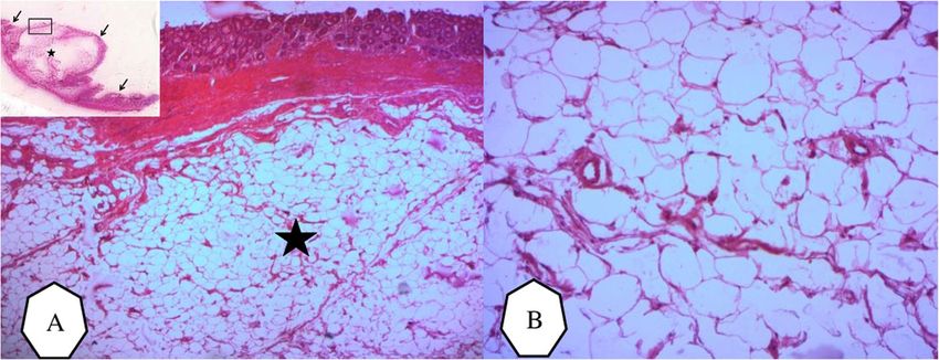
Fig. 5 Microscopic examination of small intestinal lipoma. A Low power examination showed lobules of fat separated by delicate fibrovascular
trabeculae (*). (HEx25). B A high power examination showed mature fat cells with no malignant features. (HE x 100)
duplication, polyps, and tumors) and represent 2.2–15% bleeding tend to occur when the lesion is > 2 cm in size
of cases [5, 6]. The incidence of intussusception second- [15]. Lesions < 1 cm are rarely symptomatic, while 75%
ary to PLPs is much higher in children > 2 years old of those > 4 cm in size are more likely to cause symp-
than in children < 2 years of age [7]. They are relatively toms. Small intestinal lipomas produce symptoms more
difficult to diagnose and confirm before surgery. often than colonic lipomas, irrespective of the size of the
Lipomas of the GIT are benign intestinal tumors of tumor [11].
mesenchymal origin. They are relatively rare and repre- Among children, intestinal intussusception due to lip-
sent only 2.6% of nonmalignant tumors of the GIT [8]. oma as the PLP is rare. In fact, to our knowledge, this is
Most lipomas are solitary, but they can be multiple and only the seventh reported case of its kind. Table 1 sum-
located anywhere in the GIT [9]. Lipomas are more fre- marizes the literature on pediatric small intestinal intus-
quent in the large intestine, mainly in the right colon susception induced by a lipoma.
and the cecum, and only 20–25% are found in the small For pediatric small intestinal lipomas, median age at
intestine. There are three pathological types of intestinal diagnosis is 8 years with a range from 4 to 14 years. All
lipomas: (i) intermuscular type; (ii) subserosal type; and reported cases to date have been in males. Vomiting and
(iii) submucosal type. The tumors arise from the sub- recurrent abdominal pain have been reported to be the
mucosa in 90% of cases, while the remainder are usually main symptoms. These symptoms are not specific and
subserosal [10]. Submucosal lipomas usually present as are shared with other gastrointestinal disorders. Thus,
polypoid lesions, either sessile or pedunculated [11]. the correct diagnosis may be difficult to make [11].
Lipomas of the small intestine are most likely to present dur- Pre-operative diagnosis is important because the diag-
ing the fifth to seventh decade of life [12]. In older subjects, the nosis can be difficult to make intra-operatively and a
incidence appears to be higher in females than in males [13]. correct diagnosis can avoid unnecessary excessive resec-
These tumors are slow-growing with a silent clinical tion. Simanovsky et al. [15] stated that surgery can be
course and are usually found incidentally [14]. Symp- better planned when the exact location and extent of the
toms such as abdominal pain, diarrhea, constipation, and intussusception are known from imaging [19].
Table 1 Reported pediatric cases of small intestinal lipoma in the English literature
Case/ Age Sex Diagnostic modality Pre-operative Tumor location/intussusception type Reference
year (years) imaging
1 (1991) 12 Male Vomiting + epigastric pain mimicking acute US + CT + ERCP Duodenal lipoma/duodenal-jejunal [15]
pancreatitis intussusception
2 (1997) 6 Male Recurrent abdominal pain with vomiting and CT Duodenal lipoma/duodenal-jejunal [16]
weight loss intussusception
3 (2008) 14 Male Recurrent abdominal pain and vomiting CT Ileal lipoma/ileo-colic intussusception [1]
4 (2013) 7 Male Abdominal pain US + CT Ileal lipoma/ileo-ileal intussusception [4]
5 (2014) 5 Male Abdominal pain US + CT Ileal lipoma/ileo-ileal intussusception [17]
6 (2018) 4 Male Abdominal pain and vomiting US + CT Ileal lipoma/ileo-ileal intussusception [18]
7 (2019) 8 Female Recurrent abdominal pain and vomiting US Ileal lipoma/ileo-ileal intussusception Present
case
US ultrasound, CT computed tomography scan, ERCP endoscopic retrograde cholangiopancreatography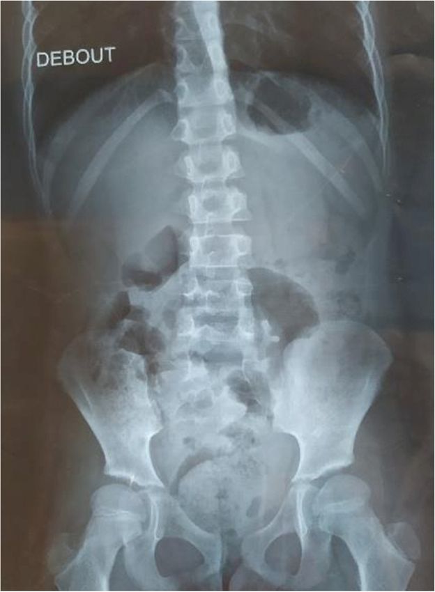
Cheikhrouhou et al. Annals of Pediatric Surgery          (2021) 17:47                                                                               Page 4 of 5

  Pre-operative auxiliary examinations such as ultra-                        Authors’ contributions
sound and computed tomography (CT) scans are very                            TC conception, design of the work, literature review, manuscript writing, and
                                                                             editing. MBD operated the patient, manuscript writing, and literature review.
helpful imaging modalities for the diagnosis of intussus-                    RM concept design and manuscript review. All authors have read and
ception and for determining the nature of the intralum-                      approved the final manuscript.
inal lesion [20]. Abdominal ultrasound will reveal a
                                                                             Funding
rounded echogenic mass typically described as the pseu-                      None.
dokidney sign [21]. This technique is generally available
in an emergency setting. In the hands of skilled and ex-                     Availability of data and materials
                                                                             None.
perienced sonographers, it has similar sensitivity and
specificity to CT scans, allowing a correct diagnosis be-                    Declarations
fore surgery.
  On CT scans, lipomas appear as well-circumscribed,                         Ethics approval and consent to participate
                                                                             None.
round, homogeneous masses with fat attenuation num-
bers (− 40 to – 120 Hounsfield units) within the lumen                       Consent for publication
of the intussusceptions [2, 22]. CT scans were not per-                      The parents of the patient have consented to use of clinical photographs for
                                                                             publication and research process.
formed in our patient because of sufficient diagnostic
data for a secondary intussusception was obtained by                         Competing interests
radiography and ultrasound, and because of the urgent                        All authors declare that they have no competing interests.
condition of the patient.
                                                                             Author details
  The treatment of lipomas depends on their symptoms.                        1
                                                                              Department of Pediatric Surgery, Hedi Chaker Hospital, 3029 Sfax, Tunisia.
                                                                             2
Surgical resection is usually the treatment of choice for                     University of Medicine of Sfax, University of Sfax, Sfax, Tunisia. 3Department
                                                                             of Pathology, Habib Bourguiba Hospital, 3029 Sfax, Tunisia.
symptomatic gastrointestinal lipomas [16]. Partial small
bowel resection can either be done by laparotomy or                          Received: 22 January 2021 Accepted: 24 June 2021
laparoscopy. Oncological principles should be main-
tained during resection unless pre-operative imaging
                                                                             References
shows a benign etiology [23]. The correct diagnosis of a                     1. Draus JM, Shelgikar CS, Buchino JJ, Bond SJ. Lipoma as a pathological lead
symptomatic, submucosal lipoma is an indication for                              point in a child with ileocolic intussusception. J Pediatr Gastroenterol Nutr.
local excision and eliminates unnecessary extensive re-                          2008;47(3):372–4. https://doi.org/10.1097/MPG.0b013e318076b489.
                                                                             2. Baron Y, Priesack W, Sötje G, Brix F, Scheunemann C. Hemorrhagic jejunal
section. In our patient, resection with a 5-cm safety mar-                       lipoma with intermittent intussusception. Eur J Radiol. 1996;22(2):123–5.
gin on each side was performed because we did not                                https://doi.org/10.1016/0720-048X(96)00753-X.
exclude malignancy.                                                          3. Triantopoulou C, Vassilaki A, Filippou D, Velonakis S, Dervenis C,
                                                                                 Koulentianos E. Adult ileocolic intussusception secondary to a submucosal
  As for the rarity of these tumors, there is a view that                        cecal lipoma. Abdom Imaging. 2004;29:426–8.
this may be due to the lack of reporting of cases or to                      4. Asaumi Y, Miyanaga T, Ishiyama Y, Hattori M, Hashizume Y. Pediatric ileoileal
false diagnoses. In most cases, the histological diagnosis                       intussusception with a lipoma lead point: a case report. Gastroenterol Rep
                                                                                 (Oxf). 2014;2(1):70–2. https://doi.org/10.1093/gastro/got032.
is made only after excision of the tumor. Therefore,                         5. Wong CWY, Chan IHY, Chung PHY, Lan LCL, Lam WWM, Wong KKY, et al.
pathological examination of all excised tissues may in-                          Childhood intussusception: 17-year experience at a tertiary referral centre in
crease the detection of this clinical entity.                                    Hong Kong. Hong Kong Med J. 2015;21(6):518–23. https://doi.org/10.12809/
                                                                                 hkmj144456.
  Treatment is usually definitive, because the lesions do                    6. Hsu W-L, Lee H-C, Yeung C-Y, Chan W-T, Jiang C-B, Sheu J-C, et al.
not often recur. No malignant transformation of lipomas                          Recurrent intussusception: when should surgical intervention be
has been reported in the literature and after resection,                         performed? Pediatr Neonatol. 2012;53(5):300–3. https://doi.org/10.1016/j.
                                                                                 pedneo.2012.07.004.
no recurrence is expected [16, 24].                                          7. Zhao L, Feng S, Wu P, Lai X-H, Lv C, Chen G. Clinical characteristics and
                                                                                 surgical outcome in children with intussusceptions secondary to pathologic
                                                                                 lead points: retrospective study in a single institution. Pediatr Surg Int. 2019;
Conclusions                                                                      35(7):807–11. https://doi.org/10.1007/s00383-019-04471-8.
                                                                             8. Ackerman NB, Chughtai SQ. Symptomatic lipomas of the gastrointestinal
Small intestinal submucosal lipoma should be consid-
                                                                                 tract. Surg Gynecol Obstet. 1975;141:565–8.
ered in case of intussusception in pediatric patients. Sur-                  9. Deeths TM, Madden PN, Dodds WJ. Multiple lipomas of the stomach and
gical resection seems sufficient in case of symptomatic                          duodenum. Am J Dig Dis. 1975;20(8):771–4. https://doi.org/10.1007/BF01
                                                                                 070835.
intestinal lipoma with low morbidity.
                                                                             10. Charalambous G, Katergiannakis V, Manouras A. Jejunojejunal lipoma
                                                                                 causing intussusception. Case Rep Gastroenterol. 2012;6(3):684–8. https://
Abbreviations                                                                    doi.org/10.1159/000345379.
CT: Computed tomography; GIT: Gastrointestinal tract; PLP: Pathologic lead   11. Janevska V, Spasevska L, Dukova B, Janevski V. Intestinal submucosal
point                                                                            lipomas. Mac J Med Sci. 2012;5(1):49–54. https://doi.org/10.3889/MJMS.1857-
                                                                                 5773.2012.0199.
                                                                             12. Fang S-H, Dong D-J, Chen F-H, Jin M, Zhong B-S. Small intestinal lipomas:
Acknowledgements                                                                 Diagnostic value of multi-slice CT enterography. World J Gastroenterol.
None.                                                                            2010;16(21):2677–81. https://doi.org/10.3748/wjg.v16.i21.2677.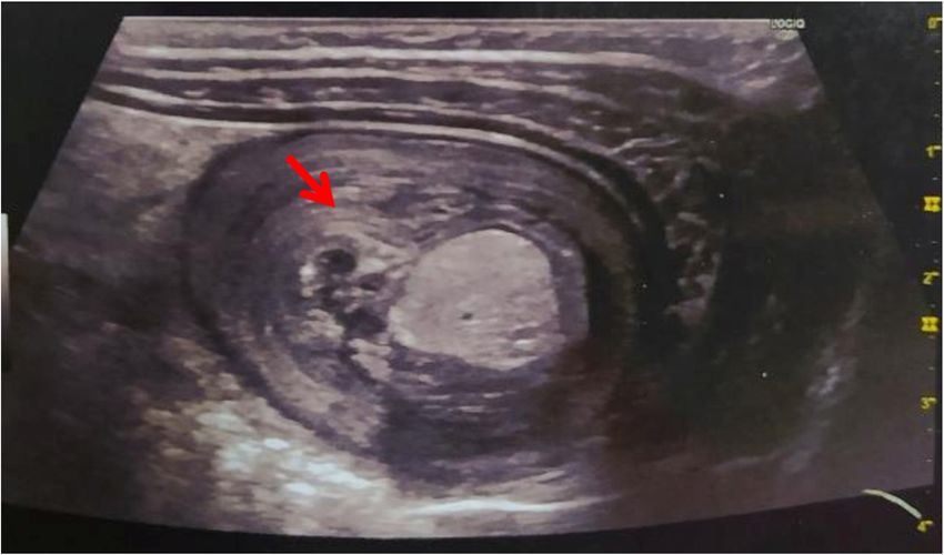
Cheikhrouhou et al. Annals of Pediatric Surgery                (2021) 17:47             Page 5 of 5

13. Manouras A, Lagoudianakis EE, Dardamanis D, Tsekouras DK, Markogiannakis
    H, Genetzakis M, et al. Lipoma induced jejunojejunal intussusception. World
    J Gastroenterol. 2007;13(26):3641–4. https://doi.org/10.3748/wjg.v13.i26.3641.
14. Turi S, Röckelein G, Dobroschke J, Wiedmann KH. Lipoma of the small
    bowel. Z Gastroenterol. 2004;42(2):147–51. https://doi.org/10.1055/s-2004-
    812837.
15. McGrath FP, Moote DJ, Langer JC, Orr W, Somers S. Duodenojejunal
    intussusception secondary to a duodenal lipoma presenting in a young
    boy. Pediatr Radiol. 1991;21(8):590–1. https://doi.org/10.1007/BF02012606.
16. Case records of the Massachusetts General Hospital. Weekly
    clinicopathological exercises. Case 24-1997. A six-year-old boy with bouts of
    abdominal pain, vomiting, and a left-sided abdominal mass. N Engl J Med.
    1997;337:329–36.
17. Destro F, Cantone N, Maffi M, Gargano T, Lima M. An interesting case of
    double compound intussusception without intestinal occlusion in a 5-year-
    old boy. Eur J Pediatr Surg Rep. 2014;2(01):20–2. https://doi.org/10.1055/s-
    0033-1361925.
18. Abdelmohsen SM, Osman MA, Hussien MT. An ileo-ileal intussusception
    secondary to polypoid lipoma in a child, a case report and review of the
    literature. Int J Surg Case Rep. 2019;57:88–90. https://doi.org/10.1016/j.ijscr.2
    019.03.003.
19. Simanovsky N, Hiller N, Koplewitz BZ, Eliahou R, Udassin R. Is non-operative
    intussusception reduction effective in older children? Ten-year experience in
    a university affiliated medical center. Pediatr Surg Int. 2007;23(3):261–4.
    https://doi.org/10.1007/s00383-006-1838-x.
20. Tan HL, Koh YX, Taufik M, Lye WK, Goh BKP, Ong HS. A clinical scoring
    system to predict the clinical sequelae of computed tomography
    diagnosed intussusception. World J Surg. 2018;42(3):682–7. https://doi.org/1
    0.1007/s00268-017-4196-z.
21. Vagholkar K, Chavan R, Mahadik A, Maurya I. Lipoma of the small intestine: a
    cause for intussusception in adults. Case Rep Surg. 2015:e856030 [Open
    Access] Available at https://www.hindawi.com/journals/cris/2015/856030/
    (accessed 17/11/20).
22. Chekan EG, Westcott C, Low VHS, Ludwig KA. Small bowel intussusception
    and laparoscopy. Surg Laparosc Endosc. 1998;8(4):324–6. https://doi.org/10.1
    097/00019509-199808000-00019.
23. Oyen TL, Wolthuis AM, Tollens T, Aelvoet C, Vanrijkel JP. Ileo-ileal
    intussusception secondary to a lipoma: a literature review. Acta Chir Belg.
    2007;107(1):60–3. https://doi.org/10.1080/00015458.2007.11680013.
24. Eisen LK, Cunningham JD, Aufses AH. Intussusception in adults: institutional
    review. J Am Coll Surg. 1999;188(4):390–5. https://doi.org/10.1016/S1072-751
    5(98)00331-7.

Publisher’s Note
Springer Nature remains neutral with regard to jurisdictional claims in
published maps and institutional affiliations.
You can also read