Seoul Orthohantavirus in Wild Black Rats, Senegal, 2012-2013 - CDC
←
→
Page content transcription
If your browser does not render page correctly, please read the page content below
DISPATCHES

        Seoul Orthohantavirus in Wild
       Black Rats, Senegal, 2012–2013
         Moussa M. Diagne, Idrissa Dieng, Laurent Granjon, Héloïse Lucaccioni, Abdourahmane Sow,
             Oumar Ndiaye, Martin Faye, Khalilou Bâ, Yamar Bâ, Mamoudou Diallo, Oumar Faye,
       Jean-Marc Duplantier, Mawlouth Diallo, Pascal Handschumacher, Ousmane Faye, Amadou A. Sall

                                                                           Southeastern Senegal has become a major trade
 Hantaviruses cause hemorrhagic fever in humans
 worldwide. However, few hantavirus surveillance cam-                 area because of urbanization and substantial im-
 paigns occur in Africa. We detected Seoul orthohanta-                provement of its road and rail networks in the late
 virus in black rats in Senegal, although we did not find             1990s (7). Within a few years, these changes led to the
 serologic evidence of this disease in humans. These                  rapid spread of a major invasive rodent species, the
 findings highlight the need for increased surveillance of            black rat (Rattus rattus [family Murinae]), which is a
 hantaviruses in this region.                                         reservoir for Seoul orthohantavirus (SEOV) (4,5,7). To
                                                                      assess the prevalence of hantaviruses in rodents, we

H     antaviruses (family Hantaviridae, genus Ortho-
      hantavirus) are RNA viruses transmitted by aero-
solized excreta from infected rodents and shrews. In
                                                                      screened for hantaviruses in R. rattus rats and com-
                                                                      mensal or peridomestic co-existing rodents in 2012–
                                                                      2013, approximately 15 years after the 1998 opening
humans, they cause hemorrhagic fever with renal                       of a tarred road in eastern Senegal.
syndrome (more often observed in Asia and Europe)
and cardiopulmonary syndrome (more common in                          The Study
the Americas) (1). Only 1 case has been confirmed                     The national ethics committee for research of Senegal
in Africa, in the Central African Republic in 1987 (2).               approved the study (authorization no. 0360-MSAS/
However, studies from 2006 through 2013 have dis-                     DPRS/DR, on October 24, 2011). During May 2012–
covered new hantaviruses in autochthonous African                     December 2013, we trapped small mammals as previ-
rodents, moles, and bats (3,4). In addition, serologic                ously described (8) inside dwelling places and their
evidence in humans and rodents in Africa suggest lo-                  surroundings (immediate and local) over periods of
cal circulation (5). For example, a study in rural areas              1–6 consecutive days.
of Senegal found 11.5% of rodents and 16.6% of hu-                         We caught 1,414 small mammals, including 403
mans had antibodies against hantaviruses (3). More                    black rats, from 10 different species (Appendix Table,
recently, serologic evidence of hantaviruses was re-                  https://wwwnc.cdc.gov/EID/article/26/10/20-
ported in domestic and peridomestic rodents from                      1306-App1.pdf). We sampled whole blood, brain,
some regions in Senegal (6).                                          and visceral organ tissues, which we then trans-
                                                                      ferred to the Institut Pasteur (Dakar, Senegal). We
Author affiliations: Institut Pasteur, Dakar, Senegal (M.M. Diagne,   triturated each solid sample in Leibovitz-15 medium
I. Dieng, A. Sow, O. Ndiaye, M. Faye, Y. Bâ, Oum. Faye, Maw.          (GIBCO-BRL, https://www.thermofisher.com) and
Diallo, Ous. Faye, A.A. Sall); Centre de Gestion des Populations,     centrifuged them to collect the suspension. To col-
Institut de Recherche pour le Développement, Montpellier,             lect serum, we centrifuged whole blood samples.
France (L. Granjon, J.-M. Duplantier); Université Paris Nanterre,     We extracted RNA from these different suspen-
Nanterre, France (H. Lucaccioni); Institut de recherche pour le       sions using the QIAamp RNA Viral Kit (QIAGEN,
développement Senegal, Dakar (K. Bâ, Mam. Diallo);                    https://www.qiagen.com) according to the man-
Sciences Economiques & Sociales de la Santé & Traitement de           ufacturer’s recommendations. To make cDNA,
l’Information Médicale, Marseille, France (P. Handschumacher);        we used avian myeloblastosis virus reverse tran-
Aix Marseille University, Marseille (P. Handschumacher); Institut     scriptase (Promega, https://www.promega.com)
National de la Santé et de la Recherche Médicale, Marseille           followed by a nested conventional PCR with Go-
(P. Handschumacher)                                                   Taq Polymerase (Promega, https://www.promega.
DOI: https://doi.org/10.3201/eid2610.201306                           com) and a highly conserved hantavirus primers

2460                         Emerging Infectious Diseases • www.cdc.gov/eid • Vol. 26, No. 10, October 2020
Seoul Orthohantavirus in Wild Black Rats, Senegal
system selective for the partial large segment protein Of the 1,414 mammals, 13 black rats tested posi-
gene (9). We sequenced amplicons using GENEWIZ tive for hantavirus RNA. We detected RNA in 14
(https://www.genewiz.com), assembled them us- samples: 9 brain homogenates, 4 multiorgan homog-
ing EMBOSS Merger software (http://www.bio- enates, and 1 serum sample. We confirmed the posi-
informatics.nl/cgi-bin/emboss/merger), and ana- tive samples using PCR with highly conserved hanta-
lyzed them with BLAST (http://blast.ncbi.nlm.nih. virus small segment primers (12). Sequence analysis
gov/Blast.cgi). We performed sequence alignment of partial large (deposited under GenBank accession
with Mafft (10) and built a maximum-likelihood nos. MT276868–81) and small (deposited under Gen-
phylogenetic tree with iQ-TREE (11), using 1,000 Bank accession nos. MT276854–67) segments revealed
replicates for bootstrapping. 99.42% identity with SEOV strain Rn-HD27 from China
Figure 1. Phylogenetic analysis
of Seoul orthohantavirus strains
from black rats (Rattus rattus
[family Murinae]; boldface) and
reference sequences, Senegal,
2012–2013. Phylogenetic
trees were generated by the
maximum-likelihood method
using the transition plus invariate
sites plus gamma 4 model of the
small segment (266 nt) (A) and
the large segment (347 nt) (B).
The numbers at each node are
bootstrap probabilities (>90%) as
determined for 1,000 iterations.
GenBank numbers are indicated
for reference sequences. Scale
bars indicate 0.01 substitutions
per nucleotide (A) and 0.1
substitutions per nucleotide (B).
Emerging Infectious Diseases • www.cdc.gov/eid • Vol. 26, No. 10, October 2020 2461DISPATCHES
(GenBank accession no. HM748799) and 99.64% iden- We did not observe signs of disease in the infected
tity with SEOV strain Hu02-529 from South Korea animals at the time of capture. Of the 4 villages that
(GenBank accession no. MF149956) (Figure 1). yielded the highest numbers of black rats in this
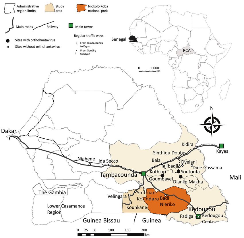
We detected SEOV RNA in 13 black rats caught study, 3 harbored rats infected with SEOV (Figure 2)
in 3 villages: Goumbayel (7 rodents), Soutouta (4 (7). High densities of black rats might contribute to
rodents), and Dianke Makha (2 rodents). These vil- the occurrence of hantavirus in these villages, espe-
lages were located ≈1 hour’s drive from the main cially because host demography might affect hanta-
road between Tambacounda and Kidira (Figure 2). virus circulation (13).
Frequent movement of goods and humans between Seasonal patterns might complicate these find-
these 3 villages might explain the low genetic diver- ings. We surveyed the villages harboring SEOV-
sity among the new SEOV strains from black rats. infected rats in February 2013, which might be a
Figure 2. Locations of trapping sites (circles) used in study of rodentborne Seoul orthohantavirus in Senegal, 2012–2013. Black circles
indicate trapping locations of Seoul orthohantavirus–infected black rats (Rattus rattus [family Murinae]). Inset shows location of Senegal
in Africa. Map created using the package maptools installed in R studio version 1.2.1335 (https://rstudio.com/products/rstudio/) and
shapefiles downloaded from the free domain of the Geographic Information System (http://www.diva-gis.org).
2462 Emerging Infectious Diseases • www.cdc.gov/eid • Vol. 26, No. 10, October 2020Seoul Orthohantavirus in Wild Black Rats, Senegal
Table. Human exposures to rodents in selected villages, Senegal, 2012–2013
No. No. (%) participants in No. distinct species
Village/town participants contact with rodents encountered Black rats Time period
Tambacounda
Youpe Hamady 87 70 (80.5) 4 No 2012 Oct 19–20
Talibadji 33 11 (33.3) 3 Yes 2012 Oct 21
Sinthiou Doube 39 37 (94.9) 4 Yes 2012 Oct 22
Ndiobene 45 20 (44.4) 2 No 2012 Oct 22
Dianke Makha 101 40 (39.6) 5 Yes 2012 Sep 10
Soutouta 89 83 (93.3) 4 Yes 2012 Sep 11
Kedougou
Kedougou 147 111 (75.5) 6 Yes 2013 Mar 9–10
Total 541 372 (68.8)
favorable period for rodent reproduction, popula- yses grouped the newly detected SEOV with strains
tion increase, and thus hantavirus circulation (13). from Asia. Exchanges between Africa and Asia can
Despite the presence of juveniles, R. rattus popula- potentially increase the opportunities for pathogens
tions had relatively high proportions of sexually ac- to expand their geographic range as previously de-
tive animals (75% in Goumbayel, 48% in Soutouta, scribed (15).
and 71% in Dianke Makha) (Appendix Figure). These In-depth phylogenetic analysis of complete ge-
data suggest that high level of interactions (male–fe- nomes would help elucidate the molecular evolution
male, adult–juvenile) occurred in these populations of this virus in Africa. This study highlights the need
during that period, possibly promoting viral circu- to improve hantavirus surveillance in Senegal and
lation. Conversely, we investigated nearby villages other countries in Africa for public health preven-
(Dieylani, Dide Gassama, Koussan, and Talibadji, tion strategies.
in which we did not find evidence of hantavirus-
infected black rats) in October 2012, at the end of the Acknowledgments
rainy season. Our investigations in May 2012 and We thank Coly Bâ, Ambroise Dalecky, Christophe Amidi
November 2013 of the Kedougou area did not detect Diagne, Philippe Gauthier, Laëtitia Husse, Mamadou
evidence of SEOV. Kane, Youssou Niang, and Aliou Sow for their
To assess potential human transmission, we outstanding work during trapping sessions. We thank
performed parallel studies of human populations Oumar Ndiaye and Magueye Ndiaye for their technical
in some villages. Participants consented to an in- laboratory support. We also thank Pierre Rollin for helping
terview about rodent exposure and gave blood to obtain ELISA reagents.
samples. During October 2012–March 2013, we re-
This work was supported by grant nos. ANR-11-CE-
cruited 541 participants with a mean age of 24 years
PL-0010 and ANR-11-JSV7-0006 (A.A.S., O.F., M.M.D.,
(range 2–91 years) (Table). Of the 541 participants,
A.G., Y.B., M.D.) from the Agence Nationale de
372 (68.8%) reported close contact with rodents. The
la Recherche.
highest rates of rodent exposure were in Soutouta
and Sinthiou Doube (Table). We performed an in-
house ELISA specific to IgG against SEOV on the About the Author
human serum samples using reagents from the US Dr. Diagne is a postdoctoral researcher at the Virology
Centers for Disease Control and Prevention (Atlan- Department of Institut Pasteur de Dakar. His research in-
ta, GA, USA). No IgG against SEOV was detected in terests include arboviruses and hemorrhagic fever viruses,
the tested human samples, regardless of whether the such as hantaviruses in animal reservoirs and humans.
participant’s village had evidence of SEOV-infected
black rats; this finding suggests a lack of human ex-
posure. The role of species diversity in virus trans- References
1. Bi Z, Formenty PB, Roth CE. Hantavirus infection: a review
mission is extremely complex (14). The relatively and global update. J Infect Dev Ctries. 2008;2:3–23.
low SEOV prevalence in black rats (Appendix Table) https://doi.org/10.3855/jidc.317
might explain the negative results of the human se- 2. Coulaud X, Chouaib E, Georges AJ, Rollin P, Gonzalez JP.
rologic survey. First human case of haemorrhagic fever with renal syndrome
in the Central African Republic. Trans R Soc Trop Med Hyg.
1987;81:686. https://doi.org/10.1016/0035-9203(87)90455-X
Conclusions 3. Witkowski PT, Klempa B, Ithete NL, Auste B, Mfune JKE,
We found SEOV, a hantavirus pathogenic to humans, Hoveka J, et al. Hantaviruses in Africa. Virus Res. 2014;
in black rats in southeastern Senegal. Phylogenic anal- 187:34–42. https://doi.org/10.1016/j.virusres.2013.12.039
Emerging Infectious Diseases • www.cdc.gov/eid • Vol. 26, No. 10, October 2020 2463DISPATCHES
4. Milholland MT, Castro-Arellano I, Suzán G, Garcia-Peña GE, fast Fourier transform. Nucleic Acids Res. 2002;30:3059–66.
Lee TE Jr, Rohde RE, et al. Global diversity and distribution https://doi.org/10.1093/nar/gkf436
of hantaviruses and their hosts. EcoHealth. 2018;15:163–208. 11. Nguyen L-T, Schmidt HA, von Haeseler A, Minh BQ.
https://doi.org/10.1007/s10393-017-1305-2 IQ-TREE: a fast and effective stochastic algorithm for
5. Saluzzo JF, Digoutte JP, Adam F, Bauer SP, McCormick JB. estimating maximum-likelihood phylogenies. Mol Biol Evol.
Serological evidence for Hantaan-related virus infection in 2015;32:268–74. https://doi.org/10.1093/molbev/msu300
rodents and in Senegal. Trans R Soc Trop Med Hyg. 12. Arthur RR, Lofts RS, Gomez J, Glass GE, Leduc JW, Childs JE.
1985;79:874–5. https://doi.org/10.1016/0035-9203(85)90145-2 Grouping of hantaviruses by small (S) genome segment
6. Diagne CA, Charbonnel N, Henttonen H, Sironen T, Brouat polymerase chain reaction and amplification of viral RNA
C. Serological survey of zoonotic viruses in invasive and from wild-caught rats. Am J Trop Med Hyg. 1992;47:210–4.
native commensal rodents in Senegal, West Africa. Vector 13. Tian H, Stenseth NC. The ecological dynamics of hantavirus
Borne Zoonotic Dis. 2017;17:730–3. https://doi.org/10.1089/ diseases: from environmental variability to disease
vbz.2017.2135 prevention largely based on data from China. PLoS Negl
7. Lucaccioni H, Granjon L, Dalecky A, Fossati O, Le Fur J, Trop Dis. 2019;13:e0006901. https://doi.org/10.1371/
Duplantier J-M, et al. From human geography to biological journal.pntd.0006901
invasions: the black rat distribution in the changing 14. Luis AD, Kuenzi AJ, Mills JN. Species diversity concurrently
southeastern of Senegal. PLoS One. 2016;11:e0163547. dilutes and amplifies transmission in a zoonotic host-
https://doi.org/10.1371/journal.pone.0163547 pathogen system through competing mechanisms. Proc Natl
8. Dalecky A, Ba K, Piry S, Lippens C, Diagne CA, Kane M, Acad Sci U S A. 2018;115:7979–84. https://doi.org/10.1073/
et al. Range expansion of the invasive house mouse Mus pnas.1807106115
musculus domesticus in Senegal, West Africa: a synthesis of 15. Simon-Loriere E, Faye O, Prot M, Casademont I, Fall G,
trapping data over three decades, 1983–2014. Mammal Rev. Fernandez-Garcia MD, et al. Autochthonous Japanese
2015;45:176–90. https://doi.org/10.1111/mam.12043 encephalitis with yellow fever coinfection in Africa. N
9. Klempa B, Fichet-Calvet E, Lecompte E, Auste B, Aniskin V, Engl J Med. 2017;376:1483–5. https://doi.org/10.1056/
Meisel H, et al. Hantavirus in African wood mouse, Guinea. NEJMc1701600
Emerg Infect Dis. 2006;12:838–40. https://doi.org/10.3201/
eid1205.051487 Address for correspondence: Moussa Moïse Diagne, Virology
10. Katoh K, Misawa K, Kuma K, Miyata T. MAFFT: a novel Department, Institut Pasteur de Dakar, 36 Avenue Pasteur,
method for rapid multiple sequence alignment based on BP.220, Dakar, Senegal; email: MoussaMoise.DIAGNE@pasteur.sn
EID Podcast
Rabbit Fever in Organ Transplant Recipients
In July 2017, three people developed tulare-
mia, or “rabbit fever,” after receiving organ
transplants from the same donor. Donated
organs are routinely screened for common
viruses, but unusual diseases like tularemia
can sometimes go undetected.
In this April 2019 EID podcast, Dr. Matthew
Kuehnert, the medical director for the nation’s
largest tissue bank, MTF Biologics, explains
how clinicians identified and diagnosed this
rare disease.
Visit our website to listen:
https://tools.cdc.gov/medialibrary/index.aspx#/media/id/397813
2464 Emerging Infectious Diseases • www.cdc.gov/eid • Vol. 26, No. 10, October 2020You can also read