Metabolomic analysis of skeletal muscle before and after strenuous exercise to fatigue - Nature
←
→
Page content transcription
If your browser does not render page correctly, please read the page content below
www.nature.com/scientificreports
OPEN Metabolomic analysis of skeletal
muscle before and after strenuous
exercise to fatigue
Hajime Ohmura*, Kazutaka Mukai, Yuji Takahashi & Toshiyuki Takahashi
Thoroughbreds have high maximal oxygen consumption and show hypoxemia and hypercapnia
during intense exercise, suggesting that the peripheral environment in skeletal muscle may be
severe. Changes in metabolites following extreme alterations in the muscle environment in horses
after exercise may provide useful evidence. We compared the muscle metabolites before and after
supramaximal exercise to fatigue in horses. Six well-trained horses ran until exhaustion in incremental
exercise tests. Biopsy samples were obtained from the gluteus medius muscle before and immediately
after exercise for capillary electrophoresis–mass spectrometry analysis. In the incremental exercise
test, the total running time and speed of the last step were 10.4 ± 1.3 (mean ± standard deviation)
min and 12.7 ± 0.5 m/s, respectively. Of 73 metabolites, 18 and 11 were significantly increased
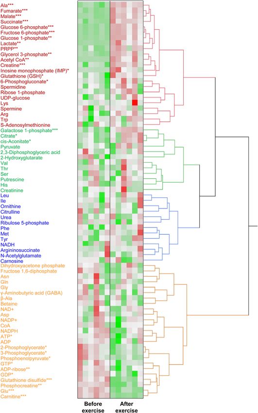
and decreased after exercise, respectively. The heat map of the hierarchical cluster analysis of
muscle metabolites showed that changes in metabolites were clearly distinguishable before and
after exercise. Strenuous exercise increased many metabolites in the glycolytic pathway and the
tricarboxylic acid cycle in skeletal muscle. Targeted metabolomic analysis of skeletal muscle may
clarify the intramuscular environment caused by exercise and explain the response of working muscles
to strenuous exercise that induces hypoxemia and hypercapnia in Thoroughbred horses.
Thoroughbred horses are considered elite athletes because their mass-specific maximal oxygen consumption
(V̇O2max) is approximately two-fold higher than that of elite human athletes1, 2. To acquire such high exercise
ability, it is thought that Thoroughbred horses need to improve their cardiopulmonary capacity and muscle adap-
tation. It has been reported that Thoroughbred horses experience severe hypoxemia (partial pressure of oxygen
[PaO2] < 75 mmHg) and hypercapnia (PaCO2 > 50 mmHg) when exercising s upramaximally1, 3–6. Therefore, it
is understandable that the muscles of the horse are subjected to extreme conditions due to strenuous exercise.
Additionally, it has been reported that horses further improve their exercise capacity under hypoxic conditions2.
Metabolome analysis, which is the method used for the comprehensive analysis of metabolites, has become
common in the field of sports science. However, few studies have performed a metabolome analysis using equine
skeletal muscle7. Therefore, further studies are warranted to understand muscle adaptations in response to exer-
cise. Currently, there is limited information on muscle metabolism under exercise conditions that induce severe
hypoxemia and hypercapnia. We hypothesized that a high-intensity exercise would induce marked changes in
the glycolytic pathway, tricarboxylic acid (TCA) cycle and high-energy phosphate compounds in skeletal muscle.
Therefore, the purpose of this study was to compare the muscle metabolomics in Thoroughbred horses before
and after supramaximal exercise to fatigue.
Results
Incremental exercise test. In the incremental exercise test, the total running distance, running time,
and speed of the last step were 4,518 ± 939 m, 10.4 ± 1.3 min, and 12.7 ± 0.5 m/s, respectively. Mean V̇O2max,
CO2 production, respiratory exchange ratio (RER), heart rate, plasma lactate concentrations, and packed cell
volume were 174 ± 10 mL/kg/min, 205 ± 17 mL/kg/min, 1.18 ± 0.07, 222 ± 11 beat/min, 20.5 ± 3.3 mmol/L, and
62.2 ± 3.3%, respectively.
Metabolome analysis. We detected 73 metabolites from the muscle samples (Supplementary Table 1,
Fig. 1). Of those, 18 and 11 metabolites were significantly increased and decreased, respectively, after exercise.
Many metabolites involved in glycolysis and the TCA cycle, such as glucose 6-phosphate (320 vs. 2,529 nmol/g),
Sports Science Division, Equine Research Institute, Japan Racing Association, 1400‑4 Shiba, Shimotsuke‑shi,
Tochigi 329‑0412, Japan. *email: hajime@equinst.go.jp
Scientific Reports | (2021) 11:11261 | https://doi.org/10.1038/s41598-021-90834-y 1
Vol.:(0123456789)www.nature.com/scientificreports/
Figure 1. A heat map of the hierarchical cluster analysis of muscle metabolites before and after exercise to
fatigue. Each column shows individual horses. Red shows a relatively high concentration of each metabolite.
Green shows a relatively low concentration of each metabolite. Asterisks show significant differences between
before and after exercise by Welch’s t-test (* < 0.05, ** < 0.01, *** < 0.001).
Scientific Reports | (2021) 11:11261 | https://doi.org/10.1038/s41598-021-90834-y 2
Vol:.(1234567890)www.nature.com/scientificreports/
acetyl coenzyme A (CoA) (0.1 vs. 4.1 nmol/g), and succinic acid (77 vs. 197 nmol/g), were increased after exer-
cise (Supplementary Table 1, Figs. 1, 2, 3). In contrast, glutamate (2488 vs. 713 nmol/g) and carnitine (5881 vs.
1474 nmol/g) were decreased after exercise (Supplementary Table 1). There were no changes in branched-chain
amino acids.
Discussion
We performed a targeted quantitative metabolomic analysis focusing on energy metabolites to investigate the
changes induced by high-intensity exercise in the glycolytic pathway, TCA cycle, pentose phosphate pathway,
amino acids, and high-energy phosphate compounds in equine skeletal muscle. The heat map of the hierarchical
cluster analysis of muscle metabolites showed that changes in metabolites were clearly distinguishable before
and after exercise. Eighteen and 11 metabolites were significantly increased and decreased, respectively, due to
strenuous exercise. Numerous metabolites involved in the TCA cycle, some metabolites of glycolysis, lactate,
and alanine were significantly increased after exercise and are classified in the same cluster. Some metabolites
of glycolysis, phosphocreatine, glutamate, and glutathione disulfide were significantly decreased after exercise
and are classified in the same cluster. The changes in several metabolites may have prevented the continuation
of strenuous exercise. Based on the intensity and duration of the bout and accumulation of lactate, the exercise
performed in this study was considered supramaximal exercise for Thoroughbreds. The observed changes in
metabolomics may vary depending on the amount, duration, and intensity of the exercise as well as the test
animal. In addition, the small n size is a limitation of this study.
For the production of energy, glycolysis is initiated from glucose 6-phosphate that is phosphorylated glucose.
In this study, the levels of glucose 6-phosphate were significantly increased (eight-fold change) after exercise.
Similarly, the levels of fructose 6-phosphate were also significantly increased after exercise. Figure 2 shows that
the concentrations of these two metabolites were relatively higher than those of other metabolites involved in the
glycolytic pathway. In contrast, the levels of metabolites from fructose 1,6-diphosphate to phosphoenolpyruvate
involved in the glycolytic pathway decreased after exercise. Fructose 1,6-diphosphate is produced by the cata-
lytic action of phosphofructokinase, which is a key regulator of the glycolytic pathway. Phosphoenolpyruvate
is the substrate of pyruvate kinase that irreversibly converts phosphoenolpyruvate to pyruvate. The concentra-
tions of three metabolites (3-phosphoglycerate, 2-phosphoglycerate, and phosphoenolpyruvate) between the
enzymes phosphofructokinase and pyruvate kinase were relatively lower than that of the two metabolites glucose-
6-phosphate and fructose-6-phosphate. Hence, they may be rapidly converted to the downstream metabolite,
and their levels subsequently decrease after the exercise. These results indicate that the exercise affected the flux
of the glycolytic pathway, and the changes in metabolites may be different at the point of enzymes that are key
regulators or react irreversibly. In addition, although there was no change in pyruvate, the levels of lactate and
acetyl CoA (products of pyruvate) were significantly increased after exercise. We believed that the increase in
metabolites of the upstream and downstream in glycolysis showed that many substrates have flowed into this
pathway. Therefore, it appears reasonable to conclude that the increased levels of glycolysis metabolites may
indicate activation of this pathway by exercise.
All metabolites we detected in the TCA cycle were significantly increased by strenuous exercise in this study
(Fig. 3). Gibala et al. reported that the TCA cycle intermediates were increased during exercise and this increase
was primarily mediated by large changes in succinate, malate, and f umarate8. We found similar changes in these
three metabolites as well as an increase in citrate. Similar to glycolysis, the observed increase in metabolites was
attributed to the increased supply of metabolites as a result of the activated TCA cycle by strenuous exercise.
It is well established that several amino acids are converted into intermediates in the TCA cycle. Following
activation of the anaplerotic process by strenuous exercise, the concentrations of amino acids involved in this
process change. In this study, glutamate was significantly decreased, whereas alanine was significantly increased
after strenuous exercise. Pyruvate and glutamate are reversibly catalyzed to alanine and α-ketoglutarate by ala-
nine transaminase. Glutamate is also reversibly catalyzed to α-ketoglutarate by glutamate dehydrogenase or a
glutamate-linked aminotransferase9. These two pathways appear to be catalyzed toward increasing the levels
of α-ketoglutarate by exercise. It has been demonstrated that the alanine aminotransferase reaction is the most
important anaplerotic process during the initial minutes of contraction in human skeletal m uscle8. In addi-
tion, studies have shown that α-ketoglutarate was the only TCA cycle intermediate to decrease at the onset of
exercise8, 10. Therefore, two reactions may supplement α-ketoglutarate during exercise. These reports are con-
sistent with our results, implying that the noted decrease in glutamate and the increase in alanine may be due
to the result of anaplerosis of the TCA cycle. However, we did not find any changes in aspartate that potentially
participates in the anaplerotic reactions. These results suggest that the changes in alanine and glutamate may be
the main indicator of the anaplerotic flux of amino acids into the TCA cycle during exercise.
Branched-chain amino acids (BCAAs) are an important source of substrates for the TCA cycle. Recently, it
was reported that the concentrations of BCAAs in blood may be related to central fatigue during e xercise11–13.
In this study, there were no changes in valine, leucine, or isoleucine in skeletal muscle before and after strenuous
exercise. It is proposed that BCAAs may not play an important role as an energy source during short duration,
high-intensity exercise and may be irrelevant to the fatigue induced by the exercise.
Phosphocreatine is reversibly catalyzed to creatine by creatine kinase and its high-energy phosphate that
can be reversibly transferred from adenosine diphosphate (ADP) to adenosine triphosphate (ATP). Guanosine
triphosphate (GTP) also contains high-energy phosphate that is reversibly transferred to ATP. In the present
study, we observed that these metabolites were significantly decreased after exercise (phosphocreatine: 0.26-
fold; ATP: 0.84-fold; GTP: 0.81-fold; guanosine diphosphate: 0.83-fold) (Fig. 4). The decrease in ADP was not
significant (0.82-fold); however, ADP and ATP were classified in the same cluster by the hierarchical cluster
analysis. The intramuscular concentration of GTP was markedly lower than that of ATP and phosphocreatine,
Scientific Reports | (2021) 11:11261 | https://doi.org/10.1038/s41598-021-90834-y 3
Vol.:(0123456789)www.nature.com/scientificreports/
Figure 2. Observed changes in metabolites mapped onto the pathways associated with glycolysis. Asterisks
show significant differences between before and after exercise by Welch’s t-test (* < 0.05, ** < 0.01, *** < 0.001).
Scientific Reports | (2021) 11:11261 | https://doi.org/10.1038/s41598-021-90834-y 4
Vol:.(1234567890)www.nature.com/scientificreports/
Figure 3. Observed changes in metabolites mapped onto the pathways associated with the TCA cycle. Asterisks
show significant differences between before and after exercise by Welch’s t-test (* < 0.05, ** < 0.01, *** < 0.001).
Scientific Reports | (2021) 11:11261 | https://doi.org/10.1038/s41598-021-90834-y 5
Vol.:(0123456789)www.nature.com/scientificreports/
Figure 4. Observed changes in metabolites mapped onto the pathways associated with pentose phosphate
pathway and high-energy phosphate compounds. Asterisks show significant differences between before and
after exercise by Welch’s t-test (* < 0.05, ** < 0.01, *** < 0.001).
and the change in GTP may not affect the energy requirements of muscle. The exercise performed in this study
was supramaximal exercise in Thoroughbreds. Therefore, it is reasonable to observe significant decreases in the
levels of phosphocreatine, ATP, and ADP. These results were consistent with the report by McGowan et al.14. It is
unclear whether the decreased high-energy phosphate in the muscle directly induced exhaustion. Nevertheless,
the decrease in phosphocreatine was marked compared with those of the other high-energy phosphates in the
muscle. As a result, we observed a significant increase (1.55-fold change) in creatine, a metabolite of phospho-
creatine, after the strenuous exercise.
Carnitine plays an important role in transporting long-chain fatty acids from the cytosol into mitochondria;
this process is termed the “carnitine shuttle.” Long-chain fatty acids in mitochondria are utilized for energy
production via β-oxidation15. It has been reported that acetylcarnitine in plasma, a metabolite of carnitine, was
increased by submaximal exercise16. In contrast, Furuichi et al. reported that the concentration of carnitine in
muscle did not increase by electric muscle contraction as heavy muscle load, whereas that of acetylcarnitine was
significantly increased17. In this study, carnitine (0.25-fold change) was one of the metabolites that showed the
largest decrease after the strenuous exercise. This may indicate that β-oxidation was also very active even during
the strenuous exercise in Thoroughbred horses. From our previous experience, the type of exercise employed
in this study results in an RER which exceeds 1.0 at e xhaustion1, 2, 18, 19. Consistent with these results, the RER
in this study was 1.18 ± 0.07. Although an RER > 1.0 indicates that carbohydrate is the predominant source of
substrates, a significant reduction in carnitine may indicate that fat is also utilized during strenuous exercise.
However, based on the RER, we hypothesized that the contribution of fat to the total energy expenditure may
be low in this type of exercise in horses.
The first two steps of the pentose phosphate pathway from glucose 6-phosphate to ribulose 5-phosphate
result in the production of two nicotinamide adenine dinucleotide phosphate (NADPH) molecules, which is a
major reductant with antioxidant activity. Glucose 6-phosphate dehydrogenase irreversibly converts glucose-
6-phosphate to 6-phosphgluconate and is a key regulator of the pentose phosphate pathway. The observed
increase in 6-phosphgluconate (1.88-fold change) indicated the activation of the pentose phosphate pathway by
the exercise (Fig. 4). Although the levels of NADPH (0.92-fold change) were maintained after exercise, it is known
that glucose 6-phosphate dehydrogenase is activated by decreasing NADPH. In this study, the concentration
of reduced glutathione (GSH) (1.51-fold change), which is deoxidized by NADPH, was significantly increased.
Reportedly, activation of the pentose phosphate pathway may be involved in the resistance against reactive oxygen
Scientific Reports | (2021) 11:11261 | https://doi.org/10.1038/s41598-021-90834-y 6
Vol:.(1234567890)www.nature.com/scientificreports/
species and amplified NADPH p roduction20. Gohil et al. reported that submaximal cycle ergometer exercise for
90 min decreased the levels of GSH, whereas those of oxidized GSH (glutathione disulfide) increased in b lood21.
This observation was inconsistent with our results. However, the differences in exercise intensity and specimens
(blood vs. muscle) analyzed may have led to different results.
Phosphoribosyl pyrophosphate (PRPP) is a substrate upstream of inosine monophosphate (IMP). As a result
of the activation of the pentose phosphate pathway, the concentrations of PRPP and IMP were significantly
increased (3.34- and 14.3-fold change, respectively) by strenuous exercise (Fig. 4). However, IMP is also gener-
ated by consuming the high-energy phosphates of ATP and ADP. Therefore, the increase in IMP may be due to
both the degradation of ADP and an increase in supply from the pentose phosphate pathway.
The majority of fibers of the gluteal muscle in Thoroughbred horses are type IIa and IIx fi bers22–24. It has also
been reported that the recruitment patterns of muscle fibers differ depending on the intensity and duration of
bers22. There-
exercise. For example, short duration/high-intensity exercise stimulated type IIx fiber and IIa/IIx fi
fore, these results may reflect the metabolomic changes in only these fast-twitch muscles after exercise in Thor-
oughbred horses. It is known that different fiber types have different mitochondrial content22. The results of the
metabolomic analysis may vary depending on the type of muscle fiber and/or pattern of exercise. Further studies
are warranted to investigate these hypotheses. The detection of changes in the muscle metabolome may help
clarify the alterations caused by exercise in the intramuscular environment and the mechanisms through which
working muscles adapt to strenuous exercise that induces hypoxemia and hypercapnia in Thoroughbred horses.
Methods
Ethics statement. This study was conducted at the Equine Research Institute, Japan Racing Association
(Tochigi, Japan), and its protocol was reviewed and approved by the institutional Animal Use and Care Commit-
tee (Approval No.: 19-23, Approval date: 14 June 2019). All experiments were performed in accordance with our
institutional guidelines. All authors confirm that the authors complied with the ARRIVE guidelines. Consent to
participate/Consent to publish is not applicable.
Horses. Six healthy Thoroughbreds (three geldings and three females; age: 6.0 ± 1.7 (mean ± SD) years;
weight: 495 ± 36 kg) were studied. The horses were trained for 1 month on an equine treadmill by running up a
6% incline twice a week prior to the experiment and veterinarians examined their health every week. The exer-
cise protocol consisted of 2-min exercise intervals at 1.7, 4.0, 7.0, 10.0, 12.0, and 13.0 m/s. On 3 days, the horses
walked at a speed of 7 km/h for 1 h in a walking machine; for the other 2 days of the week, the horses rested in
their stalls.
Incremental exercise test. Incremental exercise tests to exhaustion were performed on a treadmill
inclined to a 6% grade as a single bout for each horse. Before leading a horse onto the treadmill, a 14-gage Teflon
catheter was placed in the left jugular vein following injection of a local anesthetic agent (lidocaine; Fujisawa
Pharmaceutical Co., Osaka, Japan) and a heart rate monitor (S810; Polar, Kempele, Finland) was put to measure
the heart rate. The exercise protocol consisted of warm-up (1.7 m/s for 2 min and 3.5 m/s for 3 min), followed by
2-min exercise intervals at 1.7, 4.0, 7.0, 10.0, 12.0, and 13.0 m/s until the horse could not maintain its position at
the front of the treadmill with humane encouragement. During exercise, a mask was placed on the horse to col-
lect exhaled breath, and oxygen consumption and CO2 production were measured as previously described19, 25.
Biopsy samples for the metabolomic analysis were obtained from the same area (3 cm away from the first sam-
pling point) at the midsection of the gluteus medius muscle and from the same depth (5 cm below the skin
surface) using a 14-gage needle under local anesthesia (lidocaine; Fujisawa Pharmaceutical Co.) before and
immediately after exercise on the treadmill. Muscle samples were rapidly frozen in liquid nitrogen and stored
at − 80 °C until the metabolomic analysis. Venous blood was drawn from the jugular catheter to measure the
concentration of lactate in plasma and packed cell volume. Blood samples were centrifuged (KH120A, Kubota,
Tokyo, Japan) for 5 min (12,000×g) to measure the packed cell volume. The plasma was separated through cen-
trifugation for 5 min (1800×g) to measure the concentration of lactate using a lactate analyzer (BIOSEN C-LINE
Glucose Lactate Analyzer; EKF-diagnostic GmbH, Barleben, Germany). The running distance was calculated as
the sum of the running speed × 2 min at each running step.
Measurement of metabolites. Metabolome measurements were performed by Human Metabolome
Technologies, Inc. (Tsuruoka, Japan). Frozen muscle (~ 40 mg) was added to 750 µL of 50% acetonitrile/Milli-Q
water containing internal standards (H3304-1002; Human Metabolome Technologies, Inc.) at 0 °C to inactivate
enzymes. The muscle was homogenized (thrice at 3500 rpm for 120 s) using a tissue homogenizer (Micro Smash
MS100R; Tomy Digital Biology Co., Ltd., Tokyo, Japan), and the homogenate was centrifuged (2300×g at 4 °C
for 5 min). Subsequently, 400 µL of the upper aqueous layer was centrifugally filtered through a Millipore 5-kDa
cutoff filter (9100×g at 4 °C for 120 min) to remove proteins. The filtrate was centrifugally concentrated and re-
suspended in 50 µL of Milli-Q water for capillary electrophoresis–mass spectrometry (CE-MS) analysis.
According to the methods developed by Soga et al., cationic compounds were measured in the positive mode
of CE-time of flight MS (CE-TOFMS), while anionic compounds were measured in the positive and negative
modes of CE-MS/MS26–28. Peaks detected by CE-TOFMS and CE-MS/MS were extracted using and automatic
integration software (MasterHands; Keio University, Tsuruoka, J apan29 and MassHunter Quantitative Analysis
B.04.00; Agilent Technologies, Santa Clara, CA, USA, respectively) to obtain peak information including m/z,
migration time (MT), and peak area. The peaks were annotated with putative metabolites from the Human
Metabolome Technologies metabolite database based on their MTs in CE and m/z values determined by TOFMS
and MS/MS. The tolerance range for the peak annotation was configured at ± 0.5 min for MT and ± 10 ppm
Scientific Reports | (2021) 11:11261 | https://doi.org/10.1038/s41598-021-90834-y 7
Vol.:(0123456789)www.nature.com/scientificreports/
for m/z. In addition, the concentrations of metabolites were calculated by normalizing the peak area of each
metabolite with respect to the area of the internal standard and using standard curves, which were obtained by
three-point calibrations.
Statistical analysis. All data are presented as the mean ± standard deviation. Hierarchical cluster analysis
using a standardized value was performed by the JMP 13.1 (SAS Institute Inc., Cary, NC, USA) software. Com-
parisons were conducted using Welch’s t-test (P < 0.05).
Data availability
The datasets generated during and/or analyzed during the current study are available from the corresponding
author on reasonable request.
Received: 13 March 2021; Accepted: 17 May 2021
References
1. Ohmura, H., Matsui, A., Hada, T. & Jones, J. H. Physiological responses of young thoroughbred horses to intermittent high-intensity
treadmill training. Acta Vet. Scand. 55, 59. https://doi.org/10.1186/1751-0147-55-59 (2013).
2. Ohmura, H., Mukai, K., Takahashi, Y., Takahashi, T. & Jones, J. H. Hypoxic training increases maximal oxygen consumption in
Thoroughbred horses well-trained in normoxia. J. Equine Sci. 28, 41–45. https://doi.org/10.1294/jes.28.41 (2017).
3. Erickson, B. K. et al. Hypoxic helium breathing does not reduce alveolar-arterial PO2 difference in the horse. Respir. Physiol. 100,
253–260 (1995).
4. Ohmura, H., Hiraga, A. & Jones, J. H. Exercise-induced hypoxemia and anaerobic capacity in Thoroughbred horses. J. Phys. Fitness
Sports Med. 2, 163–168. https://doi.org/10.7600/jpfsm.2.163 (2013).
5. Wagner, P. D. et al. Effects of altered FIO2 on maximum VO2 in the horse. Respir. Physiol. 105, 123–134 (1996).
6. Wagner, P. D. et al. Mechanism of exercise-induced hypoxemia in horses. J. Appl. Physiol. 66, 1227–1233. https://doi.org/10.1152/
jappl.1989.66.3.1227 (1989).
7. Klein, D. J., McKeever, K. H., Mirek, E. T. & Anthony, T. G. Metabolomic response of equine skeletal muscle to acute fatiguing
exercise and training. Front. Physiol. 11, 110. https://doi.org/10.3389/fphys.2020.00110 (2020).
8. Gibala, M. J., MacLean, D. A., Graham, T. E. & Saltin, B. Anaplerotic processes in human skeletal muscle during brief dynamic
exercise. J. Physiol. 502(Pt 3), 703–713. https://doi.org/10.1111/j.1469-7793.1997.703bj.x (1997).
9. Cooper, A. J. L. & Kuhara, T. α-Ketoglutaramate: An overlooked metabolite of glutamine and a biomarker for hepatic encepha-
lopathy and inborn errors of the urea cycle. Metab. Brain Dis. 29, 991–1006. https://doi.org/10.1007/s11011-013-9444-9 (2014).
10. Gibala, M. J., Tarnopolsky, M. A. & Graham, T. E. Tricarboxylic acid cycle intermediates in human muscle at rest and during
prolonged cycling. Am. J. Physiol. 272, E239-244. https://doi.org/10.1152/ajpendo.1997.272.2.E239 (1997).
11. Chang, C. K. et al. Branched-chain amino acids and arginine improve performance in two consecutive days of simulated handball
games in male and female athletes: A randomized trial. PLoS ONE 10, e0121866. https://doi.org/10.1371/journal.pone.0121866
(2015).
12. Mikulski, T., Dabrowski, J., Hilgier, W., Ziemba, A. & Krzeminski, K. Effects of supplementation with branched chain amino acids
and ornithine aspartate on plasma ammonia and central fatigue during exercise in healthy men. Folia Neuropathol. 53, 377–386.
https://doi.org/10.5114/fn.2015.56552 (2015).
13. Chen, I. F., Wu, H. J., Chen, C. Y., Chou, K. M. & Chang, C. K. Branched-chain amino acids, arginine, citrulline alleviate central
fatigue after 3 simulated matches in taekwondo athletes: a randomized controlled trial. J. Int. Soc. Sports Nutr. 13, 28. https://doi.
org/10.1186/s12970-016-0140-0 (2016).
14. McGowan, C. M., Golland, L. C., Evans, D. L., Hodgson, D. R. & Rose, R. J. Effects of prolonged training, overtraining and detrain-
ing on skeletal muscle metabolites and enzymes. Equine Vet. J. Suppl. https://doi.org/10.1111/j.2042-3306.2002.tb05429.x (2002).
15. Bremer, J. Carnitine–metabolism and functions. Physiol. Rev. 63, 1420–1480. https://d oi.o
rg/1 0.1 152/p
hysre v.1 983.6 3.4.1 420 (1983).
16. Davison, G. et al. Metabolomic response to acute hypoxic exercise and recovery in adult males. Front. Physiol. 9, 1682. https://doi.
org/10.3389/fphys.2018.01682 (2018).
17. Furuichi, Y. et al. Imaging mass spectrometry reveals fiber-specific distribution of acetylcarnitine and contraction-induced carnitine
dynamics in rat skeletal muscles. Biochim. Biophys. Acta 1699–1706, 2014. https://doi.org/10.1016/j.bbabio.2014.05.356 (1837).
18. Ohmura, H., Hiraga, A. & Jones, J. H. Method for quantifying net anaerobic power in exercising horses. Equine Vet. J. Suppl. 38,
370–373 (2006).
19. Ohmura, H., Mukai, K., Matsui, A., Takahashi, T. & Jones, J. H. Cardiopulmonary function during supramaximal exercise in
hypoxia, normoxia and hyperoxia in Thoroughbred horses. J. Equine Sci. 31, 67–73. https://doi.org/10.1294/jes.31.67 (2020).
20. Kuehne, A. et al. Acute activation of oxidative pentose phosphate pathway as first-line response to oxidative stress in human skin
cells. Mol. Cell 59, 359–371. https://doi.org/10.1016/j.molcel.2015.06.017 (2015).
21. Gohil, K., Viguie, C., Stanley, W. C., Brooks, G. A. & Packer, L. Blood glutathione oxidation during human exercise. J. Appl. Phys.
64, 115–119. https://doi.org/10.1152/jappl.1988.64.1.115 (1988).
22. Yamano, S., Eto, D., Hiraga, A. & Miyata, H. Recruitment pattern of muscle fibre type during high intensity exercise (60–100%
VO2max) in thoroughbred horses. Res. Vet. Sci. 80, 109–115. https://doi.org/10.1016/j.r vsc.2005.04.006 (2006).
23. Kawai, M. et al. Muscle fiber population and biochemical properties of whole body muscles in Thoroughbred horses. Anat. Rec.
(Hoboken) 292, 1663–1669. https://doi.org/10.1002/ar.20961 (2009).
24. Yamano, S., Kawai, M., Minami, Y., Hiraga, A. & Miyata, H. Differences in muscle fiber recruitment patterns between continuous
and interval exercises. J. Equine Sci. 21, 59–65. https://doi.org/10.1294/jes.21.59 (2010).
25. Birks, E. K., Ohmura, H. & Jones, J. H. Measuring VO2 in hypoxic and hyperoxic conditions using dynamic gas mixing with a
flow-through indirect calorimeter. J. Equine Sci. 30, 87–92. https://doi.org/10.1294/jes.30.87 (2019).
26. Soga, T. & Heiger, D. N. Amino acid analysis by capillary electrophoresis electrospray ionization mass spectrometry. Anal. Chem.
72, 1236–1241. https://doi.org/10.1021/ac990976y (2000).
27. Soga, T. et al. Simultaneous determination of anionic intermediates for Bacillus subtilis metabolic pathways by capillary electro-
phoresis electrospray ionization mass spectrometry. Anal. Chem. 74, 2233–2239. https://doi.org/10.1021/ac020064n (2002).
28. Soga, T. et al. Quantitative metabolome analysis using capillary electrophoresis mass spectrometry. J. Proteome Res. 2, 488–494.
https://doi.org/10.1021/pr034020m (2003).
29. Sugimoto, M., Wong, D. T., Hirayama, A., Soga, T. & Tomita, M. Capillary electrophoresis mass spectrometry-based saliva metabo-
lomics identified oral, breast and pancreatic cancer-specific profiles. Metabolomics 6, 78–95. https://doi.org/10.1007/s11306-009-
0178-y (2010).
Scientific Reports | (2021) 11:11261 | https://doi.org/10.1038/s41598-021-90834-y 8
Vol:.(1234567890)www.nature.com/scientificreports/
Author contributions
H.O. generated the hypothesis and planned the experiment, assisted in data collection, analysis, and interpreta-
tion and wrote the first draft of the manuscript. K.M., Y.T., and T.T. assisted with data collection. All authors
reviewed the manuscript.
Funding
This research did not receive any specific grant from funding agencies in the public, commercial, or not-for-
profit sectors.
Competing interests
The authors declare no competing interests.
Additional information
Supplementary Information The online version contains supplementary material available at https://doi.org/
10.1038/s41598-021-90834-y.
Correspondence and requests for materials should be addressed to H.O.
Reprints and permissions information is available at www.nature.com/reprints.
Publisher’s note Springer Nature remains neutral with regard to jurisdictional claims in published maps and
institutional affiliations.
Open Access This article is licensed under a Creative Commons Attribution 4.0 International
License, which permits use, sharing, adaptation, distribution and reproduction in any medium or
format, as long as you give appropriate credit to the original author(s) and the source, provide a link to the
Creative Commons licence, and indicate if changes were made. The images or other third party material in this
article are included in the article’s Creative Commons licence, unless indicated otherwise in a credit line to the
material. If material is not included in the article’s Creative Commons licence and your intended use is not
permitted by statutory regulation or exceeds the permitted use, you will need to obtain permission directly from
the copyright holder. To view a copy of this licence, visit http://creativecommons.org/licenses/by/4.0/.
© The Author(s) 2021
Scientific Reports | (2021) 11:11261 | https://doi.org/10.1038/s41598-021-90834-y 9
Vol.:(0123456789)You can also read