Validation of faecal glucocorticoid analysis in cheetahs (Acinonyx jubatus)
←
→
Page content transcription
If your browser does not render page correctly, please read the page content below
Validation of faecal glucocorticoid analysis in
cheetahs (Acinonyx jubatus)

C.N. Valk
Studentnr. 3051366

Supervisors:

Prof. H. Bertschinger
Dr. A. Ganswindt
Faculty of Veterinary Science, Pretoria University, South Africa

Prof. dr. T.A.E. Stout
Faculty of Veterinary Medicine, Utrecht University, the Netherlands
Validation of faecal glucocorticoid analysis in cheetahs (Acinonyx jubatus)

Abstract
Measurement of glucocorticoid metabolites in faeces is increasingly used as a non-invasive method
for monitoring stress in wild animals. The aim of this study was to validate a non-invasive technique
for monitoring adrenocortical activity in cheetahs (Acinonyx jubatus) by measuring glucocorticoid
metabolites in the faeces before and after an adrenocorticotrophic hormone (ACTH) challenge. All
faeces were collected from five cheetahs (two female and three male) ten days before and seven
days after intramuscular injection of ACTH (50 IU). Samples were analyzed for glucocorticoid
metabolite concentrations using an enzyme immunoassay (EIA). Faecal glucocortcoid metabolites
increased 234 –715% above baseline within 20 hours of ACTH administration in all five cheetahs.
When left at ambient temperature, faecal glucocorticoid metabolites concentrations declined over
time since defaecation. Therefore it is advisable to freeze samples immediately after defaecation for
reliable monitoring of faecal glucocorticoid metabolite concentrations. Our results show that non-
invasive monitoring of faecal glucocorticoid metabolites can be a valid and useful tool for assessing
adrenal activity in cheetahs.

                                                    2
Validation of faecal glucocorticoid analysis in cheetahs (Acinonyx jubatus)
Contents
Introduction............................................................................................................................................. 4
Materials and methods ........................................................................................................................... 6
Study site and animals......................................................................................................................... 6
Sample collection ................................................................................................................................ 7
ACTH challenge.................................................................................................................................... 7
Degradation experiment ..................................................................................................................... 7
Faecal extraction and hormone analysis ............................................................................................. 7
Discussion .............................................................................................................................................. 10
Conclusion ............................................................................................................................................. 12
Literature ............................................................................................................................................... 13
3Validation of faecal glucocorticoid analysis in cheetahs (Acinonyx jubatus)
Introduction
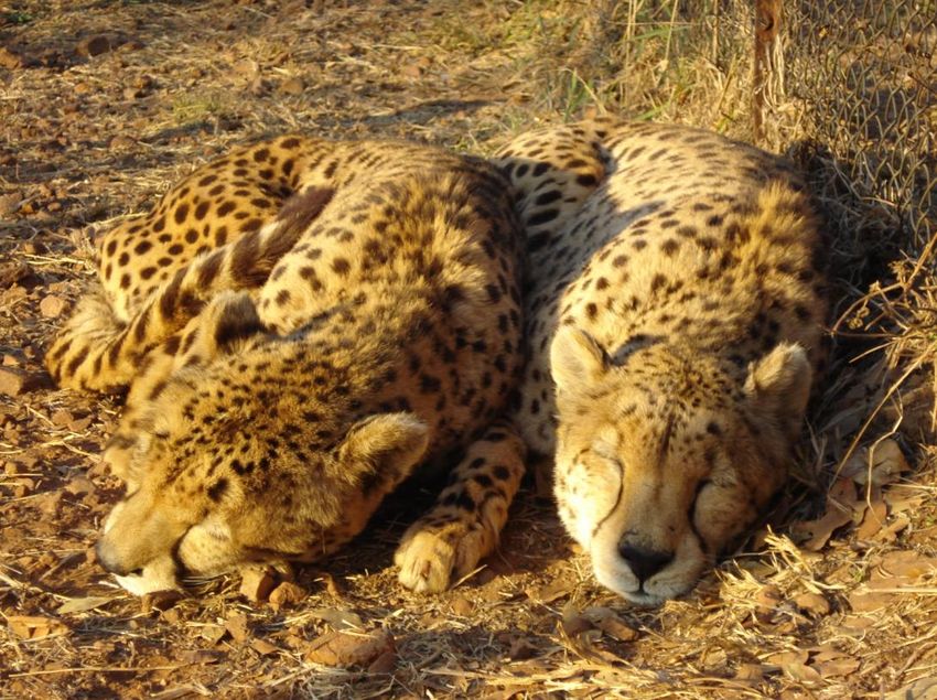
Worldwide, cheetah (Acinonyx jubatus) populations are declining, with a total number of 100,000
free-ranging cheetahs counted at the end of the 19th century to approximately 10,000 counted today.
Cheetahs are included on the International Union for Conservation of Nature (IUCN) list of vulnerable
species, as well as on the US Endangered Species Act: threatened species – Appendix I of CITES
(Convention on International Trade in Endangered Species). Cheetahs have become all but extinct in
the wild in Asia, with less than 100 cheetahs surviving in Iran. Cheetahs can, however, still be found
in a broad section of Africa, with the largest populations in Southern Africa (Namibia, Botswana) and
Western Africa (Kenya, Tanzania). Nevertheless, cheetah populations are considered viable in less
than half the countries where they still exist. Populations continue to decline from loss of habitat and
prey species, conflict with humans and loss of genetic variation. Cheetahs have a gestation of
approximately 95 days, and average litter size is 4-5 cubs. However, because wild populations are not
faring well, captive breeding programs are considered essential for the conservation of the species.
To date, however, the captive population has not been self-sustaining because of poor reproductive
success and a high prevalence of unusual diseases. Chronic stress has been suspected to be an
important contributing factor to both of these problems, which makes the development of a reliable
glucocorticoid concentration monitoring technique in cheetahs an important project. [Marker 1998;
Terio et al. 1999; Wells et al. 2004]
Stress is an imbalance in homeostasis caused by environmental stimuli (stressors). During a period of
acute stress, especially catecholamines (e.g. epinephrine, norepinephrine, and dopamine) allow
energy mobilization and may change an animal’s behaviour, better preparing it for a fight or flight
response. Prolonged elevation of glucocorticoid (e.g. cortisol, or corticosterone) concentrations due
to chronic stress however, can have several negative effects, such as suppression of the immune
system and decreased reproductive success. [Möstl & Palme 2002; Touma et al. 2005]
When a vertebrate is confronted with a stressor the hypothalamic-pituitary-adrenal (HPA) axis is
activated. Corticotrophin-releasing hormone (CRH) from the hypothalamus enhances the release of
adrenocorticotropic hormone (ACTH) from the anterior pituitary gland. ACTH in turn stimulates the
synthesis and secretion of catecholamines as well as glucocorticoids from the adrenal cortices. In this
regard, the predominant glucocorticoid produced depends primarily on the species in question, but
the most prominent endogenous glucocorticoid in most large animal species is cortisol. [Möstl &
Palme 2002; Touma et al. 2005]
The activity of the adrenal glands is one indication of a physiological stress response. A common
method of determining the activity of the adrenal glands is to measure the concentration of cortisol
in the blood. However, a blood sample represents the concentration of hormones at a given point in
time. This has the advantage that small transient changes can be detected over a short period of
time. However, because cortisol has a pulsatile secretion pattern and a clear diurnal rhythm in many
species, a lot of blood samples would need to be recovered to produce a reliable representation of
chronic changes in hormone levels. Moreover, collecting blood is an invasive approach which can
itself induce a stress response (elevated glucocorticoid concentrations) due to handling and
restraining procedures. [Millspaugh et al. 2004; Möstl & Palme 2002; Touma et al. 2005]
An alternative technique for monitoring changes in glucocorticoid output is to measure corticoid
metabolites in excreta. Circulating glucocorticoids are metabolized in the liver and excreted as
4Validation of faecal glucocorticoid analysis in cheetahs (Acinonyx jubatus)
conjugates, partly via the kidneys (in the urine) and partly via the bile into the gut, where they are
partially reabsorbed into the bloodstream via the entero-hepatic circulation. Any remaining corticoid
metabolites in the faeces will be further converted by bacterial enzymes and excreted via the faeces.
[Möstl & Palme 2002; Touma et al. 2005]
Faeces can be very easily collected without disturbing the animal. Samples can also be collected at
regular intervals over time, which makes it interesting for long-term studies. Moreover, the
concentration of glucocorticoid metabolites in the faeces reflects the total amount of production and
excretion of cortisol over a period of time, rather than at a single point in time. [Millspaugh et al.
2004; Möstl & Palme 2002; Touma et al. 2005] This makes it a more suitable for producing a reliable
representation of chronic changes in glucocorticoid secretion.
Since there are differences between species and gender, with regard to the metabolism and
excretion of glucocorticoid metabolites [Millspaugh et al. 2004; Möstl & Palme 2002; Touma et al.
2005], measurement of glucocorticoid metabolites in faecal samples must be carefully validated in
each species of interest and in both sexes before it can be concluded that a given technique can be
used as a reliable tool for monitoring adrenal activity in that species.
There are basically two ways to assess if a respective test-system detects a reliably biological signal: a
physiological and a biological validation. Physiological validation involves pharmacological induction
of a change in the concentration of glucocorticoids in the blood, followed by evaluation of faecal
glucocorticoid metabolites to see if the change is reflected in the faecal samples. The most common
way to stimulate adrenocortical activity is to conduct an ACTH challenge test. During this test an
animal will be injected with ACTH, which is expected to result in a significant increase in plasma
glucocorticoid concentrations followed by a return to baseline within several hours. Samples
(preferably faeces) are collected before and after the injection and subsequently analysed for
glucocorticoid metabolite levels. Ideally, this procedure should result in elevated concentration of
faecal glucocorticoid metabolites, with the onset of the peak delayed by the species-specific lag time
(which is affected by gut passage time). [Monfort et al. 1998; Terio et al. 1999; Touma et al. 2005;
Wasser et al. 2000] Biological validation is similar but involves collecting faecal samples before and
after a known stressful event, like capture or transportation. [Möstl & Palme 2002; Terio et al. 1999;
Touma et al. 2005; Wells et al. 2004].
Terio et al. (1999) previously validated a radioimmunoassay for quantifying faecal glucocorticoid
metabolites in cheetah. ACTH was administered to two males and two females, while seven females
were subjected to a variety of stressful events. Each resulted in increased faecal corticoid
metabolites approximately 24-72 hours later. Wells et al. (2004) measured the stress response of
cheetahs to movement between facilities. For most animals corticoid concentrations increased.
After defaecation, glucocorticoid metabolites can be further converted by bacterial enzymes. [Touma
et al. 2005] This means that the faecal samples always have to be collected at the same time post-
defecation, ideally as fresh as possible. Once collected, the samples must be frozen immediately to
stop the microbiological activity and stored at -20˚C until analysis.[Millspaugh et al. 2004; Möstl &
Palme 2002]Due to the fact that during most studies, focal animals can’t be observed 24 hours a day,
faeces are sometimes not fresh when collected. To see what happens to the concentration of
5Validation of faecal glucocorticoid analysis in cheetahs (Acinonyx jubatus)
glucocorticoid metabolites when faeces isn’t frozen immediately, a degradation experiment can be
performed.
The aim of this study was to validate a method for assessing adrenocortical activity in captive
cheetahs (Acinonyx jubatus) by measuring glucocorticoid metabolites in the faeces using an ACTH
challenge. We also measured the degradation rate of glucocorticoid metabolites in faecal samples to
further evaluate if faecal glucocorticoid metabolite measurement would be a useful tool for
monitoring stress in the cheetah.
Materials and methods
Study site and animals
The study took place at ‘the Ann van Dyk Cheetah Centre’ (also known as ‘de Wildt’), which is
situated near the Magaliesberg mountain range in the North West Province of South Africa.
The ACTH challenge was conducted on 5 cheetahs, 3 male and 2 female (Table 1). Animals ranged
from 10 to 12 years of age at the time of the study. They were housed individually in outdoor
enclosures of the size of approximately 360 m2 (Figure 1). Two sibling males (M379 and M380) were
housed together. During the day the animals were kept in the corner camp, which could be closed off
from the main camp, for easier observation and sample collection. During the night (17:00 – 7:30
hours) the animals had access to their entire enclosure. Animals were fed once a day at 7:30 hours a
mixture of Iams® and horse- and chicken-mince. For the two animals housed together, faeces were
differentiated by mixing rice (M380) and split peas (M379) in their food. Water was available ad lib.
During the experiment there was no rainfall, temperatures were approximately 20oC during the day
and approximately 5oC at night.
Cheetah Sex Age Housing
F327 Female 12 Individual
F343 Female 12 Individual
M353 Male 11 Individual
M379 Male 10 Pair (with M380)
M380 Male 10 Pair (with M379)
Table 1: Study animals
6Validation of faecal glucocorticoid analysis in cheetahs (Acinonyx jubatus)
Figure 1: Ground plan of the enclosure where the cheetahs were housed. During the day the cheetahs were
kept in the corner camp, at night the cheetahs had access to their entire enclosure.
Sample collection
The five cheetahs were observed during the day from 7:30 until 17:00 hours for 21 days from June 17
until July 7 2010. Before faecal sampling started, the cheetahs were getting used to the smaller
corner camp for four days. Homogenized faeces were collected using rubber gloves, frozen within 10
minutes after defecation, and stored at -20°C until processing and analysis. Due to logistically
restrictions, the enclosures could not be entered after 17:00 hours until 7:30 the next morning.
Therefore every morning between 7:30 and 8:00 hours the faeces from the previous night were
collected. Faecal samples were collected ten days pre ACTH administration to be able to establish
individual baseline faecal glucocorticoid metabolite concentrations, and seven days post injection.
ACTH challenge
To stimulate adrenal activity, a single dose of 50 IU ACTH (Synacthen®, Novartis RSA (Pty) Ltd,
Kempton Park South-Africa) was administered per animal into the thigh muscle between 11:00 and
11:20 hours on the 10th day of the study. Animals were physically restrained to facilitate the
administration of ACTH. The cheetahs were observed that day until 18:00 hours and the next
morning from 5:00 hours.
Degradation experiment
To determine the change in glucocorticoid concentration in samples post-defecation, two complete
faecal samples (one 19 hours after ACTH injection and one the day before injection) were separately
collected in bags and homogenized. After the zero sample was frozen immediately at -20oC, six
subsamples were created from each sample and stored at ambient temperature overnight.
Subsequently one subsample from each set was frozen 1, 2, 5, 7½, 10 and 12½ hours after the zero
sample faeces was stored at -20oC.
Faecal extraction and hormone analysis
The faecal samples were lyophilized, pulverized and sieved. Between 50 -60 mg of faecal powder was
extracted with 3 ml of 80% ethanol by vortexing for 15 minutes and subsequent centrifugation for 10
minutes at 3,300 g. The supernatant was then recovered and stored at -20°C until hormone analysis.
7Validation of faecal glucocorticoid analysis in cheetahs (Acinonyx jubatus)
The glucocorticoid metabolite concentrations were quantified using an enzyme immunoassay (EIA),
as first described in Palme & Möstl (1997), using a polyclonal antibody against 4-pregnene-11ß,21-
diol-3,20-dione -3CMO:BSA and cortisol-3-CMO-DADOO-biotin as label. The sensitivity of the assay at
90% binding was 4 pg per well. Inter- and intra-assay coefficients of variation ranged between 4.7%
and 17.8%.
Results
In total 167 faecal samples were collected, 101 baseline samples and 66 post ACTH injection samples.
Table 2 gives an overview of all the samples. Of the faecal samples 33,5% were collected fresh and
frozen immediately. All peak samples were freshly collected.
Cheetahs Total samples pre injection mean per day post injection mean per day % fresh
pre injection post injection
F327 27 18 1,8 9 1,3 7,4
F343 33 21 2,1 12 1,7 21,2
M353 31 19 1,9 12 1,7 35,5
M379 35 19 1,9 16 2,3 34,3
M380 41 24 2,4 17 2,4 34,1
Total 167 101 10,1 66 9,4 33,5
Table 2: overview of the samples collected
The ACTH injection resulted in a 234 to 715% increase in glucocorticoid metabolites concentration in
all study animals 17 to 20 hours post injection compared to pre-injection values (Figure 2 and 3). The
glucocorticoid metabolite level in the faeces increased 497-715% above baseline after ACTH injection
in females and 234-403% above baseline in males (Table 3). The peak samples in all cheetahs were
the first faecal sample after ACTH injection. The peak samples in all five cheetahs had similar delays:
17 to 20 hours after ACTH injection and returned to their baseline in the second faecal sample after
injection (between 20 and 44 hours post ACTH injection).
6 Females F327
F343
5
corticosterone (µg/g DW)
ACTH
4
3
2
1
0
-250 -200 -150 -100 -50 0 50 100
hours before and after ACTH injection
Figure 2: Glucocorticoid metabolite levels before and after ACTH injection for the two female cheetahs.
8Validation of faecal glucocorticoid analysis in cheetahs (Acinonyx jubatus)
5
Males M353
4,5
M379
4
M380
corticosterone (µg/g DW)
ACTH
3,5
3
2,5
2
1,5
1
0,5
0
-250 -200 -150 -100 -50 0 50 100
hours before and after ACTH injection
Figure 3: Glucocorticoid metabolite levels before and after ACTH injection for the three male cheetahs.
Cheetah Mean baseline ± SEM Peak concentration Elevation (%) Time of peak Decline to
(ng/g) (ng/g) (h) baseline after
injection (h)
F327 779 ± 49,5 5567 715 19 44
F343 783 ± 83,3 3888 497 17 20
M353 1112 ± 96,0 4478 403 19 44
M379 900 ± 31,7 2107 234 20 44
M380 779 ± 61,2 2883 370 18 21
Table 3: results of the enzyme immunoassay
For the degradation experiment two fresh samples were used: one baseline sample from the male
cheetah M380 and one peak sample from the female cheetah F327. Faecal glucocorticoid levels
degraded during the night (Figure 4 and Table 4). The sample of the female cheetah declined faster
(more than 20% decline at 5 hours post defaecation) than the sample of the male cheetah (more
than 20% decline at 12,5 hours post defaecation). The degradation rate for the female cheetah is
2.9% decline of faecal glucocorticoids per hour and for the male cheetah 1.9% decline per hour.
9Validation of faecal glucocorticoid analysis in cheetahs (Acinonyx jubatus)
Degradation M380
120 F327
% corticosterone (µg/g DW)
100
80
60
40
20
0
0 5 10 15
hours after defaecation
Figure 4: Degradation experiment of a fresh faecal sample of the male cheetah M380 and the peak sample of
the female cheetah F327
Degradation 1h 2h 5h 7,5 h 10 h 12,5 h
(post defaecation)
F327 -13,29% -6,82% -21,18% -25,49% -34,83% -28,19%
M380 -1,08% -7,53% +3,23% -1,08% -13,98% -26,88%
Table 4: Decline in faecal glucocorticoid metabolite levels after defaecation in % compared to the fresh zero
sample
Discussion
An increase in faecal glucocorticoid metabolites following the ACTH challenge was measured in all
five cheetahs. In this study, ACTH induced a 234-715% rise above baseline concentrations of faecal
glucocorticoid metabolites. These results are similar to other studies. Graham and Brown (1996)
found a 238-826% increase over individual baseline values in domestic cats. In the study of Schatz
and Palme (2001) the increase was 355% (median) above baseline in domestic cats and 702%
(median) in domestic dogs. Goymann et al (1999) found a 198-3748% increase above baseline in
spotted hyenas. In the study Terio et al (1999) conducted the increase was 690-4194% compared to
the baseline in four cheetahs. Young et al (2004) found a 1145-1361% increase after an ACTH
challenge in two cheetahs. They both used 400 IU ACTH, compared to the 50 IU used in this study,
which could explain the higher increase.
In this study, the glucocorticoid peak of both females (497-715% increase) was higher than the
glucocorticoid peak of the males (234-403% increase). This can be due to individual variation and/or
additional stress to the injection. The cheetahs in our study were restraint in a squeeze cage in order
to administer the ACTH injection. Terio et al (1999) found elevations of faecal glucocorticoid
concentrations in cheetahs following restraint in a squeeze cage. The two males housed together
were more used to people than the other cheetahs, which may have resulted in a lower
glucocorticoid peak due to less additional stress to the injection. The difference could also be
because of gender differences, since gender can have an effect on the glucocorticoid concentrations
found in faeces. Differences in metabolism and route of excretion of glucocorticoids are reported in
10Validation of faecal glucocorticoid analysis in cheetahs (Acinonyx jubatus)
males and females. [Millspaugh et al. 2004; Möstl & Palme 2002; Touma et al. 2005]
It has also been reported that cheetahs that were described as more ‘nervous’ by zookeepers
showed higher baseline faecal glucocorticoid metabolites than cheetahs that were described as more
‘calm’. [Jurke et al 1997; Terio et al 1999] This has also been described in clouded leopards.
[Wielebnowski et al 1999] The females in our study and one male (M353) were more nervous than
the other two males (M379 and M380). However, the glucocorticoid metabolite baselines of the
female cheetahs (F327: 779 ± 49,5 ng/g and F343: 783 ± 83,3 ng/g) were lower compared to two of
the three male cheetahs (M353: 1112 ± 96,0 ng/g and M379: 900 ± 31,7 ng/g). One male cheetah
(M380) had similar baseline concentrations as the females (779 ± 61,2 ng/g). This contradicts what
was found in the previous studies mentioned above.
Wells et al (2004) found that cheetahs with low baseline faecal glucocorticoid metabolite
concentrations had a significantly higher glucocorticoid peak after movement compared to cheetahs
that had high baseline glucocorticoid levels. They suggest that this might be due to the fact that
animals with high baseline levels might already be at maximal production, precluding further
stimulation. We found similar results in this study, since both females had lower baseline
glucocorticoid levels and higher glucocorticoid peaks compared to the other animals. Since in all
studies only small numbers of animals were used, the differences between gender and temperament
need further evaluation.
It is important to know when a glucocorticoid metabolites peak, as a reaction to a stressor, will
appear in the faeces in order to correlate the stressful event to the faecal glucocorticoid peak. The
delay between the stressor and the elevated concentration of faecal glucocorticoid metabolites (lag
time) is closely related to the gut transit time in an animal. This lag time is species-specific. [Monfort
et al. 1998; Terio et al. 1999; Touma et al. 2005; Wasser et al. 2000] In this study, all increases in
faecal glucocortcoid metabolite concentration were observed in the first faecal sample collected
after the ACTH challenge. The delay between the ACTH injection and appearance of peak
concentrations was 17-20 hours. These results are in line with previous research. Terio et al (1999)
also observed the glucocorticoid metabolite peaks in the first faecal sample after ACTH injection in
three out of four cheetahs. Similar delay times were also found in the domestic cat (25 hours, Schatz
and Palme 2001; 24-48 hours, Graham and Brown 1996), domestic dog (22 hours, Schatz and Palme
2001), African wild dog (24-30 hours, Monfort 1998) and the spotted hyena (16-50 hours, Goymann
et al 1999).
In this study 33,5% of the faecal samples were fresh on the time of collection. After defaecation
glucocorticoid metabolites can be further converted by bacterial enzymes, which can increase or
decrease the measured faecal concentrations. [Keay et al 2006; Touma et al. 2005; Möstl and Palme
2002] This can be influenced by factors as temperature and rainfall. [Washburn and Millspaugh 2002;
Palme 2005] To test the stability of faecal glucocorticoids in samples post-defaecation , a degradation
experiment was performed under the same environmental conditions as during the experiment. The
degradation experiment in our study shows that the glucocorticoid metabolite levels of faecal
samples decline over time since defaecation, when left at night. This contradicts previous data on
other animals. In a study Huber et al (2003) conducted on red deer the concentration of
glucocorticoid metabolites in faeces collected within approximately six hours from defaecation did
not differ significantly compared to faeces that were collected fresh. Rehnus et al. (2009) also found
that neither different storage conditions (10 and 25°C) nor time intervals resulted in a significant
11Validation of faecal glucocorticoid analysis in cheetahs (Acinonyx jubatus)
change in glucocorticoid concentrations in faeces of Mountain hares. Considering our results, it is
advisable to freeze faecal samples immediately after defaecation to accurately determine the levels
of glucocorticoid metabolites in the faeces.
Conclusion
The aim of this study was to evaluate a method for assessing adrenocortical activity in captive
cheetahs (Acinonyx jubatus) by measuring glucocorticoid metabolites in the faeces. An increase in
faecal glucocorticoid metabolites following the ACTH challenge was measured in all five cheetahs.
This suggests that the EIA used in this study can reliably detect stress-related changes in
glucocorticoid metabolite levels in male and female cheetahs. This can be a very useful tool for
monitoring stress in animals in captivity since it is a non-invasive technique, faecal samples can be
very easily collected and it is suitable for long-term evaluations of adrenal activity.
Since our results show a decline in faecal glucocorticoid metabolites when left at ambient
temperature, due to bacterial degradation, it is important to freeze samples immediately after
defaecation for reliable monitoring of faecal glucocorticoid metabolite levels using this EIA in
cheetahs.
Acknowledgements
I thank everyone at the Ann van Dyk Cheetah Centre (de Wildt) for allowing us to do the project
there and for their assistance with sample collection. I would also like to thank my supervisors Prof.
Henk Bertschinger, Dr. Andre Ganswindt and Prof. Dr. Tom Stout for their help and guidance.
I thank Stefanie Muenscher for help with the laboratory procedures. At last I would like to thank
Kirsten in ‘t Ven for doing this project with me.
12Validation of faecal glucocorticoid analysis in cheetahs (Acinonyx jubatus)
Literature
CITES 2011, The CITES Appendices. www.cites.org
IUCN 2011, IUCN Red List of Threatened Species, Version 2011.2. www.iucnredlist.org
Goymann, W; E Möstl; T van ’t Hof; ML East; H Hofer. Noninvasive Fecal Monitoring of
Glucocorticoids in Spotted Hyenas, Crocuta crocuta. General and Comperative Endocrinology 114:
340-348 (1999).
Graham, LH; JL Brown. Cortisol metabolism in the domestic cat and implications for non-invasive
monitoring of adrenocortical function in endangered felids. Zoo Biol. 15: 71-82 (1996).
Huber, S; R Palme; W Arnold. Effects of season, sex, and sample collection on concentrations of fecal
metabolites in red deer (Cervus elaphus). General and Comparative Endocrinology 130: 48-54
(2003).
Jurke, HM; NM Czekala; DG Lindburg; SE Millard. Fecal corticoid metabolite measurement in the
cheetah (Acinonyx jubatus). Zoo Biol. 16: 133-147 (1997).
Keay, JM; J Singh; MC Gaunt; T Kaur. Fecal glucocorticoids and their metabolites as indicators of
stress in various mammalian species: a literature review. Journal of Zoo and Wildlife Medicine
37(3): 234-244 (2006).
Marker, L. Current status of the cheetah (Acinonyx jubatus). Proceedings of a symposium on cheetahs
as game ranch animals, Onderstepoort, 23&24 October 1998.
Millspaugh, JJ; BE Washburn. Use of fecal glucocorticoid metabolite measures in conservation biology
research: considerations for application and interpretation. General and Comperative
Endocrinology 138 (2004) 189-199.
Monfort, SL; KL Mashburn; BA Brewer; SR Creel. Evaluating adrenal activity in African wild dogs
(lycaon pictus) by fecal corticosteroid analysis. Journal of Zoo and Wildlife Medicine 29(2): 129-
133 (1998).
Möstl, E.; JL Maggs; G Schrötter; U Besenfelder; R Palme. Measurement of cortisol metabolites in
faeces of ruminants. Veterinary Research Communications, 26 (2002) 127-139.
Möstl, E; R Palme. Hormones as indicators of stress. Domestic Animal Endocrinology 23 (2002) 67-74.
Palme, R; E Möstl. Measurement of cortisol metabolites in faeces of sheep as a parameter of cortisol
concentrations in blood. Int J Mamm Biol 62:192-197 (1997).
Palme, R. Measuring fecal steroids: guidelines for practical application. Ann N Y Acad Sci 1046: 75-80
(June 2005).
Rehnus, M; K Hackländer; R Palme. A non-invasive method for measuring glucocorticoid metabolites
(GCM) in Mountain hares (Lepus timidus). Eur J Wildl Res 55: 615-620 (2009).
13Validation of faecal glucocorticoid analysis in cheetahs (Acinonyx jubatus)
Schatz, S; R Palme. Measurement of faecal cortisol metabolites in cats and dogs: a non-invasive
method for evaluating adrenocortical function. Veterinary Research Communications, 25(2001)
271-287.
Terio, KA; SB Citino; JL Brown. Fecal cortisol metabolite analysis for noninvasive monitoring of
adrenocortical function in the cheetah (acinonyx jubatus). Journal of Zoo and Wildlife Medicine
30(4): 484-491 (1999).
Touma, C; R Palme. Measuring fecal glucocorticoid metabolites in mammals and birds: the
importance of validation. Ann. N.Y. Acad. Sci. 1046: 54-74 (2005).
Washburn, BE; JJ Millspaugh. Effects of simulated environmental conditions on glucocorticoid
metabolite measurements in white-tailed deer feces. General and Comparative Endocrinology
127: 217-222 (2002).
Wasser, SK; KE Hunt; JL Brown; K Cooper; CM Crockett; U Bechert; JJ Millspaugh; S Larson; SL
Monfort. A generalized fecal glucocorticoid assay for use in a diverse array of nondomestic
mammalian and avian species. General and Comparative Endocrinology 120, 260–275 (2000).
Wells, A; KA Terio; MH Ziccardi; L Munson. The stress response to environmental change in captive
cheetahs (Acinonyx Jubatus). Journal of Zoo and Wildlife Medicine 35(1), 8-14 (2004).
Wielebnowski, N; J Busso; J Brown. Adrenal activity in relation to subjective temperament assessment
in clouded leopards, Neofelis nebulosa. In: DE Wildt, JD Mellen, J Brown, editors. Felid taxon
advisory group action plan, report. Lake Buena Vista, FL: Disney’s Animal Kingdom Publ: 1999 p74-
5.
Young, KM; SL Walker; C Lanthier; WT Waddell; SL Monfort; JL Brown. Noninvasive monitoring of
adrenocortical activity in carnivores by fecal glucocorticoid analyses. General and Comparative
Endocrinology 137: 148-165 (2004).
14You can also read