Propofol abolishes torsade de pointes in different models of acquired long QT syndrome - Nature
←
→
Page content transcription
If your browser does not render page correctly, please read the page content below
www.nature.com/scientificreports
OPEN Propofol abolishes torsade de
pointes in different models
of acquired long QT syndrome
Christian Ellermann1,2*, Hilke Könemann1, Julian Wolfes1, Benjamin Rath1, Felix K. Wegner1,
Kevin Willy1, Dirk G. Dechering1, Florian Reinke1, Lars Eckardt1 & Gerrit Frommeyer1
There is conflicting evidence regarding the impact of propofol on cardiac repolarization and the risk of
torsade de pointes (TdP). The purpose of this study was to elucidate the risk of propofol-induced TdP
and to investigate the impact of propofol in drug-induced long QT syndrome. 35 rabbit hearts were
perfused employing a Langendorff-setup. 10 hearts were perfused with increasing concentrations of
propofol (50, 75, 100 µM). Propofol abbreviated action potential duration (APD90) in a concentration-
dependent manner without altering spatial dispersion of repolarization (SDR). Consequently, no
proarrhythmic effects of propofol were observed. In 12 further hearts, erythromycin was employed
to induce prolongation of cardiac repolarization. Erythromycin led to an amplification of SDR
and triggered 36 episodes of TdP. Additional infusion of propofol abbreviated repolarization and
reduced SDR. No episodes of TdP were observed with propofol. Similarly, ondansetron prolonged
cardiac repolarization in another 13 hearts. SDR was increased and 36 episodes of TdP occurred.
With additional propofol infusion, repolarization was abbreviated, SDR reduced and triggered
activity abolished. In this experimental whole-heart study, propofol abbreviated repolarization
without triggering TdP. On the contrary, propofol reversed prolongation of repolarization caused by
erythromycin or ondansetron, reduced SDR and thereby eliminated drug-induced TdP.
Up to 22% of patients treated on intensive care units experience ventricular arrhythmias and have a higher
mortality compared to patients without heart rhythm disturbances1. Many risk factors have been identified over
the past decades and include individual characteristics of each patient but also external factors such as use of
pharmacological agents that impair repolarization reserve. Since propofol is commonly used in anaesthesia and
intensive care medicine, the impact of propofol alone or in combination with other drugs that influence cardiac
electrophysiology is of great interest. As a consequence, some studies have already investigated the electrophysi-
ological effects of propofol in vivo and vitro:
Intravenous application of propofol has direct impact on several ion currents including I Na, some potassium
channels (IKs, IK,to, IK,1), and I Ca,L2–5. However, previous clinical and experimental studies report conflicting
data regarding its impact on ventricular repolarization and potential proarrhythmic effects. Higashijima et al.
described a significant abbreviation of QTc interval during anaesthetic induction mediated by p ropofol6. In
contrast, a recent study demonstrated an increase in ventricular repolarization duration calculated by Fridericia-
corrected QT interval with propofol7. Additionally, Tpeak to Tend interval (Tpeak-Tend) was significantly amplified in
the presence of propofol in this study. An increased Tpeak-Tend interval is a surrogate for an amplified transmural
dispersion of repolarization which in turn represents a major risk factor for drug-induced arrhythmias8. In
contrast to this study, no significant changes of QTc or T peak-Tend have been reported during propofol infusion
in children9.
Some other experimental studies have investigated effects of propofol in different models of long QT syn-
drome (LQTS): In healthy and transgenic LQT2 and LQT3 rabbits, propofol administration resulted in an
increase of QT index resulting in arrhythmia-related death in two LQT2 rabbits5. In another study employ-
ing a model of (drug-induced) LQTS, propofol reduced the action potential duration increase mediated by
erythromycin10.
1
Department of Cardiology II (Electrophysiology), University Hospital Münster, Albert‑Schweitzer‑Campus
1, 48149 Münster, Germany. 2Klinik für Kardiologie II – Rhythmologie, Universitätsklinikum Münster,
Albert‑Schweitzer Campus 1, 48149 Münster, Germany. *email: christian.ellermann@ukmuenster.de
Scientific Reports | (2020) 10:12133 | https://doi.org/10.1038/s41598-020-69193-7 1
Vol.:(0123456789)
www.nature.com/scientificreports/
In conclusion, existing studies report conflicting data concerning propofol-induced changes in repolarization
duration and heterogeneity and provocation of arrhythmias. Therefore, the purpose of the present study was to
elucidate propofol’s impact in a sensitive model of repolarization disorders. Previous experimental studies have
solely investigated the effect of propofol infusion on ventricular repolarization duration and other ECG markers
(e.g. Tpeak-Tend) but did not investigate other proarrhythmic mechanisms such as dispersion of repolarization
or action potential shape. Thus, this study aimed at elucidating further potential mechanisms in arrhythmia
initiation induced by propofol.
Methods
All experimental protocols were approved by the local animal care committee (Landesamt für Natur, Umwelt
und Verbraucherschutz Nordrhein-Westfalen, Germany) and were carried out in accordance with the Guide for
the Care and Use of Laboratory Animals published by the US National Institutes of Health (NIH Publication No.
852-3, revised 1996). Since hearts served as their own control, no randomization of the hearts was performed.
The experimental setting of the antegradely-perfused Langendorff-heart has been described earlier
extensively11. In short, 35 hearts of female New Zealand white rabbits were explanted and mounted to a Lan-
gendorff apparatus. Spontaneously beating hearts were perfused by a warmed, oxygenated (95% O 2, 5% CO2)
modified Krebs–Henseleit buffer (NaCl 118 mM, NaHCO3 24.88 mM, d-glucose 5.55 mM, KCl 4.70 mM, Na-
pyruvate 2 mM, CaCl2 1.80 mM, KH2PO4 1.18 mM, MgSO4 0.83 mM) at a constant flow (52 mL/min) with a
pressure around 90 mmHg. Monophasic action potentials (MAP) were acquired by eight specifically designed
MAP catheters that were placed endo- and epicardially. Hearts were immersed in a warmed tissue bath, thereby
enabling recording of a volume-conducted 12-lead ECG. Spontaneously beating hearts were mechanically AV
node-ablated using surgical tweezers in order to perform the following stimulation protocol.
Hearts were stimulated at seven different cycle lengths (900–300 ms), thus obtaining cycle-lengths dependent
QT interval and action potential duration (APD90). APD90 was measured between the fastest upstroke and 90%
of repolarization. Premature extra-stimuli ( S2 and S 3) were delivered to the hearts in order to assess ventricular
vulnerability and to determine effective refractory periods (ERP) at different basic cycle-lengths (900–300 ms,
see Fig. 1). In case sustained ventricular arrhythmias occurred after short-coupled extra-stimuli, hearts were
defibrillated, and the pacing protocol was halted for 5 min to assure recovery of the hearts. Post-repolarization
refractoriness (PRR) was calculated as the difference between ERP and APD90. Spatial dispersion of repolarization
was determined by the difference of maximum and minimum of the A PD90 of the eight MAPs. Configuration
PD90/APD5012,13.
of action potentials is displayed by the ratio of A
Hearts were divided up into three different groups: In the first group, propofol in ascending concentrations
(50, 75, 100 µM) was infused after generating baseline data and the protocol was repeated for each concentration.
In this experimental arm, premature extra-stimuli were solely delivered after pacing the hearts at a basic cycle-
length of 500 ms in order to abridge the experimental protocol. The second group was perfused with 300 µM
erythromycin after generating baseline data. Afterwards, hearts were additionally treated with 75 µM propofol.
The last group was infused with 5 µM ondansetron and thereafter 75 µM propofol was added. Before continuing
the experimental protocol with a new drug or concentration, hearts were equilibrated for 15 min.
Statistics. Electrograms and action potentials were recorded on a multi-channel recorder and digitalized at
a rate of 1 kHz with a 12-bit resolution. Variables are shown as mean ± standard deviation. Statistical analyses
were performed using SPSS Statistics for Windows (version 24.0). Drug effects on APD90, QT interval, disper-
sion of repolarization, action potential configuration ( APD90/APD50), ERP and PRR were analysed employing
Wilcoxon signed rank test. P values < 0.05 were considered to be statistically significant. Data are expressed as
mean ± standard deviation.
Results
Propofol effects on ventricular repolarization and arrhythmia induction. Infusion of propofol
abbreviated APD90 in a concentration-dependent manner (baseline: 158 ± 25 ms; 50 µM: 142 ± 21 ms, p < 0.01
compared to baseline; 75 µM: 129 ± 19 ms, p < 0.01 compared to baseline; 100 µM: 129 ± 23 ms, p < 0.01 compared
to baseline; Fig. 2) while QT interval remained relatively stable at the lowest propofol concentration and was
slightly abbreviated under the influence of 75 and 100 µM propofol (baseline: 242 ± 30 ms; 50 µM: 243 ± 35 ms,
p = ns; 75 µM: 241 ± 32 ms, p = 0.07; 100 µM: 239 ± 27 ms, p < 0.01; each p compared to baseline).
ERP were increased after propofol treatment in a concentration-dependent manner (baseline: 196 ± 65 ms;
50 µM: 196 ± 44 ms, p = ns; 75 µM: 201 ± 33 ms, p = ns; 100 µM: 222 ± 44 ms, p = ns), resulting in a significant
amplification of PRR (baseline: 38 ± 43 ms; 50 µM: 58 ± 43 ms, p = ns; 75 µM: 76 ± 27 ms, p < 0.02; 100 µM:
86 ± 51 ms, p = ns).
Spatial dispersion of repolarization was not significantly altered at lowest or highest propofol concentration
but was decreased with 75 µM propofol (baseline: 43 ± 15 ms; 50 µM: 45 ± 14 ms, p = ns; 75 µM: 36 ± 13 ms,
p < 0.05; 100 µM: 39 ± 15 ms, p = ns).
The action potential shape which can be expressed by the ratio of APD90/APD50 was not significantly changed
in the presence of 50 or 75 µM propofol (baseline: 1.48 ± 0.13, 50 µM: 1.44 ± 0.11, p = ns; 75 µM: 1.47 ± 0.11,
p = ns). However, with the highest concentration tested (100 µM), a further rectangulation of action potential
shape could be observed (1.42 ± 0.14, p = 0.01).
3 episodes of ventricular tachycardia or fibrillation were inducible by programmed ventricular stimulation
(S2 and S3) under baseline conditions. No episodes occurred with 50 µM propofol (p = ns) while 2 episodes of
VT/VF were inducible under the influence of 75 µM propofol (p = ns). With the highest propofol concentration
(100 µM) 6 episodes of VT/VF were inducible (p = ns).
Scientific Reports | (2020) 10:12133 | https://doi.org/10.1038/s41598-020-69193-7 2
Vol:.(1234567890)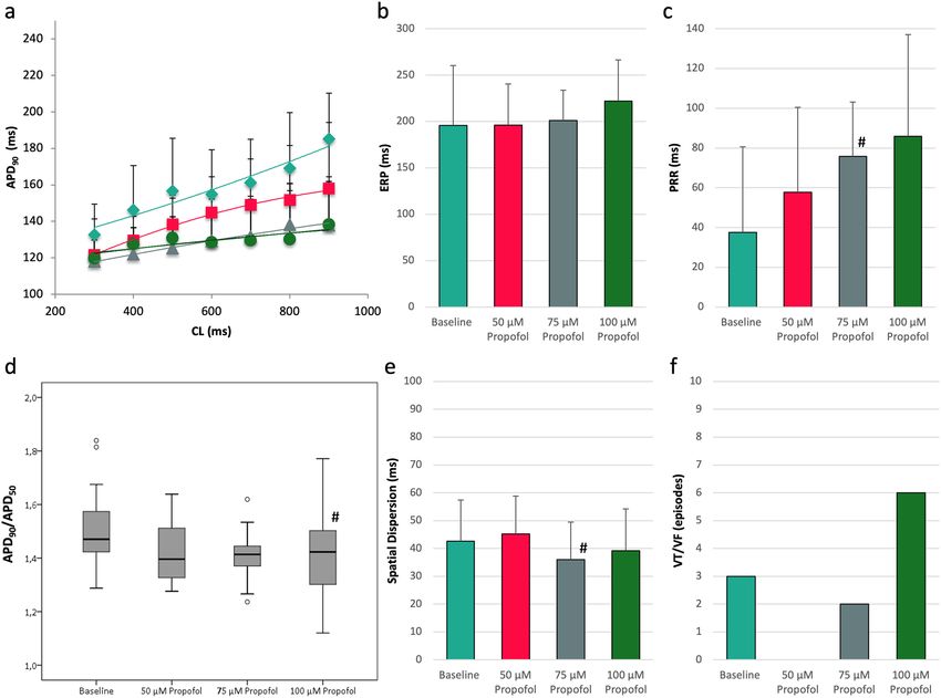
www.nature.com/scientificreports/
Figure 1. Determination of effective refractory periods (MAP = monophasic action potential).
Erythromycin. Treatment with 300 µM erythromycin prolonged QT interval from 246 ± 27 ms to
275 ± 30 ms (p < 0.01; Fig. 3) while A PD90 was just slightly increased from 168 ± 18 ms to 171 ± 28 ms (p = ns).
Propofol reversed these effects and abbreviated QT interval to 267 ± 18 ms (p < 0.01) and APD90 to 158 ± 23 ms
(p < 0.01). Spatial dispersion of repolarization was significantly amplified in the presence of erythromycin (base-
line: 40 ± 16 ms; erythromycin: 48 ± 18 ms, p < 0.01) and reduced by the additional treatment with propofol to
42 ± 12 ms (p = 0.01 compared to erythromycin).
There was a trend towards an increase of APD90/APD50 after infusion of erythromycin from 1.49 ± 0.12 to
1.52 ± 0.19 (p = 0.13), representing a triangulation of action potential shape. With propofol, a non-significant
decrease of the APD90/APD50 ratio was observed (1.48 ± 0.10, p = 0.21).
No torsade de pointes occurred under baseline conditions. However, 36 episodes of torsade de pointes spon-
taneously occurred after treatment with erythromycin (p < 0.05 compared to baseline) and were completely
eliminated in the presence of propofol (0 episodes, p < 0.05).
Scientific Reports | (2020) 10:12133 | https://doi.org/10.1038/s41598-020-69193-7 3
Vol.:(0123456789)www.nature.com/scientificreports/
Figure 2. (a) Cycle-length dependent APD90 under baseline conditions (filled rhombus) and after treatment
with 50 µM (filled square), 75 µM (filled traingle) or 100 µM (filled circle) propofol. (b) Impact of propofol
on effective refractory periods (ERP). (c) Concentration-dependent effect of propofol on post-repolarization
refractoriness (#p < 0.05 compared to baseline conditions). (d) Box plots of the ratio of action potential
duration at 90% of repolarization (APD90) and action potential duration at 50% of repolarization ( APD50). A
decrease in A
PD90/APD50 represents a rectangulation of action potential. (e) Influence of propofol treatment
on repolarization heterogeneity as indicated by spatial dispersion of repolarization (#p < 0.05 compared to
baseline conditions). (f) Occurrence of ventricular fibrillation (VF) tachycardia (VT) induced by programmed
ventricular fibrillation.
Ondansetron. With ondansetron, an increase of QT interval from 252 ± 46 ms to 309 ± 68 ms (p = ns; Fig. 4)
and of APD90 from 162 ± 28 to 174 ± 32 ms (p < 0.01) was observed. Additional infusion of propofol abbreviated
QT interval (to 289 ± 61 ms, p = ns) as well as A PD90 (to 150 ± 23 ms, p < 0.01). Spatial dispersion of repolariza-
tion was amplified after ondansetron infusion (from 40 ± 20 ms to 59 ± 24 ms, p < 0.01) and substantially reduced
under the additional influence of propofol (37 ± 16 ms, p < 0.01).
The APD90/APD50 ratio was significantly increased in the presence of ondansetron from 1.53 ± 0.18 to
1.56 ± 0.15 (p < 0.05). Propofol did not significantly alter the A
PD90/APD50 ratio (1.53 ± 0.19; p = ns).
No episodes of torsade de pointes occurred in the spontaneously beating, AV-blocked hearts under baseline
conditions. With ondansetron, 36 episodes of torsade de pointes were observed (p < 0.02, Fig. 5). Again, propofol
treatment eliminated torsade de pointes in each heart (0 episodes, p < 0.02).
Discussion
To our best knowledge, this is the first experimental whole-heart study investigating propofol’s effects on cardiac
electrophysiology in different models of acquired long QT syndrome. This study demonstrates that sole propo-
fol infusion slightly abbreviates ventricular repolarization without triggering torsade de pointes. Furthermore,
administration of propofol on top of proarrhythmic agents such as erythromycin or ondansetron reduces repo-
larization and spatial dispersion of repolarization and thereby eliminates torsade de pointes.
Impact of propofol on cardiac electrophysiology. In the present study, propofol induced a significant
abbreviation of cardiac repolarization as indicated by APD90 and QT interval. This is in line with the majority
of former clinical studies investigating repolarization duration under the influence of propofol14. Previous data
Scientific Reports | (2020) 10:12133 | https://doi.org/10.1038/s41598-020-69193-7 4
Vol:.(1234567890)www.nature.com/scientificreports/
Figure 3. (a,b) Cycle-length dependent APD90 and QT interval under baseline conditions (filled rhombus),
after treatment with 300 µM erythromycin (filled square) and after additional infusion of 75 µM propofol (filled
triangle). (c) Box plots of the ratio of APD90 to APD50. (d) Impact of erythromycin and propofol on spatial
dispersion of repolarization (#p < 0.05 compared to baseline conditions; *p < 0.05 compared to sole erythromycin
infusion). (e) Occurrence of torsade de pointes under baseline conditions, with erythromycin and with the
combination of erythromycin and propofol (#p < 0.05 compared to baseline conditions; *p < 0.05 compared to
sole erythromycin treatment).
concerning propofol’s influence on T peak-Tend is equivocal. While a recent clinical study showed a prolonged
Tpeak-Tend interval with propofol7, no changes were observed in another trial with a paediatric study cohort9.
Tpeak-Tend interval has been proposed as a surrogate for transmural dispersion of cardiac repolarization15. In
contrast to the QT interval that mediocrely predicts occurrence of torsade de pointes, an increased transmural
dispersion of repolarization is a good indicator for drug-induced arrhythmias8,13. This study clearly indicates a
stable dispersion of repolarization during propofol-treatment even at supratherapeutic concentrations. A stable
dispersion of repolarization (even in the presence of a prolonged cardiac repolarization) is linked to a safe elec-
trophysiologic profile of several antiarrhythmic drugs16.
Furthermore, the shape of action potential duration was transformed by the highest concentration of propofol
to a more rectangular shape as indicated by a decrease in A PD90/APD50. A rectangulation of action potential
reduces the risk of arrhythmias and is mediated by an acceleration of phase 3 repolarization which reduces
the time in the window voltage for calcium channel reactivation and subsequent triggered a ctivity12. Thus, no
arrhythmias were observed in bradycardic hearts even with the highest concentration of propofol used. As a con-
sequence, this study highlights a good safety profile of propofol. With propofol, post-repolarization refractoriness
was significantly lengthened. Prolongation of PRR protects the myocardium against premature beats, is therefore
antiarrhythmic11,17 and a common pharmacological property of class I antiarrhythmic drugs. Consequently,
ventricular vulnerability as tested by programmed ventricular stimulation was not increased with propofol.
In this study, supratherapeutic concentrations of propofol have been employed to determine adverse drug
effects. Mean propofol concentration during anesthesia induction is 11.7 (± 5.0) µg/mL which equals approxi-
mately 65.6 µM18. However, since genetic polymorphisms in hepatic metabolizing enzymes (e.g. CYP2C9) may
further increase propofol concentrations during anaesthesia18, higher plasma concentrations might be achieved.
Therefore, concentrations of up to 100 µM have been employed in this study.
Scientific Reports | (2020) 10:12133 | https://doi.org/10.1038/s41598-020-69193-7 5
Vol.:(0123456789)www.nature.com/scientificreports/
Figure 4. (a,b) Cycle-length dependent APD90 and QT interval under baseline conditions (filled rhombus),
after treatment with 5 µM ondansetron (filled square) and after additional infusion of 75 µM propofol (filled
triangle). (c) Box plots of the ratio of APD90 to APD50 (#p < 0.05 compared to baseline conditions) (d) Influence
of ondansetron and propofol on spatial dispersion of repolarization (#p < 0.05 compared to baseline conditions;
*p < 0.05 compared to sole ondansetron administration). (e) Occurrence of torsade de pointes under baseline
conditions, with ondansetron and with the combination of ondansetron and propofol (#p < 0.05 compared to
baseline conditions; *p < 0.05 compared to sole ondansetron treatment).
Models of acquired long QT syndrome. With erythromycin, a marked prolongation of repolarization
duration, an amplification of spatial dispersion of repolarization and a trend towards a triangulation of action
potential shape were observed. This is in line with previous studies in which the IKr inhibitor erythromycin was
employed to simulate LQT2 s yndrome11. Similar results have been achieved for ondansetron which also inhibits
hERG (human Ether-a-go-go Related Gene) potassium c hannels19. Accordingly, ondansetron augmented repo-
larization duration and amplified spatial dispersion of repolarization20.
Erythromycin and ondansetron changed the shape of the action potential to a more triangular shape which
can be explained by an inhibition of I Kr (Fig. 6). This leads to a slowing of phase 3 repolarization which in turn
prolongs the time frame in which early afterdepolarizations and subsequent torsade de pointes can be generated12.
Consequently, early afterdepolarizations and torsade de pointes were observed with both drugs.
In contrast, infusion of propofol reversed the changes induced by erythromycin or ondansetron. To be more
precise, propofol abbreviated repolarization and reduced spatial dispersion of repolarization in both groups.
Previous studies demonstrated that a decrease of spatial dispersion of repolarization is a crucial antiarrhythmic
mechanism in acquired long QT s yndrome11,17. Recently, Bossu and c olleagues21 elegantly demonstrated that
reduction of spatial dispersion of repolarization induced by the INa,L inhibitor GS967 predominantly inhibits
perpetuation of torsade de pointes in the chronic atrioventricular block dog. It is noteworthy that early afterdepo-
larization which are regarded as initiating mechanism were just slightly suppressed. Consequently, the prevention
of perpetuation instead of prevention of the initiation of the arrhythmia can be regarded as the antiarrhythmic
mechanism of GS967 in this s tudy21. Similarly, the crucial antiarrhythmic action of propofol in this study might
not be inhibition of triggered activity but rather prevention of perpetuation of torsade de pointes tachycardia.
There was a non-significant trend towards a rectangular action potential shape with additional propofol treat-
ment. As a consequence, triggered activity (early afterdepolarizations or torsade de pointes) occurred neither
Scientific Reports | (2020) 10:12133 | https://doi.org/10.1038/s41598-020-69193-7 6
Vol:.(1234567890)www.nature.com/scientificreports/
Figure 5. (a) Representative example of early afterdepolarizations induced by erythromycin
(MAP = monophasic action potential). (b) Spontaneously occurring polymorphic ventricular tachycardia
resembling torsade de pointes after ondansetron treatment.
in the erythromycin nor in the ondansetron group after additional propofol treatment. These results are in line
with a previous study in which propofol reversed the prolongation of repolarization induced by erythromycin.
However, no further mechanistic investigations were performed, and no arrhythmias were recorded due to the
experimental setup10. These findings were confirmed in a clinical setting in which propofol reversed QT interval
prolongation induced by s evoflurane22.
Surprisingly, in transgenic LQT2 rabbits, propofol prolonged repolarization and subsequently triggered tor-
sade de p ointes5. Even though above-mentioned studies indicated different effects of propofol in LQTS-linked
arrhythmias, one would actually expect similar results since inhibition of IKr either by erythromycin or ondan-
setron is likely to result in similar electrophysiologic effects as observed in LQT2.
Of note, the electrophysiologic effects of propofol in acquired LQTS observed in this study are comparable
to those obtained for the sodium current inhibitor m exiletine17.
Limitations
The present study was conducted in isolated rabbit hearts. Therefore, a direct extrapolation to humans is not
possible. However, previous studies indicate that the rabbit heart is a reasonable model for studying cardiac ion
channel function and especially for investigating cardiac repolarization disorders23. Furthermore, the rabbit
heart is particularly suitable for studying complex ventricular arrhythmias like ventricular fibrillation due to its
effective size, which relates the size of the heart to the wavelength of the a rrhythmia24. Following this concept
as proposed by P anfilov24, the effective size of the rabbit heart is similar to the human heart leading to a similar
arrhythmia pattern in both species.
However, this model does not allow precise statements concerning direct effects on ion channels. Reduction
of repolarization duration induced by propofol can probably be explained by the predominant inhibition of
sodium and calcium channels that overrides the effects of potassium channel block. Accordingly, distinct effects
of propofol on different human cardiac channels have been described and this multi-channel inhibition most
likely explains the results observed in this study: To be more precise, previous patch clamp studies reported that
propofol inhibits human L-type calcium c urrents25, human sodium3 as well as human potassium channels26.
Scientific Reports | (2020) 10:12133 | https://doi.org/10.1038/s41598-020-69193-7 7
Vol.:(0123456789)www.nature.com/scientificreports/
Figure 6. Illustrative example of action potential and ECG tracings under baseline conditions (a) and after
administration of erythromycin (b) in spontaneously beating bradycardic hearts under hypokalemic conditions.
With erythromycin, action potentials are substantially prolonged and triangulated. Of note, spatial dispersion
of repolarization (as determined by the duration differences between MAP 5 and MAP 3 in (b)) is amplified.
(MAP = monophasic action potential).
Conclusion
The present study demonstrates a safe electrophysiologic profile of propofol even at high concentrations. Propofol
abbreviated cardiac repolarization and did not bear the risk of proarrhythmia. Quite the contrary, propofol abbre-
viated repolarization duration in different models of acquired long QT syndrome, reduced spatial dispersion of
repolarization and thereby eliminated drug-induced torsade de pointes. As a consequence, propofol might even
be beneficial in drug-induced QT prolongation by reducing the risk of torsade de pointes.
Data availability
The datasets generated during and analysed during the current study are available from the corresponding author
on reasonable request.
Received: 24 March 2020; Accepted: 7 July 2020
References
1. Artucio, H. & Pereira, M. Cardiac arrhythmias in critically ill patients: Epidemiologic study. Crit. Care Med. 18, 1383–1388 (1990).
2. Yang, C.-Y., Wong, C.-S., Yu, C.-C., Luk, H.-N. & Lin, C.-I. Propofol inhibits cardiac L-type calcium current in guinea pig ven-
tricular myocytes. Anesthesiology 84, 626–635 (1996).
3. Stoetzer, C. et al. Inhibition of the cardiac Na+ channel α-subunit Nav1. 5 by propofol and dexmedetomidine. Naunyn Schmiede-
bergs Arch. Pharmacol. 389, 315–325 (2016).
4. Buljubasic, N. et al. Differential effects of etomidate, propofol, and midazolam on calcium and potassium channel currents in
canine myocardial cells. Anesthesiology 85, 1092–1099 (1996).
5. Odening, K. E. et al. Pharmacogenomics of anesthetic drugs in transgenic LQT1 and LQT2 rabbits reveal genotype-specific dif-
ferential effects on cardiac repolarization. Am. J. Physiol. Heart Circ. Physiol. 295, H2264–H2272 (2008).
6. Higashijima, U. et al. A comparison of the effect on QT interval between thiamylal and propofol during anaesthetic induction.
Anaesthesia 65, 679–683 (2010).
7. Wutzler, A. et al. Effects of propofol on ventricular repolarization and incidence of malignant arrhythmias in adults. J. Electrocardiol.
51, 170–174 (2018).
Scientific Reports | (2020) 10:12133 | https://doi.org/10.1038/s41598-020-69193-7 8
Vol:.(1234567890)www.nature.com/scientificreports/
8. Antzelevitch, C. Role of transmural dispersion of repolarization in the genesis of drug-induced torsades de pointes. Heart Rhythm.
2, S9–S15 (2005).
9. Hume-Smith, H. V., Sanatani, S., Lim, J., Chau, A. & Whyte, S. D. The effect of propofol concentration on dispersion of myocardial
repolarization in children. Anesth. Analg. 107, 806–810 (2008).
10. Morey, T. E. et al. Ionic basis of the differential effects of intravenous anesthetics on erythromycin-induced prolongation of ven-
tricular repolarization in the guinea pig heart. Anesthesiology 87, 1172–1181 (1997).
11. Ellermann, C. et al. Antiarrhythmic effect of antazoline in experimental models of acquired short-and long-QT-syndromes.
Europace 20, 1699–1706 (2018).
12. Milberg, P. et al. Divergent proarrhythmic potential of macrolide antibiotics despite similar QT prolongation: Fast phase 3 repo-
larization prevents early afterdepolarizations and torsade de pointes. J. Pharmacol. Exp. Ther. 303, 218–225 (2002).
13. Frommeyer, G. & Eckardt, L. Drug-induced proarrhythmia: Risk factors and electrophysiological mechanisms. Nat. Rev. Cardiol.
13, 36 (2016).
14. Staikou, C., Stamelos, M. & Stavroulakis, E. Impact of anaesthetic drugs and adjuvants on ECG markers of torsadogenicity. Br. J.
Anaesth. 112, 217–230 (2013).
15. Yan, G.-X. & Antzelevitch, C. Cellular basis for the normal T wave and the electrocardiographic manifestations of the long-QT
syndrome. Circulation 98, 1928–1936 (1998).
16. Frommeyer, G. et al. Electrophysiological profile of vernakalant in an experimental whole-heart model: The absence of proar-
rhythmia despite significant effect on myocardial repolarization. Europace 16, 1240–1248 (2014).
17. Frommeyer, G. et al. Broad antiarrhythmic effect of mexiletine in different arrhythmia models. Europace 20, 1375–1381 (2017).
18. Khan, M. S. et al. Pharmacogenetics, plasma concentrations, clinical signs and EEG during propofol treatment. Basic Clin. Phar-
macol. Toxicol. 115, 565–570 (2014).
19. Kuryshev, Y. A., Brown, A. M., Wang, L., Benedict, C. R. & Rampe, D. Interactions of the 5-hydroxytryptamine 3 antagonist class
of antiemetic drugs with human cardiac ion channels. J. Pharmacol. Exp. Ther. 295, 614–620 (2000).
20. Frommeyer, G. et al. Severe proarrhythmic potential of the antiemetic agents ondansetron and domperidone. Cardiovasc. Toxicol.
17, 451–457 (2017).
21. Bossu, A. et al. Selective late sodium current inhibitor GS-458967 suppresses Torsades de Pointes by mostly affecting perpetuation
but not initiation of the arrhythmia. Br. J. Pharmacol. 175, 2470–2482 (2018).
22. Terao, Y. et al. The effects of intravenous anesthetics on QT interval during anesthetic induction with sevoflurane. J. Anesth. 30,
929–934 (2016).
23. Clauss, S. et al. Animal models of arrhythmia: Classic electrophysiology to genetically modified large animals. Nat. Rev. Cardiol.
16, 457–475 (2019).
24. Panfilov, A. V. Is heart size a factor in ventricular fibrillation? Or how close are rabbit and human hearts?. Heart Rhythm. 3, 862–864
(2006).
25. Fassl, J., High, K. M., Stephenson, E. R., Yarotskyy, V. & Elmslie, K. S. The Intravenous anesthetic propofol inhibits human L-type
calcium channels by enhancing voltage-dependent inactivation. J. Clin. Pharmacol. 51, 719–730 (2011).
26. Yang, L., Liu, H., Sun, H.-Y. & Li, G.-R. Intravenous anesthetic propofol inhibits multiple human cardiac potassium channels.
Anesthesiology 122, 571–584 (2015).
Acknowledgements
This work was supported by the Hans-and-Gertie Fischer Foundation and by the German Cardiac Society (to
G.F.). We acknowledge support from the Open Access Publication Fund of the University of Muenster.
Author contributions
C.E., L.E. and G.F. designed the study. C.E., H.K.., P.N., J.W., B.R., F.K.W. and K.W. performed the experiments.
C.E., D.G.D. and F.R. analyzed the data. C.E., L.E. and G.F. wrote the paper.
Competing interests
The authors declare no competing interests.
Additional information
Correspondence and requests for materials should be addressed to C.E.
Reprints and permissions information is available at www.nature.com/reprints.
Publisher’s note Springer Nature remains neutral with regard to jurisdictional claims in published maps and
institutional affiliations.
Open Access This article is licensed under a Creative Commons Attribution 4.0 International
License, which permits use, sharing, adaptation, distribution and reproduction in any medium or
format, as long as you give appropriate credit to the original author(s) and the source, provide a link to the
Creative Commons license, and indicate if changes were made. The images or other third party material in this
article are included in the article’s Creative Commons license, unless indicated otherwise in a credit line to the
material. If material is not included in the article’s Creative Commons license and your intended use is not
permitted by statutory regulation or exceeds the permitted use, you will need to obtain permission directly from
the copyright holder. To view a copy of this license, visit http://creativecommons.org/licenses/by/4.0/.
© The Author(s) 2020
Scientific Reports | (2020) 10:12133 | https://doi.org/10.1038/s41598-020-69193-7 9
Vol.:(0123456789)You can also read