Association of severity of primary open-angle glaucoma with serum vitamin D levels in patients of African descent
←
→
Page content transcription
If your browser does not render page correctly, please read the page content below
Molecular Vision 2019; 25:438-445                                         © 2019 Molecular Vision
         Received 16 December 2016 | Accepted 7 August 2019 | Published 9 August 2019

Association of severity of primary open-angle glaucoma with
serum vitamin D levels in patients of African descent
     Radha Ayyagari,1 Yii-der I. Chen,2 Linda M. Zangwill,1 Matt Holman,1 Keri Dirkes,1 Yang Hai,2 Zorayr
     Arzumanyan,2 Rigby Slight,1 Naama Hammel,1 Christopher A. Girkin,3 Jeffrey M. Liebmann,4 Robert
     Feldman,5 Harvey Dubiner,6 Kent D. Taylor,2 Jerome I. Rotter,2 Xiuqing Guo,2 Robert N. Weinreb,1 for the
     ADAGES III Genomics Study Group

     (The first two authors contributed equally to this work.)

     1
      Viterbi Family Department of Ophthalmology, Shiley Eye Institute, Hamilton Glaucoma Center, UC San Diego, La Jolla, CA;
     2
      Institute for Translational Genomics and Population Sciences, Los Angeles Biomedical Research Institute and Department of
     Pediatrics, Harbor-UCLA Medical Center, Torrance, CA; 3Department of Ophthalmology, University of Alabama at Birmingham,
     Birmingham, AL; 4Bernard and Shirlee Brown Glaucoma Research Laboratory, Harkness Eye Institute, Columbia University
     Medical Center, New York, New York; 5Ruiz Department of Ophthalmology, University of Texas Health Science Center, Houston,
     TX; 6Eye Care Center Management, Inc., Marrow, GA

         Purpose: To study the relationship between primary open-angle glaucoma (POAG) in a cohort of patients of African
         descent (AD) and serum vitamin D levels.
         Methods: A subset of the AD and glaucoma evaluation study III (ADAGES III) cohort, consisting of 357 patients with
         a diagnosis of POAG and 178 normal controls of self-reported AD, were included in this analysis. Demographic infor-
         mation, family history, and blood samples were collected from all the participants. All the subjects underwent clinical
         evaluation, including visual field (VF) mean deviation (MD), central cornea thickness (CCT), intraocular pressure (IOP),
         and height and weight measurements. POAG patients were classified into early and advanced phenotypes based on the
         severity of their visual field damage, and they were matched for age, gender, and history of hypertension and diabetes.
         Serum 25-Hydroxy (25-OH) vitamin D levels were measured by enzyme-linked immunosorbent assay (ELISA). The
         association of serum vitamin D levels with the development and severity of POAG was tested by analysis of variance
         (ANOVA) and the paired t-test.
         Results: The 178 early POAG subjects had a visual field MD of better than −4.0 dB, and the 179 advanced glaucoma
         subjects had a visual field MD of worse than −10 dB. The mean (95% confidence interval [CI]) levels of vitamin D of the
         subjects in the control (8.02 ± 6.19 pg/ml) and early phenotype (7.56 ± 5.74 pg/ml) groups were significantly or marginally
         significantly different from the levels observed in subjects with the advanced phenotype (6.35 ± 4.76 pg/ml; p = 0.0117
         and 0.0543, respectively). In contrast, the mean serum vitamin D level in controls was not significantly different from
         that of the subjects with the early glaucoma phenotype (p = 0.8508).
         Conclusions: In this AD cohort, patients with advanced glaucoma had lower serum levels of vitamin D compared with
         early glaucoma and normal subjects.

     Glaucoma is the leading cause of irreversible blind-                      to be higher in black compared with white and mixed-race
ness in the world [1]. The disease can be characterized as a                   populations [2]. Similarly, the mean IOP was reported to be
progressive optic neuropathy, where gradual visual field loss                  highest in black compared with mixed-race or white patients
eventually leads to blindness. Primary open-angle glaucoma                     [3]. Currently, no reliable methods or biomarkers for the early
(POAG) is the most common type of glaucoma; it is typi-                        detection of glaucoma are available.
cally asymptomatic at the early stages, and it is often not                         In recent years, findings have suggested that serum
diagnosed until irreversible loss of the visual field transpires.              25-OH vitamin D levels are associated with glaucoma. In
Well-known risk factors are elevated intraocular pressure                      a study on a South Korean population, participants with
(IOP), advanced age, positive family history, and African                      low serum 25-OH vitamin D levels were found to be at a
ancestry [1]. The prevalence of glaucoma has been reported                     significantly elevated risk of open-angle glaucoma (OAG) [4].
                                                                               More recently, in a French case-control study, POAG cases
Correspondence to: Robert N. Weinreb, Shiley Eye Institute,                    had a lower mean serum 25-OH vitamin D concentration
University of California, San Diego 9415 Campus point drive, Room              than controls did, as well as a greater prevalence of vitamin
251; La Jolla, CA 92037; Phone: (858) 534-8824 | Fax: (858) 534-               D insufficiency [5]. Past findings have also revealed asso-
1625: email: rweinreb@ucsd.edu                                                 ciations between vitamin D levels and various other ocular

                                                                         438
Molecular Vision 2019; 25:438-445 © 2019 Molecular Vision
conditions. Serum 25-OH vitamin D deficiency was associ- 25-OH vitamin D levels were associated with lower all-cause
ated with a reduced ganglion cell complex in a cohort of older mortality in white Americans, but they were less strongly
Caucasians [6]. Moreover, a lower serum 25-OH vitamin D associated with all-cause mortality in black Americans [10].
concentration was associated with a high risk of myopia in However, there is limited information on the effects of 25-OH
a population of young adults in Australia [7]. In addition, a vitamin D levels on ocular abnormalities in African Ameri-
poorer quality of overall visual acuity was associated with cans. The aim of this study was investigating the possible
vitamin D insufficiency in older adults [8]. Although the association between vitamin D levels and the development
prevalence of glaucoma has been observed to be high in of POAG, as well as the severity of POAG in a cohort of
patients of African descent (AD), information is not avail- patients of AD.
able on the relationship between serum vitamin D levels and
POAG in this population. METHODS
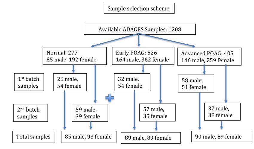
There is abundant evidence that, compared with Ameri- Subjects: A subset of 357 POAG patients and 178 control
cans with European ancestry, African Americans display subjects of AD participating in the “Contribution of Geno-
significantly lower levels of 25-OH vitamin D. Lower serum type to Glaucoma phenotype in African Americans” study
levels of 25-OH vitamin D in African American women (Figure 1), commonly referred to as the AD and glaucoma
has been attributed to a reduction in dermal synthesis [9]. evaluation study III (ADAGES III), were included in this
Contrasting health outcomes have been observed in these study. The research was conducted following the tenets of
studies when comparing 25-OH vitamin D levels between the Declaration of Helsinki and with the approval of the
white and African Americans. For example, higher serum institutional review boards of the participating institutions.
levels of 25-OH vitamin D were associated with lower To be considered to have POAG, patients were required
cardiovascular disease (CVD) risk in white Americans, while to have glaucomatous visual field damage or an optic disc
higher serum 25-OH vitamin D in black Americans was asso- photograph or clinical drawing clearly indicating POAG in
ciated with a slightly higher CVD risk. In addition, higher their medical charts. Control subjects had IOP ≤ 21 mmHg,
Figure 1. Sample selection scheme.
439Molecular Vision 2019; 25:438-445 © 2019 Molecular Vision
no evidence of glaucomatous optic neuropathy based on a among the three POAG groups, and a post hoc Tukey’s test
clinical exam, and no family history of POAG in first-degree was used for pairwise comparisons. We also compared the
relatives. The exclusion criteria included the following: 1) demographic data among the three groups, using ANOVA
ocular pathology making it difficult to determine whether for continuous data (age, BMI, MD, IOP, CCT) and the chi-
there was characteristic visual field damage, 2) closed or square test for binary traits (gender, diabetes, hypertension)
occluded angles, 3) secondary glaucoma, 4) a history of and categorical data (family history of glaucoma). To test the
human immunodeficiency virus or hepatitis C infection, and correlations between free vitamin D and the demographic
5) non-African or European descent. data, we applied the Pearson correlation for continuous data
(age, BMI, MD, IOP, CCT), t-test for binary data (gender,
For this report, POAG patients were classified into
diabetes, hypertension), and ANOVA for categorical data
“early” and “advanced” phenotypes based on the severity of
(family history of glaucoma). Most of the quantitative traits
their visual field damage at the time of blood sample collec-
in this study were normally distributed; the exception was the
tion; early POAG patients had a visual field MD better than
free vitamin D levels, which had a skewness level of 2.2973
−4 dB, and advanced POAG had visual field MD values ≤
and kurtosis of 6.2872. Thus, we applied log transformation
−10 dB. Glaucoma patients with visual field MD between −4
only to the free vitamin D levels (Appendix 1).
and −10 dB were excluded. The three groups were matched
for age, sex, and self-reported history of hypertension and
diabetes. RESULTS
The demographic, clinical, and ocular characteristics of the
Ocular measurements: Central corneal thickness (CCT), the
early POAG, advanced POAG, and controls without POAG
mean deviation (MD) of the visual field closest to the blood
study groups are presented in Table 1. As patients were
draw, and the highest IOP recorded were extracted from each
matched for age, sex, history of systemic hypertension,
patient’s medical record. The mean values of both eyes were
and diabetes, no significant differences were found in these
included in the analysis. IOP measurements were obtained
characteristics. BMI was significantly lower in the advanced
using a Goldman Applanation Tonometer.
POAG patients compared with the controls and early POAG
Body mass index: Each participant’s height and weight were (p = 0.0334 and 0.0041). There were no significant differ-
measured, and the body mass index (BMI) was calculated ences between controls and early POAG regarding BMI (p
according to standard protocols. Height and weight measure- = 0.7142). Most subjects had eye measurements performed
ments were performed at each study center using standardized on the same day as the blood draw. Specifically, the median
protocols and equipment. A model 213 stadiometer (Seca, (interquartile range) difference in years between the blood
Chino, CA) was used to measure the height to the eighth of an draw and 1) visual field testing and 2) IOP measurements
inch. A Professional Remote Digital Scale (Health-o-Meter, was small, at 0.4 (1.1) years and 0.0 (0.1) years, respectively.
McCook, IL) was used to measure weight after the removal
Advanced glaucoma patients had a significantly higher
of jackets or bulky sweaters, as described previously [11].
“highest IOP” recorded (pMolecular Vision 2019; 25:438-445 © 2019 Molecular Vision
Table 1. Demographic, clinical and ocular characteristics by study groups.
Control Early POAG Advanced POAG P
Information Trait (n=178) (n=178) (n=179) value*
Age at blood draw (SD;
MIN,MAX) 67.2 (9.09; 27.8, 95.5) 65.29 (10.47; 33.3, 92.4) 67.51 (11.28; 32.2, 91.4) 0.0934
Gender (% male) 32.6 32.6 33 0.9962
BMI (SD; MIN,MAX) 31.0 (6.7; 17.9, 54.5) 31.6 (6.3; 19.2, 54.4) 29.2 (6.3; 16.9, 49.4) 0.0039
Demographic Self-reported diabetes (%) 31.5 33.7 31.3 0.8615
and clinical Self-reported hypertension
73 67.4 65.9 0.3217
information (%)
** −2.39 (3.99; −21.99, 1.2) −1.77 (1.47; −3.98, 1.85) −21.65 (6.53; −35.3, −10.1)Molecular Vision 2019; 25:438-445 © 2019 Molecular Vision
Table 3. Correlation between Gender, Diabetes and Hypertension.
Mean pg/ Median Std Dev Minimum
Trait N P value* Category N ml pg/ml pg/ml pg/ml
Gender 519 0.0002 Female 349 7.9 6.28 5.97 0.77
Male 170 6.09 4.87 4.62 0.59
Diabetes 519 0.0886 No 355 7.01 5.63 5.33 0.59
Yes 164 7.96 6.02 6.18 1.14
Hypertension 515 0.2724 No 163 6.93 5.43 5.26 0.59
Yes 352 7.52 5.99 5.8 0.77
Family History
321 0.0041 No 214 6.99 5.44 5.05 0.59
Glaucoma
Yes 107 5.54 4.5 3.72 0.77
* t test p value
pathologies, including osteoporosis, CVD, diabetes, cancer, to the difference in the severity of the two glaucoma groups
autoimmune diseases, and depression in several popula- included in the analysis. For example, in the current study, we
tions, including subjects of AD [12,15]. A strong association compared vitamin D levels in early (better than −4 dB) and
between vitamin D levels and various ophthalmic conditions, advanced POAG (worse than −10 dB), while the French study
such as age-related macular degeneration, cataract, and dry compared vitamin D levels in moderate (better than −12 dB)
eye syndrome has been reported [8]. Specifically, the associa- and advanced glaucoma (worse than −12 dB). In addition, the
tion of vitamin D levels with a risk of glaucoma has been underlying genetic variation between the French population
reported in Chinese, Korean, French, and Caucasian popula- and patients of AD may also contribute to the differences
tions [4-6,13]. observed. Analysis of a larger cohort of patients from both
In the present study on individuals of African descent, populations using a common set of diagnostic criteria may
advanced glaucoma patients had significantly lower levels provide a better understanding of the association between
of serum vitamin D (pMolecular Vision 2019; 25:438-445 © 2019 Molecular Vision
Figure 2. Analysis of variance
(ANOVA) analysis of serum free
vitamin D levels in the three study
groups. The y-axis gives the mean
log free vitamin D levels in each
of the three study groups. The p
value on the right of the figure was
obtained from the ANOVA test
when comparing the three groups
of subjects. On the top of the figure
are p values for each pairwise
comparison.
factors influencing inflammation contribute to the severity of APPENDIX 1. A SCATTER PLOT SHOWING
neurodegeneration [19]. Studies on vitamin D have suggested THE RELATIONSHIP BETWEEN DIFFERENT
that it has a key role in regulating the physiological processes OCULAR AND CLINICAL TRAITS.
involved in the inflammation and degeneration of neuronal To access the data, click or select the words “Appendix 1.”
tissue [20]. Therefore, it is likely that low levels of vitamin
D may not result in development of glaucoma; however, the ACKNOWLEDGMENTS
insufficiency of vitamin D may affect the severity of glau- Financial Support: National Eye Institute: EY023704,
coma in patients as a result of increased inflammation and P30EY022589, EY110008, EY019869, EY021818, EY021237.
Supported in part by the National Center for Advancing
neurodegeneration [21]. The design of the current study does
Translational Sciences, CTSI grant UL1TR001881, and the
not provide information on the rate of glaucoma progression, National Institute of Diabetes and Digestive and Kidney
and therefore, the effect of vitamin D status on the rate of Disease Diabetes Research Center (DRC) grant DK063491
progression is unknown. Gaining an in-depth understanding to the Southern California Diabetes Endocrinology Research
of the molecular mechanisms underlying glaucoma and the Center, Supported in part by Research to Prevent Blindness
influence of vitamin D on disease pathology may provide (New York, NY). Conflict of Interest: LMZ: Carl Zeiss
Meditec, Heidelberg Engineering, Optovue Inc.,Topcon
insight into the pathophysiology of glaucoma and the effect of
Medical System Inc. (financial support) CAG: National
vitamin D insufficiency. Furthermore, as serum vitamin D is Eye Institute, EyeSight Foundation of Alabama, Research
a modifiable factor, the findings of this study could be helpful to Prevent Blindness, Carl Zeiss Meditec, Heidelberg Engi-
in managing the severity of glaucoma by targeting vitamin D. neering, SOLX (financial support) JML: Alcon, Allergan,
443Molecular Vision 2019; 25:438-445 © 2019 Molecular Vision
Bausch & Lomb, Carl Zeiss Meditec, Heidelberg Engineering, systems-based approach to investigating clinical practice,
Reichert, Valeant Pharmaceuticals (Consultant); Bausch & research, and public health - expert panel meeting report.
BMC Proc 2018; 12:6-[PMID: 30044889].
Lomb, Carl Zeiss Meditec, Heidelberg Engineering, National
Eye Institute, Optovue, Reichert, Topcon (financial support) 11. Zangwill L, Ayyagari R, Liebmann JM, Girkin CA, Feldman
RF: None FAM: Alcon, Allergan, Bauch + Lomb, Carl Zeiss R, Dubiner H, Dirkes KA, Holmann M, Williams-Steppe
E, Hammel N, Saunders LJ, Vega S, Sandow K, Roll K,
Meditec, Heidelberg Engineering, Merck, Reichert, Sensimed Slight R, Auerbach D, Samuels BC, Panarelli JF, Mitchell
and Topcon (financial support); Alcon, Allergan, Carl Zeiss JP, Al-Aswad LA, Park SC, Tello C, Cotliar J, Bansal R,
Meditec, National Eye Institute, and Reichert (research Sidoti PA, Cioffi GA, Blumberg D, Ritch R, Bell NP, Blieden
support); Allergan, Carl Zeiss Meditec, Novartis (consultant) LS, Davis G, Medeiros FA, Ng MCY, Das SK, Palmer ND,
RNW: Alcon, Allergan, Bausch & Lomb, Carl Zeiss Meditec, Divers J, Langefeld CD, Freedman BI, Bowden DW, Chris-
topher MA, Chen YI, Guo X, Taylor KD, Rotter JI, Weinreb
Topcon (Consultant); Heidelberg Engineering, Carl Zeiss
RN. African Descent and Glaucoma Evaluation Study III
Meditec, Genentech, Optovue, Topcon (Financial support) Genomics Study Group. Ophthalmology 2019; 126:156-70.
[PMID: 29361356].
12. Wilkins CH, Birge SJ, Sheline YI, Morris JC. Vitamin D defi-
REFERENCES
ciency is associated with worse cognitive performance and
1. Coleman AL, Brigatti L. The glaucomas. Minerva Med 2001; lower bone density in older African Americans. J Natl Med
92:365-79. [PMID: 11675580]. Assoc 2009; 101:349-54. [PMID: 19397226].
2. Leske MC, Connell AM, Schachat AP, Hyman L. The 13. Lv Y, Yao Q, Ma W, Liu H, Ji J, Li X. Associations of vitamin
Barbados Eye Study. Prevalence of open angle glaucoma. D deficiency and vitamin D receptor (Cdx-2, Fok I, Bsm I
Arch Ophthalmol 1994; 112:821-9. [PMID: 8002842]. and Taq I) polymorphisms with the risk of primary open-
3. Leske MC, Connell AM, Wu SY, Hyman L, Schachat AP. angle glaucoma. BMC Ophthalmol 2016; 16:116-[PMID:
Distribution of intraocular pressure. The Barbados Eye Study. 27435453].
Arch Ophthalmol 1997; 115:1051-7. [PMID: 9258228]. 14. Tsuprykov O, Chen X, Hocher CF, Skoblo R, Lianghong Y,
4. Yoo TK, Oh E, Hong S. Is vitamin D status associated with Hocher B. Why should we measure free 25(OH) vitamin
open-angle glaucoma? A cross-sectional study from South D? J Steroid Biochem Mol Biol 2018; 180:87-104. [PMID:
Korea. Public Health Nutr 2014; 17:833-43. [PMID: 29217467].
24476947]. 15. Holick MF. Vitamin D deficiency. N Engl J Med 2007;
5. Goncalves A, Milea D, Gohier P, Jallet G, Leruez S, Baskaran 357:266-81. [PMID: 17634462].
M, Aung T, Annweiler C. Serum vitamin D status is asso- 16. Perez-Lopez FR, Chedraui P, Fernandez-Alonso AM.
ciated with the presence but not the severity of primary Vitamin D and aging: beyond calcium and bone metabolism.
open angle glaucoma. Maturitas 2015; 81:470-4. [PMID: Maturitas 2011; 69:27-36. [PMID: 21429678].
26059919].
17. Amer M, Qayyum R. Relation between serum 25-hydroxyvi-
6. Uro M, Beauchet O, Cherif M, Graffe A, Milea D, Annweiler tamin D and C-reactive protein in asymptomatic adults (from
C. Age-Related Vitamin D Deficiency Is Associated with the continuous National Health and Nutrition Examination
Reduced Macular Ganglion Cell Complex: A Cross-Sectional Survey 2001 to 2006). Am J Cardiol 2012; 109:226-30.
High-Definition Optical Coherence Tomography Study. [PMID: 21996139].
PLoS One 2015; 10:e0130879-[PMID: 26090872]. 18. Grimm MOW, Thiel A, Lauer AA, Winkler J, Lehmann J,
7. Yazar S, Hewitt AW, Black LJ, McKnight CM, Mountain JA, Regner L, Nelke C, Janitschke D, Benoist C, Streidenberger
Sherwin JC, Oddy WH, Coroneo MT, Lucas RM, Mackey O, Stötzel H, Endres K, Herr C, Beisswenger C, Grimm
DA. Myopia is associated with lower vitamin D status in HS, Bals R, Lammert F, Hartmann T. Vitamin D and Its
young adults. Invest Ophthalmol Vis Sci 2014; 55:4552-9. Analogues Decrease Amyloid-beta (Abeta) Formation and
[PMID: 24970253]. Increase Abeta-Degradation. Int J Mol Sci 2017; 18:18-
8. Beauchet O, Milea D, Graffe A, Fantino B, Annweiler C. [PMID: 29257109].
Association between serum 25-hydroxyvitamin D concen- 19. Jiao KP, Li SM, Lv WY, Jv ML, He HY. Vitamin D3 repressed
trations and vision: a cross-sectional population-based study astrocyte activation following lipopolysaccharide stimulation
of older adults. J Am Geriatr Soc 2011; 59:568-70. [PMID: in vitro and in neonatal rats. Neuroreport 2017; 28:492-7.
21391957]. [PMID: 28430709].
9. Aloia JF, Talwar SA, Pollack S, Yeh J. A randomized controlled 20. Briones TL, Darwish H. Vitamin D mitigates age-related
trial of vitamin D3 supplementation in African American cognitive decline through the modulation of pro-inflamma-
women. Arch Intern Med 2005; 165:1618-23. [PMID: tory state and decrease in amyloid burden. J Neuroinflam-
16043680]. mation 2012; 9:244-[PMID: 23098125].
10. Brown LL, Cohen B, Tabor D, Zappala G, Maruvada P, 21. Mowry EM, Pelletier D, Gao Z, Howell MD, Zamvil SS,
Coates PM. The vitamin D paradox in Black Americans: a Waubant E. Vitamin D in clinically isolated syndrome:
444Molecular Vision 2019; 25:438-445 © 2019 Molecular Vision
evidence for possible neuroprotection. Eur J Neurol 2016; 23:327-32. [PMID: 26518224].
Articles are provided courtesy of Emory University and the Zhongshan Ophthalmic Center, Sun Yat-sen University, P.R. China.
The print version of this article was created on 9 August 2019. This reflects all typographical corrections and errata to the
article through that date. Details of any changes may be found in the online version of the article.
445You can also read