Comparative characteristics of myohistogenesis of musculoskeletal tissue in hens and quails - E3S Web of Conferences
←
→
Page content transcription
If your browser does not render page correctly, please read the page content below
E3S Web of Conferences 254, 09020 (2021)                                    https://doi.org/10.1051/e3sconf/202125409020
FARBA 2021

         Comparative characteristics of
         myohistogenesis of musculoskeletal tissue in
         hens and quails
         G.R. Shakirova, E.N. Borkhunova, G.V. Kondratov, and V.V. Stepanishin*
         Federal State Budgetary Educational Institution of Higher Education "Moscow State Academy of
         Veterinary Medicine and Biotechnology – MVA by K.I. Skryabin", Department of Animal Anatomy
         and Histology named after Professor A.F. Klimov, Moscow, Russia

                        Abstract. A comprehensive comparative microscopic and morphometric
                        study of the superficial pectoral (SPM) and quadriceps femoral (QFM)
                        muscles in hens and quails in the early stages after hatching was
                        performed. In the muscles, diameter of the muscle fiber, area of the muscle
                        tissue, representation of the muscle and connective tissue components were
                        determined. A set of general regularities and specific features of
                        postembryonic myogenesis of the studied structures in hens and quails has
                        been established. The peculiarities of SPM and QFM ultrastructure in
                        quails of the Manchzhurskaya zolotistaya (Manchurian gold) breed were
                        revealed. Representatives of both studied groups of birds are characterized
                        by an increase in the muscle tissue area, the diameter of muscle fibers and
                        their bundles. The difference is a more significant development of the
                        quadriceps femoris in hens, and the superficial pectoral muscle in quails. It
                        was found that quails, unlike hens, had thickening of the endomysium and
                        perimysium in both muscles.

         1 Introduction
             Poultry farming is an actively developing branch of agriculture. At the same
         time, much attention is paid to the breeding of meat and egg breeds of not only
         hens and turkeys, but also quails.
             The quail farming industry is a promising direction in agriculture, due to the fact that
         this type of poultry is characterized by a set of important criteria for productivity and
         profitability of cultivation technology [1, 9]. First of all, this concerns the speed of growth
         processes, which are 5 times higher than similar indicators in broiler chickens, while it is
         worth noting that the earlier period of the egg laying onset. Moreover, the high nutritional
         value of eggs and meat of these birds is well known.
             The study of the development of quail skeletal muscles is of particular practical interest
         [7, 8, 11, 12]. Skeletal musculature is represented by muscle fibers, which are characterized
         by a number of distinctive features, one of which is the type of energy exchange. Muscle

         *
             Corresponding autor: stepanishin.victor@yandex.ru

   © The Authors, published by EDP Sciences. This is an open access article distributed under the terms of the Creative
   Commons Attribution License 4.0 (http://creativecommons.org/licenses/by/4.0/).
E3S Web of Conferences 254, 09020 (2021)                           https://doi.org/10.1051/e3sconf/202125409020
FARBA 2021

         fibers are divided into oxidative or red fibers, and glycolytic or white muscle fibers, which
         can be identified by selective staining [5].
             In the available literature, there is insufficient information concerning the
         morphological features of the muscular system of quails and hens [7, 8, 12]. All this
         determines the relevance of research.
             The study of muscles structure in birds and mammals is covered in a large number of
         scientific papers [3-8, 11-14, 17, 19]. According to macro- and microscopic data, the
         internal structure of the myogaster of different muscles differs in terms of the ratio of
         muscle and connective tissue, so they have different functional characteristics. They are
         divided into static muscles, which have completely lost their muscle fibers and become
         ligamentous muscles; static-dynamic muscles with a predominance of static elements,
         which have largely lost their muscle fibers; static-dynamic muscles with a predominance of
         dynamic elements; dynamic muscles with a predominant development of the muscle part
         and a weakly represented tendon. When studying the innervation of the muscles on the
         thoracic limbs in various animals, Yu.F. Yudichev found that the thickness of nerve
         branches to muscles depends on the degree of muscle part development; with a decrease in
         their muscle mass, the thickness of the nerve branches decreases in which the latter branch
         out [13].
             According to the Russian histologist A.A. Zavarzin (1952), the development of skeletal
         muscles is a restructuring of components from the cellular level of organization to the
         symplast, which represents the highest stage of differentiation of a certain histological
         structure.
             R.K. Danilov in his studies showed that the nodal points of muscle tissue development
         are: at the cellular level, the choice of the path of differentiation by promyoblasts; tissue
         level - the development of myotubules and their partial death, the appearance of
         myosatelitocytes; organ level - the stage of myotubules myohistogenesis, the formation of
         neuromuscular connections, the formation of the vascular system, which determines the
         organization nature of energy apparatus of myotubes [4]. At the same time, the higher the
         considered level of muscular system organization, the more complex and diverse are the
         forms of tissue components interaction that make up the organ. In the development of
         skeletal muscle tissue, the formation of myosatellitocytes with cambial properties occurs,
         due to which the resistance of skeletal muscle tissue to unfavorable conditions for the body
         development and regeneration when it is damaged is ensured.
             From the stage of late myotubules and immature muscle fibers the structuring of the
         contractile apparatus of the fiber comes with a pronounced ordering of thick and thin
         myofilaments [18, 20].
             According to ultrastructural studies of M.D. Schmerling et al., the most important
         criterion for determining the variety of fiber types is the quantitative and qualitative
         characteristics of the mitochondrial apparatus distribution [14]. In red-type fibers, the
         relative representation of mitochondria exceeds that of white-type fibers [15].
             The literature data indicate that the distribution of mitochondria in different types of
         muscle fibers is heterogeneous. Thus, in the diaphragmatic muscle fibers of the red type,
         under the sarcolemma, there are grouped mitochondria in the central areas, while in the
         white type fibers, such an organization of mitochondria is extremely rare [16].
             In the results of a number of studies, it was noted that the pectoral and pelvic muscles
         are characterized by different representation of muscle fibers. White fibers occupy from 70
         to 98% of the representation in the pectoral muscles in hens, while the pelvic limb muscles
         contain a greater number of red-type muscle fibers [2].
             The results of studying the features of the structural design of skeletal muscles, as the
         most promising in the consumer aspect, were reflected in a number of our works [7, 8]. It is
         worth noting that the studies present key aspects of the skeletal muscle tissue

                                                      2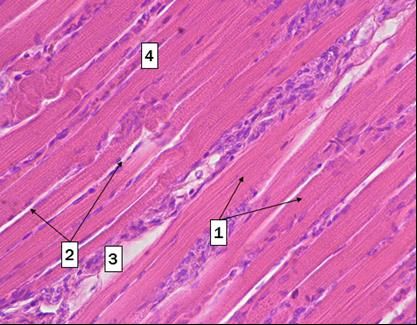
E3S Web of Conferences 254, 09020 (2021)                            https://doi.org/10.1051/e3sconf/202125409020
FARBA 2021

         morphogenesis and its micromorphological parameters. At the same time, such works
         devoted to the study of quails in this matter are extremely few, which reflects the relevance
         of considering the features of myogenesis and micromorphometric indicators of skeletal
         muscles in ontogenesis in this type of poultry, as well as conducting a comparative analysis
         according to the methodological scheme proposed by us [7, 8, 12].
            The purpose of the work is to present comparative morphological and morphometric
         data on the structure of the quadriceps femoris and the superficial pectoral muscle in hens
         and quails of meat and egg breeds.

         2 Materials and Methods
         The work was carried out on the basis of the Department of Animal Anatomy and
         Histology named after Professor A.F. Klimov Moscow SAVMB and on the basis of the
         JSC Technology Department of the FSC ARRTPI RAS.
             The object of the study was quail chickens of the Manchurian golden breed and
         chickens of hen of Smena 8 "Plymouthrock" cross.
             Samples of the superficial pectoral muscle (hereinafter – SPM) and the straight head of
         the quadriceps femoris (hereinafter – QFM) were taken from quails on the 1st, 8th, 21st,
         35th, 42nd day after hatching, from hen chickens on the 1st, 8th, 20th, 29th, and 50th day
         after hatching. In each age group, 6 individuals were examined.
             The material was fixed in formalin, followed by filling in paraffin according to
         generally accepted methods. Histological sections were made on a rotary automated
         microtome HM-325 (Microm international GmbH, Germany). The resulting material was
         stained with hematoxylin and eosin, according to van Gieson and Mallory. Histological
         sections and microphotography were studied using a Jenamed 2 light microscope (Carl
         Zeiss, Jena, Germany) combined with the ImageScope C digital microscopy system
         (Systems for Microscopy and Analysis, LLC). In hen chickens and quails, morphometric
         studies of muscle fibers were performed in terms of thickness, their bundles, endomysium,
         perimysium, and the ratio of muscle and connective tissues at all stages of myohistogenesis
         in SPM and QFM.
             For an in-depth study of the quail muscles structure, ultrastructural studies of SPM and
         QFM samples were performed on the 50th day of development. During electronic
         microscopic examination, the tissue samples were fixed in 2.5% glutaraldehyde (pH 7.2-
         7.4), followed by additional fixation in 1% osmium solution on the corresponding buffer.
         The material was poured into Epon 812. Ultrathin sections were prepared on the LKB – 3
         ultratome (Sweden), contrasted with a 2% aqueous solution of uranyl acetate, lead citrate
         according to Reynolds (Weekley B., 1975) and studied on a JEM CX 2 transmission
         microscope (JEOL Ltd., Japan) at magnifications from 4000 to 35000.

         3 Results and discussion

         3.1 Histological and morphometric characteristics of skeletal muscles of
         hens of Smena 8 "Plymouthrock" cross

         When studying the age-related structural and micromorphometric features of the muscles in
         hens, it was found that their structure was formed at the time of hatching, differing from
         that of adult individuals by morphometric indicators and some morphological features of
         muscle fibers. Thus, the muscle fibers of a day-old chicken have a transverse striation, but
         are very thin, and the nuclei in the myosymplast are located both in the center and on the
         periphery, which indicates insufficient development of the contractile apparatus. At the

                                                      3
E3S Web of Conferences 254, 09020 (2021) https://doi.org/10.1051/e3sconf/202125409020
FARBA 2021
same time, the nuclei are large, light, and numerous, which indicates an active course of
biosynthetic processes aimed at forming the muscle fiber structure. Connective tissue in this
period of life is abundantly represented in the muscle, while the endomysium forms thin
layers, and the paratenon, on the contrary, very wide layers of loose connective tissue
(Figure 1).
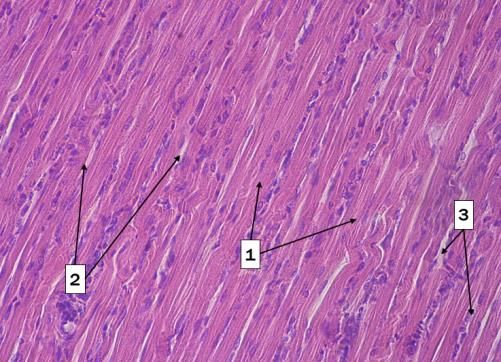
Fig. 1. Longitudinal section of the quadriceps Fig. 2. Longitudinal section of the quadriceps
femoris muscle in a day old chicken of femoris muscle in 50-day old chicken of
cross Smena 8 - Plymouthrock. cross Smena 8 - Plymouthrock.
The transverse striation in the muscle fibers is 1 – muscle fibers, 2 - endomysium, 3 -
clearly visible. 1 - muscle fibers, perimysium, 4 - bundle of muscle fibers.
2 - endomysium, 3 - perimysium, Staining with hematoxylin and eosin, ob. 40, oc.
4 - bundles of muscle fibers. 10.
Staining with hematoxylin and eosin, ob. 40, oc.
10.
From 1 to 50 days (Figure 2) of chicken growth and development during muscle
development, the following trends are observed: the area of muscle tissue, the diameter of
muscle fibers and their bundles increase, the thickness of the endomysium does not change,
the thickness of the perimysium decreases (Table 1).
Table 1. Morphometric characteristics of the quadriceps femoris and the superficial pectoral
muscle of chickens of hens of Smena 8 "Plymouthrock" cross of meat and egg productivity direction
1st day after hatching
Indicators Quadriceps Superficial pectoral
femoris muscle
Area of muscle tissue, %* 75±1.45 80±2.13
Thickness of muscle fibers, microns 7.68±0.54 6.81±0.43
Thickness of muscle fiber bundles, microns 82±7.84 78±6.76
Endomysium thickness, microns 4.43±0.23 3.28±0.76
Perimysium thickness, microns 30.7±3.15 20.2±2.41
Number of fibers per HPF** 13±1 15±1
8th day after hatching
Area of muscle tissue, %* 79±2.26 84±2.51
Thickness of muscle fibers, microns 10.91±0.23 8.22±0.53
Thickness of muscle fiber bundles, microns 112±7.12 121±8.44
Endomysium thickness, microns 3.87±0.78 3.36±1.12
Perimysium thickness, microns 29.1±5.13 24.2±3.56
Number of fibers per HPF** 15±1 17±1
20th day after hatching
Area of muscle tissue, %* 81±2.83 87±2.14
Thickness of muscle fibers, microns 13.1±0.23 11.2±0.21
Thickness of muscle fiber bundles, microns 132±6.43 144±7.5
4E3S Web of Conferences 254, 09020 (2021) https://doi.org/10.1051/e3sconf/202125409020
FARBA 2021
Endomysium thickness, microns 3.1±0.56 2.95±0.66
Perimysium thickness, microns 31.5±1.57 24.2±6.13
Number of fibers per HPF** 14±1 16±1
29th day after hatching
Area of muscle tissue, %* 85±2.61 91±2.15
Thickness of muscle fibers, microns 14.7±0.42 12.75±0.23
Thickness of muscle fiber bundles, microns 164±7.1 171±6.34
Endomysium thickness, microns 3.24±0.12 3.01±0.6
Perimysium thickness, microns 28±2.54 23±1.43
Number of fibers per HPF** 12±1 14±1
50th day after hatching
Area of muscle tissue, %* 89±1.93 92±2.06
Thickness of muscle fibers, microns 36.2±3.1 27.9±1.77
Thickness of muscle fiber bundles, microns 292±14.1 305±8.2
Endomysium thickness, microns 4.7±1.1 3.96±0.13
Perimysium thickness, microns 21±3.44 17.5±2.4
Number of fibers per HPF** 5±1 6±1
* comparative morphometric analysis was performed at a magnification 400.
**calculation was carried out in the standard HPF of the microscope at a magnification 1000.
The morphometric analysis of the muscle tissue parameters of a day-old hen chicken
showed that the area of the muscle tissue and the number of muscle fibers are larger in the
SPM, and the diameter of the muscle fiber is smaller compared to the QFM. This feature
persists in the subsequent periods of hen development, and the most significant increase in
the diameter of the muscle fiber is observed in the period from 29 to 50 days of
development, which can be considered a critical stage in hen myohistogenesis.
Based on the analysis of the digital material, it can be noted that from the 1st to the 50th
day of development, the area of muscle tissue increases in QFM by 18.6%, in SPM - by
15%, the diameter of muscle fibers increases (in QFM by 4.7 times, in SPM by 4.1 times)
and the diameter of muscle fiber bundles (in QFM by 3.5 times, in SPM by 3.9 times).
Against this background, the thickness of the perimysium decreases in QFM by 30%, in
SPM by 12.5%.
3.2 Histological and morphometric characteristics of skeletal muscles of the
Manchurian golden quail
To identify the general patterns and specific features of myogenesis, similar studies were
conducted on the material obtained from quails. The muscles of quail chickens by the 1st
day after hatching appear to be fully formed: the muscle fibers have a pronounced
transverse striation, they form bundles. The connective tissue component of muscle forms
thin layers of endomysium and quite powerful ones layers - perimysium (Figure 3).
By the 42nd day, the thickness of both the muscle fiber itself and its bundles, as well as
endo- and perimysium, increases. Light-optical studies have shown the presence of
numerous neurovascular bundles (Figures 4, 5, 6) located in the perimysium. In Mallory
staining, there are differences in the basophily of muscle fibers, reflecting their
heterogeneity and belonging to different types.
5E3S Web of Conferences 254, 09020 (2021) https://doi.org/10.1051/e3sconf/202125409020
FARBA 2021
Fig. 3. Longitudinal section of the quadriceps Fig. 4. Longitudinal section of the quadriceps
femoris in a day-old Manchurian golden quail femoris in a 42-day-old Manchurian golden quail
chicken. The transverse striation of the muscle chicken. The transverse striation of the muscle
fibers is clearly visible. 1 - muscle fibers, 2 - fibers is clearly visible. 1 - muscle fibers, 2 -
endomysium, 3 - perimysium. endomysium, 3 - perimysium.
Staining with hematoxylin and eosin, ob. 40, oc. Staining with hematoxylin and eosin, ob. 40, oc.
10. 10.
Fig. 5. Bundles of muscle fibers and bundles of Fig. 6. Muscle fibers in the superficial pectoral
nerve fibers between them in the superficial muscle and the neurovascular bundle between
pectoral muscle of the Manchurian golden quail them in the Manchurian golden quail on the 50th
on the 50th day after hatching. day after hatching. Cross-section.
Mallory staining, ob. 40, oc. 10. Van Gieson staining, ob. 40, oc. 10.
Morphometric studies found that at all observation periods, the area of muscle tissue,
the diameter of muscle fibers and their bundles in quails, in contrast to hens, is greater in
QFM, and the number of muscle fibers - in SPM (Table 2).
Table 2. Morphometric parameters of the quadriceps femoris and superficial pectoral
muscles in chickens and adult quails of the Manchurian golden breed
1st day after hatching
Indicators Musculus quadriceps Superficial pectoral
femoris muscle
Area of muscle tissue, %* 78±3 74±2
Thickness of muscle fibers, microns 4.19±0.32 4.28±0.52
Thickness of muscle fiber bundles, microns 66.1±4.54 43.1±4.65
Endomysium thickness, microns 2.12±0.18 1.76±0.54
Perimysium thickness, microns 8.47±2.65 7.89±7.27
Number of fibers per HPF** 22±2 23±3
6E3S Web of Conferences 254, 09020 (2021) https://doi.org/10.1051/e3sconf/202125409020
FARBA 2021
8th day after hatching
Area of muscle tissue, %* 84±3.21 82±2.14
Thickness of muscle fibers, microns 7.98±0.31 6.84±1.18
Thickness of muscle fiber bundles, microns 88.7±7.65 70.1±11.3
Endomysium thickness, microns 3.59±0.83 1.88±2.01
Perimysium thickness, microns 8.02±2.12 5.82±3.15
Number of fibers per HPF** 13±1 14±1
21st day after hatching
Area of muscle tissue, %* 85±2 82±3
Thickness of muscle fibers, microns 10.1±0.23 9.04±0.41
Thickness of muscle fiber bundles, microns 144±5.12 159±3.65
Endomysium thickness, microns 4.15±1.24 3.05±1.51
Perimysium thickness, microns 15.3±3.21 11.1±2.04
Number of fibers per HPF** 15±1 16±2
35th day after hatching
Area of muscle tissue, %* 87±2 85±2
Thickness of muscle fibers, microns 12.6±1.23 10.6±2.04
Thickness of muscle fiber bundles, microns 151±4.41 162±4.12
Endomysium thickness, microns 2.18±0.34 2.68±1.63
Perimysium thickness, microns 26.4±3.38 21.1±3.24
Number of fibers per HPF** 14±2 12±2
42nd day of the hatching
Area of muscle tissue, %* 88±4 86±3
Thickness of muscle fibers, microns 21.7±1.24 18.3±1.05
Thickness of muscle fiber bundles, microns 169±5.13 189±5.32
Endomysium thickness, microns 3.28±1.42 2.52±1.42
Perimysium thickness, microns 21.5±3.15 23.2±3.32
Number of fibers per HPF** 12±1 13±2
* comparative morphometric analysis was performed at a magnification 400.
**calculations were carried out in the standard HPF of the microscope at a magnification 1000.
According to the data obtained, the general trends in the formation of the structure of
the studied quail muscles are as follows. From day 1 to day 42 of development, the area of
muscle tissue increases in QFM by 12%, in SPM - by 16% (in chickens, on the contrary,
more active growth was noted in QFM), the diameter of muscle fibers increases (in QFM
by 5 times, in SPM by 4.2 times) and the diameter of muscle fiber bundles (in QFM by 2.6
times, in SPM by 4.4 times). At the same time, the perimysium thickness, in contrast to
hens, does not decrease, but increases in QFM by 2.5 times, and in SPM – by 2.8 times.
3.3 Results of electronic microscopic studies of the muscles of the
Manchurian golden quail on the 50th day after hatching
The muscles in quails on the 50th day after hatching are a highly differentiated structure of
muscle fibers, in which the transverse striation is clearly defined, due to the developed
contractile apparatus in the form of myofibrillas consisting of sarcomeres (Figure 7). The
muscle fibers contain many nuclei, which can be located both on the periphery under the
sarcolemma, and in the fiber thickness among the myofibrillas. The nuclei are
7E3S Web of Conferences 254, 09020 (2021) https://doi.org/10.1051/e3sconf/202125409020
FARBA 2021
morphofunctionally active, contain 2-3 nucleoli and an abundance of RNP granules in the
karyoplasm, which ensures a high level of metabolism of the protein synthesis apparatus in
the fiber (Figure 8). In some fibers, nuclei with a strongly altered shape were observed,
which allows to intensify the metabolism between nucleus and cytoplasm. It is noteworthy
that the Z-lines stretch through all the fiber myofibrillas. There are the cisterns of the
sarcoplasmic reticulum, mitochondria, and glycogen grains between the myofibrillas.
Glycogen grains are better represented in the SPM of quail and are often found near the Z-
lines. In SPM, a larger diameter of myofibrillas was noted, compared with QFM, as well as
separation from each other due to wide layers of sarcoplasm. Mitochondria are an important
component of muscle fibers. In the SPM of quails, they are small, round, oval or irregular
in shape, located at a distance from each other, as well as at a distance from the
myofibrillas. Blood capillaries are often found between the muscle fibers (Figure 7).
In QFM, myofibrillas are narrow and form bundles of 3-4 elements located close to
each other (Figure 8). Between these bundles there are wide layers of sarcoplasm, which
contain large rod-shaped mitochondria, the length of which corresponds to the length of 5-6
sarcomeres. Thus, the most pronounced differences in muscle tissue in SPM and QFM are
manifested in the distribution of myofibrillas, as well as the size, number and localization
of mitochondria in the fiber. In QFM, the muscle fibers are red, and they are characterized
by the presence of peculiar mitochondrial complexes under the sarcolemma (Figure 9).
Another feature of the ultrastructure of quail QFM is the presence of clearly visible cisterns
of the sarcoplasmic reticulum, which forms groups of 3-5 rounded vesicles near the Z-lines
of the myofibrilla.
In both muscles, small destructive changes in individual mitochondria were observed in
the form of matrix enlightenment and cristae length shortening. We believe that these
changes are due to structural transformations of muscle fibers. Between the muscle fibers in
the connective tissue framework there is a large number of hemocapillaries, in the structure
of which the nuclear apparatus is detected, with a predominance of euchromatin,
ribonucleoproteins and nucleoli.
Fig. 7. Ultrastructural design of the superficial Fig. 8. Ultrastructural organization of the
pectoral muscle in quail on the 50th day after superficial pectoral muscle in quail on the 50th
hatching. The blood capillary is located between day after hatching. In the muscle fiber between
the muscle fibers. the myofibrillas, there are wide layers of
sarcoplasm.
8E3S Web of Conferences 254, 09020 (2021) https://doi.org/10.1051/e3sconf/202125409020
FARBA 2021
Fig. 9. Ultrastructural organization of the Fig. 10. Ultrastructural organization of the
superficial pectoral muscle in quail on the 50th quadriceps femoris muscle in quail on the 50th
day after hatching. Euchromatin and RNP day after hatching. In myofibrillas, transverse
granules are observed in the nucleus. striation is visible. Large mitochondria are
Mitochondria of oval or irregular shape are located in the central part of the fiber.
located in the central part of the fiber.
Fig. 11. Ultrastructural organization of the Fig. 12. Ultrastructural organization of the
quadriceps femoris muscle in quail on the 50th quadriceps femoris muscle in quail on the 50th
day after hatching. The mitochondrial complex day after hatching. In the muscle fiber,
is located on the periphery of the fiber. myofibrillas are narrow, located close to each
other.
4 Conclusions
Thus, a comparative morphological and morphometric assessment of the studied skeletal
muscles of hens and quails allowed to establish general patterns of their growth.
Representatives of both groups of birds studied with an increase in the area of muscle
tissue, the diameter of muscle fibers and their bundles, and the percentage of increase in
these indicators is very similar. The difference is a more significant development of QFM
in hens, and SPM – in quails. It is noteworthy that quails, unlike hens, had thickening of the
endomysium and perimysium in both muscles.
On the basis of the conducted studies, we conducted a comprehensive study of the
superficial pectoral muscle and quadriceps in hens of the Smena 8 "Plymouthrock" crosses
and in quail of the Manchurian golden breed at the early stages of bird ontogenesis. The
study of morphometric indicators showed that the general pattern is an increase in the area
of muscle tissue and the diameter of muscle fibers with the age of chickens and quails,
while simultaneously reducing their number in the field of view, as well as a decrease in the
ratio between muscle and connective tissues. The quail has a higher level of vascularization
and innervation in the studied muscles compared to the hen muscles. In the muscles of
birds, the contractile apparatus is characterized by a strictly ordered structure.
9E3S Web of Conferences 254, 09020 (2021) https://doi.org/10.1051/e3sconf/202125409020
FARBA 2021
Ultrastructural analysis of SPM and QFM in adult quails showed that muscle fibers
contain the largest representation of myofibrillas and mitochondria, which are characterized
by close morphofunctional connections. At the same time, differences in the structural
arrangement of myofibrillas, the location and shape of mitochondria, and the cisterns of the
sarcoplasmic reticulum were revealed between QFM and SPM.
References
1. A.N. Belogurov, Morphofunctional adaptation of the internal organs of the Japanese
quail in technological traumatism in industrial poultry farming (experimental and
clinical studies), autoref. dis ... dr. vet. sc., 50 (M., 2013)
2. V.F. Vrakin, M.V. Sidorova, Anatomy and histology of poultry, 426 (Moscow,
KolosS, 2001)
3. P.A. Glagolev, Bulletin of TAA, 4, 155-170 (1959)
4. R.K. Danilov, Differentiation of myosatellitocytes and muscle fibers in embryogenesis
and reparative histogenesis, Dis. doc. med. sc. (Kuibyshev, 1982)
5. R.K. Danilov, Essays on the histology of muscle tissues 50 (Ufa, Bashkortostan, 1994)
6. V.I. Ippolitova, K.S. Zablotskaya, TAA reports, 164, 146-152 (1970)
7. G.V. Kondratov, E.N. Borkhunova, Morphology, 3, 40 (2016)
8. G.V. Kondratov, Features of skeletal muscle histogenesis in chickens of different
productivity trends, Autoref. diss ... c. b. sc., 26 (M., 2016)
9. I.I. Kochish, N.A. Slesarenko, L.P. Troyanovskaya, A.N. Belogurov, Quail farming:
problems and ways to solve them, 157 (M., 2015)
10. T.V. Chernysheva, Age-related structure features of organs of movement in chickens:
autoref, Dis ... cand. biol. sc. Moscow, TAA, 15 (1974).
11. G.R. Shakirova, N.A. Mufazalova, S.M. Shakirova, Success of modern natural studies,
2, 20-21 (2009)
12. G.R. Shakirova, V.A. Bolshunov, S.M. Shakirova, Issues of regulatory and legal
regulation in veterinary medicine, 2, 131-134 (2019)
13. Yu.F. Yudichev, G.I. Barabanshchikova, Questions of morphology, physiology and
nutrition of farm animals and fur-bearing animals: Scientific works of the Omsk vet.
Institute, 35 (1), 3-9 (1978)
14. M.D. Shmerling, E.E. Filyushina, I.I. Buzuyeva et al., Skeletal muscle. Structural and
functional aspects of adaptation, 121 (Novosibirsk: Nauka, Siberian Branch, 1991)
15. B.R. Eisenberg, / J. Ultrastruct, 54, 76-88 (1976)
16. G.F. Gauthier, H.A. Padycyla, Ibid, 28 (2), 333-354 (1966)
17. M. Luxey, B. Berki, W. Heusermann, S. Fischer, P. Tschopp, Developmental Biology,
458 (2), 133-140 (2020)
18. G. Schippel, K. Schippel, K. Welt, Anat. Ans., 140 (4), 400-404 (1976)
19. J.R. Torrella, V. Fouces, J. Palomeque, G. Viscor, Journal of Anatomy, 192 (2), 211-
222 (1998)
20. S. Webb, J. Pathol., 106 (4), 221-228 (1972)
10You can also read