Breast Cancer Atlas for Radiation Therapy Planning: Consensus Definitions
←
→
Page content transcription
If your browser does not render page correctly, please read the page content below
Breast Cancer Atlas for Radiation
       Therapy Planning:
     Consensus Definitions

                                    1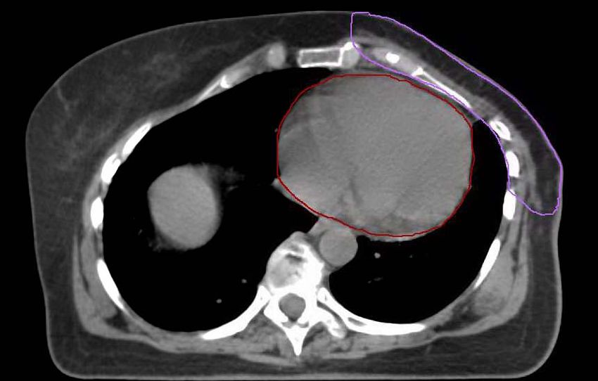
Collaborators

Julia White1, An Tai1, Douglas Arthur2, Thomas Buchholz3,
Shannon MacDonald4, Lawrence Marks5, Lori Pierce6,
Abraham Recht7, Rachel Rabinovitch8, Alphonse Taghian4,
Frank Vicini9, Wendy Woodward3, X. Allen Li1

1Medical  College of Wisconsin, 2Virginia Commonwealth University, 3M.D.
Anderson Cancer Center, 4Massachusetts General Hospital, 5University of North
Carolina, 6University of Michigan, 7Beth Israel Deaconess Medical Center
Hospital, 8University of Colorado, 9 William Beaumont Hospital

                                                                            2
                                                                            2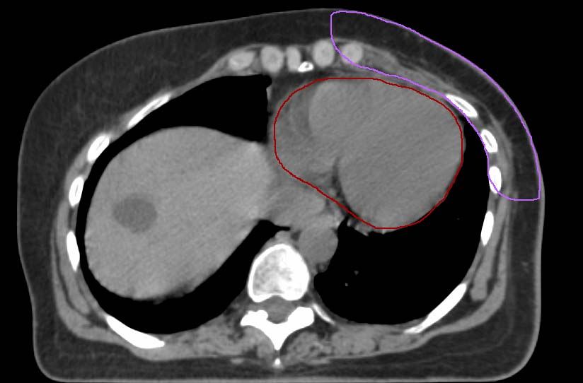
Content
→ Overlying principles: slides 4 - 6
→ Consensus definitions of anatomical boundaries:
      slides 7 - 12
→ Illustrative cases:
  – A: Stage I intact post-lumpectomy left breast
              (slides 13 - 30)
  – B: Stage III post-mastectomy left breast
              (slides 32 - 51)
  – C: Stage III intact post-lumpectomy right
      breast (slides 54 - 71)
                                                    3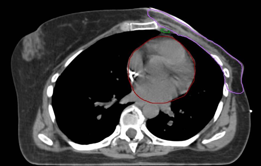
Overlying principles: Breast Contour
Breast CTV:
   – Considers referenced clinical breast at time of
     CT
   – Includes the apparent CT glandular breast
     tissue
   – Incorporates consensus definitions of
     anatomical borders (see table)
   – Includes the lumpectomy CTV
Lumpectomy GTV: Includes seroma and surgical
  clips when present
                                                 4     4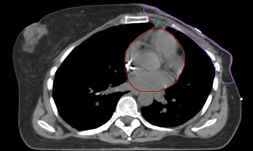
Overlying principles: Chestwall Contour

Chestwall CTV:
  – Considers referenced clinical chestwall at time
    of CT
  – Incorporates consensus definitions of
    anatomical borders (see table)
  – Includes the mastectomy scar (may not be feasible
    for occasional cases where the scar extends beyond the typical
    borders of the chestwall)

                                                              5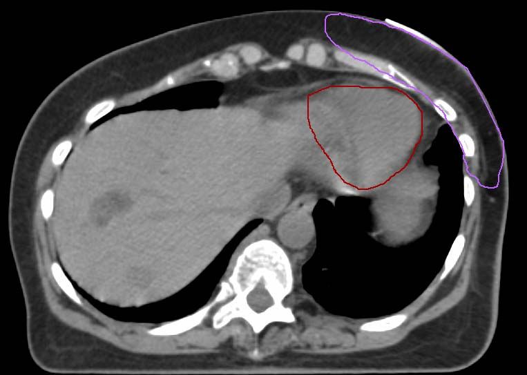
Overlying principles: Nodal volumes
Regional nodal CTV:
  – Nodal volumes contoured for targeting will
    depend on the specific clinical case
  – Considers consensus definitions of anatomical
    borders (see table)
  – The three levels of the axilla can overlap
    caudal to cranial
  – “Axillary apex” was considered level III
     of the axilla
                                              6
                                                  6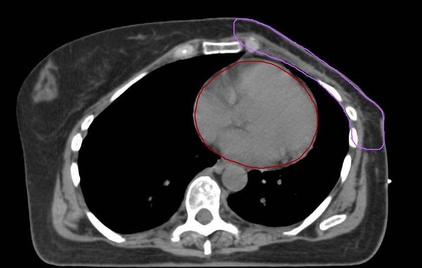
Breast and Chestwall Contour: Anatomical Boundaries
             Cranial        Caudal        Anterior    Posterior         Lateral          Medial
                                                                         Clinical
                                                                       Reference +
              Clinical      Clinical                  Excludes         mid axillary
             Reference    reference +                 pectoralis                         Sternal-
                                                                      line typically,
 Breast1     + Second      loss of CT       Skin      muscles,
                                                                        excludes
                                                                                           rib
                 rib        apparent                  chestwall
                                                                        latissimus      junction c
             insertiona      breast                  muscles, ribs
                                                                      (Lat.) dorsi m.
                                                                             b

                                                       Includes
 Breast +                                             pectoralis
               Same          Same          Same       muscles,            Same            Same
Chestwall2                                            chestwall
                                                     muscles, ribs

                             Clinical                Rib-pleural         Clinical
              Caudal                                  interface.        Reference/
                           reference+
             border of                                (Includes        mid axillary      Sternal-
                           loss of CT
Chestwall3       the                        Skin      pectoralis      line typically,      rib
                            apparent
              clavicle                                 muscles,         excludes        junction b
                          contralateral               chestwall       lattismus dorsi
                head
                              breast                 muscles, ribs)         ma             7
                                                                                               7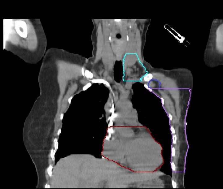
Contouring Comments:
              Breast and Chestwall
1. Breast: After appropriate lumpectomy for breast only
     treatment
   a. Cranial border is highly variable depending on breast

        size and patient position. The lateral aspect can be
        more cranial then the medial aspect depending on
        breast shape and patient position.
   b. Lateral border is highly variable depending on breast

        size and amount of ptosis.
   c. Medial border is highly variable depending on breast

        size and amount of ptosis. Clinical reference needs to
        be taken into account. Should not cross midline.
                                                          8
Contouring Comments:
                 Breast and Chestwall
2. Breast-Chestwall:   CTV after appropriate lumpectomy for more
      locally advanced cases includes those:
    – With clinical stage IIb, III who receive neoadjuvant
         chemotherapy and lumpectomy
    – Who have sufficient risk disease to require post-mastectomy
         radiation had mastectomy done
3. Chestwall: CTV after appropriate mastectomy:

    a. Lateral border meant to estimate the lateral border of the previous

         breast. Typically extends beyond the lateral edge of the
         pectoralis muscles but excluded the latissimus dorsi muscle
    b. Clinical reference marks need to be taken into account. The

         chestwall typically should not cross midline. Medial extent of
         mastectomy scar should typically be included                9
Regional Nodal Contours: Anatomical Boundaries
              Cranial            Caudal            Anterior          Posterior          Lateral            Medial
                                                                                      Cranial: lateral
                                 Junction of      Sternocleido                         edge of SCM
               Caudal to        brachioceph.-                       Anterior aspect                        Excludes
  Supra-                        axillary vns./
                                                    mastoid                                  m.
              the cricoid                                           of the scalene                        thyroid and
clavicular                       caudal edge         (SCM)                                Caudal:
               cartilage                                                  m.          junction 1st rib-     trachea
                               clavicle head a    muscle (m.)
                                                                                          clavicle

                Axillary        Pectoralis         Plane defined       Anterior                             Lateral
              vessels cross                         by: anterior                         Medial
 Axilla-     lateral edge of
                               (Pec.) major                           surface of                           border of
                               muscle insert      surface of Pec.                     border of lat.
 Level I     Pec. Minor m.                         Maj. m. and      subscapularis                         Pec. minor
                                                                                        dorsi m.
                                into ribs b        Lat. Dorsi m.          m.                                  m.

                Axillary                                                                 Lateral            Medial
                               Axillary vessels
             vessels cross                          Anterior           Ribs and
 Axilla-                        cross lateral                                           border of          border of
              medial edge
                                edge of Pec.      surface Pec.        intercostal
 level II    of Pec. Minor                                                             Pec. Minor         Pec. Minor
                                Minor m.
                                           c       Minor m.            muscles
                  m.                                                                       m.                 m.

                               Axillary vessels                                          Medial
             Pec. Minor                            Posterior           Ribs and
 Axilla-                        cross medial                                            border of          Thoracic
             m. insert on       edge of Pec.
                                                  surface Pec.        intercostal
level III                                                                              Pec. Minor            inlet
               cricoid           Minor m. d.       Major m.            muscles
                                                                                           m.

 Internal      Superior
                               Cranial aspect
             aspect of the                             - e.              - e.               - e.              - e.
mammary      medial 1st rib.    of the 4th rib
Contouring Comments:
Regional Nodal Volumes
a. Supraclavicular caudal border meant to approximate the
superior aspect of the breast/ chestwall field border
b. Axillary level I caudal border is clinically at the base of
the anterior axillary line
c. Axillary level II caudal border is the same as the cranial
border of level 1
d. Axillary level III caudal border is the same as the
cranial border of level II
e. Internal Mammary lymph nodes: encompass the
internal mammary/ thoracic vessels
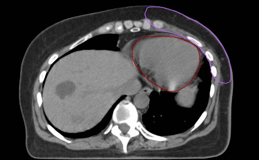
11Case A- Intact post lumpectomy breast
• Stage I ( T1c, N0, M0) Left breast cancer
• Surgery: Lumpectomy and sentinel node biopsy
• Radiation: Breast
• Six surgical clips placed at lumpectomy site
• External markers placed at time of CT:
– BB at AP set-up point
– 4 wire markers for clinical estimate of cranial, caudal,
medial, and lateral extent of anticipated tangents
– Wire extending from 9-3 o’clock around the infra-
mammary fold
– Wire over the lumpectomy scar
1213
14
15
16
 16
17
18
19
20
21
22
23
24
25
26
27
28
29
30
Case B: Post-mastectomy, Stage III
• Stage IIIB (T-3, N-3, M-0) left breast cancer, tumor size
  7 cm, 11/15 nodes positive
• Surgery: total mastectomy and axillary done dissection
• Radiation: chestwall + regional lymph nodes
• External wires present on CT:
   – Wire on mastectomy scar
   – BB on AP set-point at clinically estimated level of the
     match for the supraclavicular + axilla with the
     chestwall + IMC fields
   – Wires at lateral and inferior clinically estimated extent
     of the chestwall                                    31
32
33
34
35
     35
36
37
38
39
40
40
41
41
42
42
43
44
45
46
47
48
49
50
51
Case C: Stage III- Intact breast
                 post lumpectomy
• Stage IIIA (T-2, N-2, M-0) right breast cancer, tumor size 3 cm,
  4/18 nodes positive
• Surgery: Lumpectomy and axillary node dissection
• Radiation: Breast, chestwall + regional lymph nodes
• External wires present on CT:
   – Wire on lumpectomy scar
   – BB on AP set-point at clinically estimated level of the match for
     the supraclavicular + axilla with the chestwall + IMC fields
   – Wire extending from 9-3 o’clock around the infra-mammary fold
   – Wires at lateral and inferior clinically estimated extent of the
     chestwall

                                                               52
                                                                    52
53
54
55
56
57
58
59
60
61
62
63
64
65
66
67
68
69
70
71
You can also read


