Cancer Association of South Africa (CANSA) Fact Sheet on Paget's Disease of the Breast
←
→
Page content transcription
If your browser does not render page correctly, please read the page content below
Cancer Association of South Africa (CANSA)
Fact Sheet
on
Paget’s Disease
of the Breast
Introduction
The breast is the tissue overlying the chest (pectoral)
muscles. Women's breasts are made of specialised
tissue that produces milk (glandular tissue) as well as
fatty tissue. The amount of fat determines the size
of the breast.
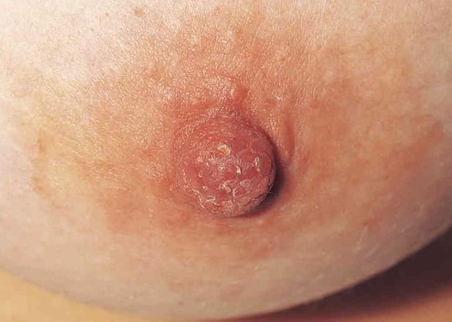
[Picture Credit: Paget’s Disease of the Breast I]
The milk-producing part of the breast is organised
into 15 to 20 sections, called lobes. Within each lobe
are smaller structures, called lobules, where milk is
produced. The milk travels through a network of tiny
tubes called ducts. The ducts connect and come together into larger ducts, which eventually exit the skin
in the nipple. The dark area of skin surrounding the nipple is called the areola.
Connective tissue and ligaments provide support to the breast and give it its shape. Nerves provide
sensation to the breast. The breast also contains blood vessels, lymph vessels, and lymph nodes.
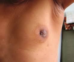
[Picture Credit: Paget’s Disease Male Breast]
Breast Conditions
• Breast cancer - malignant (cancer) cells
multiplying abnormally in the breast, eventually
spreading to the rest of the body if untreated.
Breast cancer occurs almost exclusively in
women, although men can be affected. Signs of
breast cancer include a lump, bloody nipple
discharge, or skin changes.
• Ductal carcinoma in situ (DCIS) - breast
cancer in the duct cells that has not invaded
deeper or spread through the body. Women
diagnosed with DCIS have a high likelihood of
being cured.
Researched and Authored by Prof Michael C Herbst
[D Litt et Phil (Health Studies); D N Ed; M Art et Scien; B A Cur; Dip Occupational Health; Dip Genetic Counselling; Diagnostic
Radiographer; Dip Audiometry and Noise Measurement; Medical Ethicist]
Approved by Ms Elize Joubert, Chief Executive Officer [BA Social Work (cum laude); MA Social Work]
April 2021 Page 1
•    Lobular carcinoma in situ (LCIS) - although called a carcinoma LCIS, which occurs in the milk-
     producing lobule cells, does not invade or spread and is not a true cancer. However, women with
     LCIS have an increased likelihood of developing invasive breast cancer in the future.
•    Invasive ductal carcinoma - breast cancer that begins in the duct cells but then invades deeper into
     the breast, carrying the potential of spreading to the rest of the body (metastasising). Invasive ductal
     carcinoma is the most common type of invasive breast cancer.
•    Invasive lobular carcinoma - breast cancer that begins in the milk-producing lobule cells, but then
     invades deeper into the breast, carrying the potential of spreading to the rest of the body
     (metastasising). Invasive lobular carcinoma is an uncommon form of breast cancer.
•    Simple breast cyst – a benign (noncancerous), fluid-filled sac that commonly develops in women in
     their 30s or 40s. Breast cysts may cause tenderness and may be drained.
•    Breast fibroadenoma - a very common noncancerous solid tumour of the breast. A typical
     fibroadenoma creates a painless, mobile lump in the breast and most commonly occurs in women in
     their 20s or 30s.
•    Fibrocytic breast disease - a common condition in which noncancerous breast lumps may become
     uncomfortable and change in size throughout the menstrual cycle.
•    Usual hyperplasia of the breast - a breast biopsy may show normal-appearing, noncancerous ductal
     cells multiplying abnormally. The presence of usual hyperplasia may slightly increase a woman's
     lifetime risk of breast cancer.
•    Atypical hyperplasia of the breast - abnormal-appearing cells multiplying either in the breast ducts
     (atypical ductal hyperplasia) or lobules (atypical lobular hyperplasia), sometimes discovered by a
     breast biopsy. Although the condition is noncancerous, women with atypical hyperplasia are at four
     to five times higher risk of developing breast cancer compared to women with no breast
     abnormalities.
•    Intraductual papilloma - a noncancerous, wart-like breast mass that grows inside the breast ducts.
     Intraductal papillomas may be felt as a lump or cause clear or bloody fluid to leak from the nipple.
•    Adenosis of the breast: A noncancerous enlargement of the breast lobules. Adenosis can look like
     breast cancer on mammograms, so a biopsy may be needed to rule out breast cancer.
•    Phyllodes tumour - a rare, usually large, rapidly growing breast tumour that looks like a
     fibroadenoma on ultrasound. Phyllodes tumours may be benign or malignant and most commonly
     develop in women in their 40s.
•    Fat necrosis - in response to an injury in the fatty part of the breast, a lump of scar tissue may
     develop. This mass can seem like breast cancer on examination or in mammograms.
•    Mastitis - inflammation of the breast, causing redness, pain, warmth, and swelling. Nursing mothers
     are at higher risk for mastitis, which is usually the result of infection.
•    Breast calcifications - calcium deposits in the breast are a common finding on mammograms. The
     pattern of calcium might suggest cancer, leading to further tests or a biopsy.
•    Gynaecomastia overdevelopment of male breasts. Gynecomastia can affect newborns, boys, and
     men.

Paget’s Disease of the Breast
Paget's disease of the breast is a rare form of breast cancer. Paget's disease of the breast starts on
the nipple and extends to the dark circle of skin (areola) around the nipple. It is not related to
Paget's disease of the bone, which is a metabolic bone disease.

Researched and Authored by Prof Michael C Herbst
[D Litt et Phil (Health Studies); D N Ed; M Art et Scien; B A Cur; Dip Occupational Health; Dip Genetic Counselling; Diagnostic
Radiographer; Dip Audiometry and Noise Measurement; Medical Ethicist]
Approved by Ms Elize Joubert, Chief Executive Officer [BA Social Work (cum laude); MA Social Work]
April 2021                                                                                                              Page 2
Paget's disease of the breast occurs most often in women older than age 50. Most women with
Paget's disease of the breast have underlying ductal breast cancer, either in situ or, less commonly,
invasive breast cancer. Only in rare cases is Paget's disease of the breast confined to the nipple itself.
Paget's disease is much more frequent in women but can also occur in men.

Moore, S.A., Notgrass, H.M., Vandergriff, T.W. & Sahoo, S. 2020.
“Mammary Paget's disease is rare and comprises about 0.62% of all breast cancer cases, only 1.65%
of which occur in male patients. This case report involves a 76-year-old man who presented to his
primary care physician with an itching, scaly, unilateral lesion involving the nipple skin. He
underwent wide local excision of the lesion for a diagnosis of Bowen's disease (squamous cell
carcinoma in situ). Histologic examination of the specimen revealed mammary Paget's disease with
ductal carcinoma in situ in the underlying breast tissue. A panel of immunohistochemical stains
revealed the Paget cells to be positive for cytokeratin 7, MUC1, GATA3, and androgen receptor and
negative for cytokeratins 5/6, p63, SOX10, and MART-1/Melan-A. Paget cells were also negative for
estrogen receptor and progesterone receptor, and positive for HER2/neu. However, the underlying
ductal carcinoma in situ was positive for both estrogen receptor and progesterone receptor and
negative for HER2/neu. This discordance, supported by the current literature, suggests an alternative
etiology for Paget's disease in certain cases that cannot be explained by the well-established
epidermotropic and transformative theories of Paget's disease evolution.”

Yasir, M. & Lotfollahzadeh, S. 2020.
“Sir James Paget, in 1874, identified 15 female patients with chronic nipple lesions; all of them
developed breast cancers later on. These lesions were described as eczematous ulcerative or
vesicular lesions with clear yellowish exudate. Initially, these lesions were considered benign in
nature, but it was subsequently discovered that these epidermal lesions, which are usually present
over nipple and areola, had malignant cells. This condition was later described as mammary Paget
disease (MPD) or Paget disease of the breast (PDB). A similar disease process was identified in
female and male external genitalia, known as extramammary Paget's disease. The histological
features of both conditions are the same, but the pathogenesis is different.”

Incidence of Paget’s Disease of the Breast
The National Cancer Registry (2017) does not provide any information regarding the incidence of
Paget’s Disease of the Breast.

Ooi, P.S., Draman, N., Yusoff, S.S.M., Zain, W.Z.W., Ganasagaran, D. & Chua, H.H. 2019.
“Mammary Paget's disease is clinically defined as skin inflammation of the nipple area and is an
adenocarcinoma of the epidermis of the nipple. The pathogenesis of mammary Paget's disease is
relatively unknown; nonetheless, there are two popular theories that support the underlying
carcinoma and de novo carcinogenesis. For the attending medical practitioner, mammary Paget's
disease poses a diagnostic and therapeutic dilemma, especially in the absence of a clinically palpable
breast mass. We report a rare case of a 48-year-old Malay woman who presented at Hospital
Universiti Sains Malaysia, Kelantan, Malaysia with the symptom of skin erosion on the left nipple and
unresponsiveness to multiple topical treatments. A full evaluation and assessment of the patient
were conducted, and mammary Paget's disease was diagnosed.”

Researched and Authored by Prof Michael C Herbst
[D Litt et Phil (Health Studies); D N Ed; M Art et Scien; B A Cur; Dip Occupational Health; Dip Genetic Counselling; Diagnostic
Radiographer; Dip Audiometry and Noise Measurement; Medical Ethicist]
Approved by Ms Elize Joubert, Chief Executive Officer [BA Social Work (cum laude); MA Social Work]
April 2021                                                                                                              Page 3
Causes and Risk Factors for Paget’s Disease of the Breast
Doctors do not know what causes Paget's disease of the breast. The most widely accepted theory is
that the disease results from an underlying ductal breast cancer. The cancer cells from the original
tumour then travel through milk ducts to the nipple and its surrounding skin.

Another theory is that the disease can develop independently in the nipple.

Risk factors that affect one’s likelihood of developing Paget's disease of the breast are the same
factors that affect one’s risk of developing any other type of breast cancer.

Some factors that make you more susceptible to breast cancer include:

  •    Age. One’s chances of developing breast cancer increase as one gets older.
  •    A personal history of breast cancer. If someone has had breast cancer in one breast, he/she
       has an increased risk of developing cancer in the other breast.
  •    A personal history of breast abnormalities. If one has had lobular carcinoma in situ or atypical
       hyperplasia, the risk of developing breast cancer is higher. Certain benign breast conditions
       also are associated with a slightly increased risk.
  •    Family history. If one has a mother, sister or daughter with breast or ovarian cancer or both,
       or even a father or brother with breast cancer, one also has a greater chance of developing
       breast cancer.
  •    An inherited gene mutation that increases the risk of breast cancer. Defects in one of several
       genes, especially BRCA1 or BRCA2, puts one at greater risk of developing breast cancer as well
       as ovarian and other cancers. Such defects account for fewer than 1 out of 10 breast cancers.
  •    Dense breast tissue. Women with dense breast tissue, as seen on a mammogram, face a
       higher risk of breast cancer.
  •    Radiation exposure. If one received radiation treatments to one’s chest as a child or young
       adult to treat another cancer, one is more likely to develop breast cancer later in life.
  •    Excess weight. Weighing more than is healthy for age and height increases the risk of breast
       cancer - especially after menopause and if one has gained weight as an adult.
  •    Hormone replacement. Taking oestrogen after menopause increases the risk of breast cancer
       for some women.
  •    Race. White women are more likely to develop breast cancer than black or Hispanic women,
       but black women are more likely to die of the disease.
  •    Alcohol. Alcohol consumption increases the risk of developing breast cancer.

Having one or more risk factors does not
necessarily mean one will develop breast
cancer. Most women with breast cancer have
no known risk factors.

            [Picture Credit: Paget’s Disease of the Breast II]

Symptoms and Diagnosis of Paget’s Disease of
the Breast
Paget disease of the breast (also known as
Paget disease of the nipple and mammary
Paget disease) is a rare type of cancer involving

Researched and Authored by Prof Michael C Herbst
[D Litt et Phil (Health Studies); D N Ed; M Art et Scien; B A Cur; Dip Occupational Health; Dip Genetic Counselling; Diagnostic
Radiographer; Dip Audiometry and Noise Measurement; Medical Ethicist]
Approved by Ms Elize Joubert, Chief Executive Officer [BA Social Work (cum laude); MA Social Work]
April 2021                                                                                                              Page 4
the skin of the nipple and, usually, the darker circle of skin around it, which is called the
areola. Paget's disease is much more frequent in women but can occur in men.
Paget's disease causes the skin on and around the nipple to become red, sore, and flaky, or scaly. At
first, these symptoms tend to come and go.
Over time, symptoms of Paget's disease usually worsen and may include:
• itching, tingling, and/or a burning sensation
• pain and sensitivity
• scaling and thickening of the skin
• flattening of the nipple
• yellowish or bloody discharge from the nipple
Because Paget's disease of the nipple is rare, doctors often mistake it for eczema (severe skin rash
and inflammation), an infection or injury, or some other skin condition. For many people, it can take
several months to get a correct diagnosis. If you have any of the above symptoms and they persist in
spite of treatment, get them checked out by a breast specialist. In most cases, Paget's disease affects
one breast, not both.
Diagnosis and Treatment of Paget’s Disease of the Breast
Diagnosing Paget's disease usually involves the following steps:
• A physical examination of the breasts, with special attention paid to the area around the nipple.
The doctor may be able to feel a lump or mass in the breast.
• A mammogram to check the nipple area and also to look for evidence of cancer in other areas of
the breast.
• Ultrasound and/or breast MRI to create additional images of the breast and check for other
areas of cancer.
• Biopsy of the nipple and areola. A breast surgeon will perform minor surgery to remove a small
piece of tissue from the nipple and areola area and examine it under a microscope. If there is
unusual discharge from the breast, the doctor will take a sample of that for examination as well.
For some women with Paget’s disease of the nipple, radiotherapy is almost always recommended,
and may be the only treatment needed after biopsy and breast conserving surgery. Radiotherapy is
sometimes also recommended after mastectomy.
For many years, mastectomy, with or without the removal of lymph nodes under the arm on the
same side of chest (known as axillary lymph node dissection), was regarded as the standard surgery
for Paget disease of the breast. This type of surgery was done because patients with Paget disease of
the breast were almost always found to have one or more tumours inside the same breast. Even if
only one tumour was present, that tumour could be located several centimetres away from the
nipple and areola and would not be removed by surgery on the nipple and areola alone.
Studies have shown, however, that breast-conserving surgery that includes removal of the nipple
and areola, followed by whole-breast radiation therapy, is a safe option for people with Paget
Researched and Authored by Prof Michael C Herbst
[D Litt et Phil (Health Studies); D N Ed; M Art et Scien; B A Cur; Dip Occupational Health; Dip Genetic Counselling; Diagnostic
Radiographer; Dip Audiometry and Noise Measurement; Medical Ethicist]
Approved by Ms Elize Joubert, Chief Executive Officer [BA Social Work (cum laude); MA Social Work]
April 2021 Page 5disease of the breast who do not have a palpable lump in their breast and whose mammograms do
not reveal a tumour.
The prognosis, or outlook, for people with Paget disease of the breast depends on a variety of
factors, including the following:
• Whether or not a tumour is present in the affected breast
• If one or more tumours are present in the affected breast, whether those tumours
are ductal carcinoma in situ or invasive breast cancer
• If invasive breast cancer is present in the affected breast, the stage of that cancer is staged
the same as breast cancer
The presence of invasive cancer in the affected breast and the spread of cancer to nearby lymph
nodes are associated with reduced survival.
Li, C., Yang, X., Wang, P., We, L. & Wang, X. 2020.
“Mammary Paget's disease (MPD) is a rare breast carcinoma represented with an eczematoid
cutaneous manifestation. Imitating inflammatory or infectious diseases, it makes early diagnosis and
prompt treatment difficult. Non-invasive imaging examinations such as reflectance confocal
microscopy (RCM) could extend assistance in making a diagnosis because of its near-cellular
resolution in skin diseases. Herein, the RCM feature of two cases of MPD, and the corresponding
dermoscopy, ultrasonography examination and immunohistochemistry staining results were
described.”
Treatment of Paget’s Disease of the Breast
Yao, Y., Sun, L., Meng, Y., Zhuang, Y., Zhao, L., Yu, Q. & Si, C. 2019.
Background: We aimed to analyze the association between Paget's disease (PD) and breast cancer
(BC) subtypes and compare the effect of breast-conserving surgery (BCS) as a local treatment with
mastectomy for PD.
Materials and methods: Data of patients with histologic type International Classification of Diseases-
0-3 8540-8543 who were treated from 1973 to 2014 were retrieved from the Surveillance,
Epidemiology, and End Results database of the National Cancer Institute. A chi-square test was used
to identify differences in categorical data among different groups. Overall survival (OS) was analyzed
using the Kaplan-Meier method, log-rank test, Cox proportional hazards models, sequential
landmark analysis, and propensity score-matched analysis.
Results: The study cohort included 5398 patients. Triple-negative BC accounted for the fewest
patients with PD-only (1/22, 4.54%), Paget's disease-ductal carcinoma in situ (PD-DCIS) (3/48,
6.25%), and Paget's disease-invading ductal carcinoma (PD-IDC) (23/352, 6.53%). According to the
results of the log-rank test and Cox analysis, the 10-year OS rates were similar for the BCS and
mastectomy subgroups among patients with PD-DCIS or PD-IDC. Furthermore, there were no
significant differences in survival benefits among the different surgeries after propensity score
matching. Landmark analyses for OS of patients with PD-DCIS or PD-IDC surviving more than 1, 3,
and 5 y showed no significant differences in survival. There were statistical differences in 10-year OS
rates for patients with PD-DCIS or PD-IDC who underwent radiation therapy, or not, following BCS
(both, P < 0.001).
Conclusions: For patients with PD-DCIS or PD-IDC, breast conservation therapy with lumpectomy
and radiation is an effective local treatment strategy, compared with mastectomy.
Researched and Authored by Prof Michael C Herbst
[D Litt et Phil (Health Studies); D N Ed; M Art et Scien; B A Cur; Dip Occupational Health; Dip Genetic Counselling; Diagnostic
Radiographer; Dip Audiometry and Noise Measurement; Medical Ethicist]
Approved by Ms Elize Joubert, Chief Executive Officer [BA Social Work (cum laude); MA Social Work]
April 2021 Page 6Medical Disclaimer This Fact Sheet is intended to provide general information only and, as such, should not be considered as a substitute for advice, medically or otherwise, covering any specific situation. Users should seek appropriate advice before taking or refraining from taking any action in reliance on any information contained in this Fact Sheet. So far as permissible by law, the Cancer Association of South Africa (CANSA) does not accept any liability to any person (or his/her dependants/ estate/heirs) relating to the use of any information contained in this Fact Sheet. Whilst CANSA has taken every precaution in compiling this Fact Sheet, neither it, nor any contributor(s) to this Fact Sheet can be held responsible for any action (or the lack thereof) taken by any person or organisation wherever they shall be based, as a result, direct or otherwise, of information contained in, or accessed through, this Fact Sheet. Sources and References Consulted or Utilised Adams, S.J. & Kanthan, R. 2016. Paget's disease of the male breast in the 21st century: A systematic review. Breast. 2016 Oct;29:14-23. doi: 10.1016/j.breast.2016.06.015. Epub 2016 Jul 6. BreastCancer.Org http://www.breastcancer.org/symptoms/types/pagets/sympt_diag Cancer Research UK http://www.cancerresearchuk.org/about-cancer/breast-cancer/stages-types-grades/types/pagets-disease-breast Dubar, S., Boukrid, M., Bouquet de Joliniere, J., Guillou, L., Vo, Q>D>, Major, A., Alin N.B. Khomsi, F. & Feki, A. 2017. Paget's Breast Disease: A Case Report and Review of the Literature. Front Surg. 2017 Oct 23;4:51. doi: 10.3389/fsurg.2017.00051. eCollection 2017. Gaurav, A., Gupta, V., Koul, R., Dabas, S., Sareen, R., Geeta, K., Arora, V., Parikh, P.M. & Aggarwal, S. 2018. Practical consensus recommendatons for Paget's disease in breast cancer. South Asian J Cancer. 2018 Apr-Jun;7(2):83-86. doi: 10.4103/sajc.sajc_107_18. Godbole C, Mehta J, Methil B, Palep R, Bhuta P. 2017. Perianal Paget's Disease-a Case Report and a Review of Current Diagnosis and Management. Indian J Surg Oncol. 2017 Dec;8(4):619-621. doi: 10.1007/s13193-016-0548-7. Epub 2016 Sep 1. Li, C., Yang, X., Wang, P., We, L. & Wang, X. 2020. Mammary Paget’s disease diagnosed with reflectance confocal microscopy. Photodiagnosis Photodyn Ther. 2020 Dec;32:102069. Mayo Clinic https://www.mayoclinic.org/diseases-conditions/pagets-disease-of-the-breast/symptoms-causes/syc-20351079 Moore, S.A., Notgrass, H.M., Vandergriff, T.W. & Sahoo, S. 2020. Mammary Paget’s disease of the male breast: a rare case with an unusual immunohistochemical profile. Int J Surg Pathol. 2020 Apr;28(2):210-215. doi: 10.1177/1066896919874878. Epub 2019 Sep 12. National Cancer Institute https://www.cancer.gov/types/breast/paget-breast-fact-sheet#q6 Ooi, P.S., Draman, N., Yusoff, S.S.M., Zain, W.Z.W., Ganasagaran, D. & Chua, H.H. 2018. Mammary Paget's Disease of the Nipple: Relatively Common but Still Unknown to Many. Korean J Fam Med. 2018 Nov 29. doi: 10.4082/kjfm.17.0143. [Epub ahead of print] Researched and Authored by Prof Michael C Herbst [D Litt et Phil (Health Studies); D N Ed; M Art et Scien; B A Cur; Dip Occupational Health; Dip Genetic Counselling; Diagnostic Radiographer; Dip Audiometry and Noise Measurement; Medical Ethicist] Approved by Ms Elize Joubert, Chief Executive Officer [BA Social Work (cum laude); MA Social Work] April 2021 Page 7
Paget’s Disease Male Breast
http://www.bioline.org.br/pdf?cr10021

Paget’s Disease of the Breast I
http://www.pathologyoutlines.com/topic/breastmalignantpaget.html

Paget’s Disease of the Breast II
https://radiopaedia.org/articles/paget-disease-of-the-breast-1

Radiopaedia
https://radiopaedia.org/articles/paget-disease-of-the-breast-1

RSNA RadioGraphics
http://pubs.rsna.org/doi/full/10.1148/rg.317115070

Uthamalingam, M. & Periyasamy, K. 2016. Paget’s Disease of Nipple in Male Breast with Cancer. J Clin Diagn Res, 2016
Feb; 10(2): PD14-PD16.

WebMD
https://www.webmd.com/women/picture-of-the-breasts#1

Yao, Y., Sun, L., Meng, Y., Zhuang, Y., Zhao, L., Yu, Q. & Si, C. 2019. Breast-conserving surgery in patients with mammary
Paget’s disease. J Surg Res. 2019 Sep;241:178-187. doi: 10.1016/j.jss.2019.03.025. Epub 2019 Apr 24.

Yasir, M. & Lotfollahzadeh, S. 2020. Mammary Paget disease. In: StatPearls [Internet]. Treasure Island (FL): StatPearls
Publishing; 2020 Jan. 2020 Dec 1.

Researched and Authored by Prof Michael C Herbst
[D Litt et Phil (Health Studies); D N Ed; M Art et Scien; B A Cur; Dip Occupational Health; Dip Genetic Counselling; Diagnostic
Radiographer; Dip Audiometry and Noise Measurement; Medical Ethicist]
Approved by Ms Elize Joubert, Chief Executive Officer [BA Social Work (cum laude); MA Social Work]
April 2021                                                                                                              Page 8
You can also read