A multiparametric MRI score for prostate cancer detection: Performance in patients with and without endorectal coil
←
→
Page content transcription
If your browser does not render page correctly, please read the page content below
RESEARCH PAPER
A multiparametric MRI score for prostate
cancer detection: Performance in
patients with and without endorectal
coil
Purpose: To assess the diagnostic performance of multiparametric MRI (mpMRI), in the detection of prostate cancer in two different
coil setting: endorectal coil (ERC) versus phased array coil alone (PAC).
Materials and Methods: The study included 302 out of 395 consecutive patients with PSA values between 2.5-4 ng/ml and an
abnormal Digital Rectal Examination (DRE), or patients with PSA values between 4-10ng/ml, independently from DRE. Each patient
provided informed consent to undergo at serum free/total PSA ratio (f/t PSA) assay, morphological MRI (mMRI), DWI, MRS, and Trans
Rectal Ultrasonography (TRUS) biopsy. The MRI data sets were scored singularly and then mMRI, DWI and MRS data were combined in
a single score (cMRI score). cMRI score was correlated to negative biopsies and Gleason score biopsies. ROC curve and McNemar tests
were performed.
Results: cMRI score showed high value of sensitivity and NPV for both coil setting (84% and 93% respectively using ERC, 87% and 87%
respectively using PAC). cMRI score using ERC did not show statistical superiority compared with cMRI score using PAC alone (p value
at McNemar test > 0.05). A significant correlation was obtained comparing the cMRI score to the Gleason score for both coil conditions.
Conclusions: Pelvic phased array coil imaging of the prostate produces high quality images and the overall performance in prostate
cancer detection is equal than those obtained with endorectal coil imaging.
KEYWORDS: prostate cancer magnetic resonance imaging spectroscopy diffusion weighted imaging gleason score
Introduction results in greater visibility of anatomical details
and more accurate prostate cancer staging. The Roberta Fusco*,
Prostate cancer diagnosis still represents a
use of both a PAC and an ERC (a combination Antonella Petrillo,
clinical challenge, as currently available diagnostic
referred to hereafter as ERC+PAC) improves Sergio V Setola,
methods remain suboptimal [1,2]. On the other
side, there are both over-diagnosis and over- prostate imaging by providing greater signal- Vincenza Granata ,
treatment risks [3,4] Magnetic resonance imaging to-noise ratio (SNR) and thus increased spatial Nicola Raiano, Paola
(MRI) provides excellent high-contrast, high- resolution. However, the insertion of the ERC Delprete, Giuseppe
resolution images of the prostate. In recent years, causes patient discomfort, is expensive, and can Quarto, Alessandro Izzo
functional techniques have been applied in order to lead to complications such as proctitis [15]. Sisto Perdona
improve the performance of MRI in the diagnosis Recently, the availability of higher field strength Division of Radiology, Department of
magnets, increased numbers of phased array Diagnostic Imaging, Radiant and
of prostate cancer [5-8]. Magnetic resonance Metabolic Therapy, “Istituto Nazionale
spectroscopy (MRS) measures prostate metabolites receiver coils or multi-channels phased array Tumori Fondazione Giovanni Pascale–
concentrations, particularly choline and citrate, hat (8 channels or more), and improved pulse- IRCCS”, Via Mariano Semmola, Naples,
Italy
are respectively increased and reduced in cancer [6]. sequence techniques has generated interest in
*Author for correspondence:
Diffusion Weighted Image (DWI) provides water the possibility of performing prostate MRI
r.fusco@istitutotumori.na.it
diffusion properties based imaging, with reduced using only a PAC.
water diffusion in highly cellular cancer tissues [5]. The aim of this study was to evaluate
Early promising data suggest that multiparametric the diagnostic performance of mpMRI in
MRI (mpMRI), including morphologic sequences the detection of prostate cancer, including
and functional MR approaches, may be of morphological (mMRI), DWI and MRS
additional value for the localization of prostate obtained with an ERC and a PAC compared to
cancer and its local staging [8-12].
mpMRI obtained with only a PAC.
Literature studies have found that, compared
to the use of a phased array coil (PAC) alone, the Material and Methods
use of both an endorectal coil (ERC) and a PAC
to acquire T2-weighted fast spin-echo images
Patient selection
of the prostate at 1.5T [13-16] and 3.0T [15] From 2009 to 2011, 395 consecutive male
ISSN 1755-5191 Imaging Med. (2018) 10(1) 21
RESEARCH PAPER Fusco, Petrillo, Setola, Granata , Raiano, et al.
patients were screened to be enrolled in a single- were: TR/TE, 690/120 ms; flip angle, 90◦. The
center prospective observational study. The volume of Interest (VOI) was composed by 16
inclusion criteria were: PSA values between × 16 × 16 voxel of 6.25 × 6.25 × 6.25 mm3.
2.5-4ng/ml and an abnormal DRE, or PSA The transverse echo-planar DWI pulse sequence
values between 4-10ng/ml, independently from parameters were: TR/TE, 2700/83ms; slice
DRE. The exclusion criteria were: inability to thickness, 3.56 mm; flip angle, 90◦, acquisition
give informed consent; prior history of prostate matrix; 160x102 and FOV, 136x160 mm2; b
cancer excluded, prior pelvic irradiation, value=0, 50, 100, 150, 300, 600, 800 s/mm2.
previous hormonal or surgical therapy; MRI Antispasmodic drug was not used.
contraindications (cardiac pacemakers, surgical
clips, metallic hip implant); recent rectal surgery, Image analysis
latex allergy. All mpMRI findings were graded by two
All eligible patients underwent morphological radiologists with over 10-years of experience in
and functional MRI and TRUS prostate prostate MRI and blinded to both clinical and
biopsies. A register included every single patient biochemical data of the patients. A consensus
that decided to be enrolled in this approved evaluation method for scoring MRI findings
study. Any patient who decided to undergo the was adopted in order to reach more reliability
MRI examination signed an explicit informed in image interpretation. The scoring system
consent. was evaluated on a per-patient basis being not
achievable a reasonably accurate evaluation on a
MRI data acquisition per-site basis. A 5 mm diameter was considered
as the inferior threshold for including lesions.
The MRI was performed before prostate
The mMRI was scored using a 0-3 scale
biopsies. The MRI protocol included insertion
for peripheral gland (0=No abnormality;
of an endorectal coil (ERC) (Medrad, Pittsburg,
1=Geometric hypointense area, with low cancer
PA, USA), inflated with 60-90ml of air, and
suspicion; 2=Diffuse non nodular hypointense
subsequent imaging acquisition using both a
area, with intermediate cancer suspicion; 3=
1.5T MRI system (Siemens Symphony Tim,
nodular hypointense area, with high cancer
Erlangen, Germany) coupled to a phased-array
suspicion) and by using a 0-2 scale for the central
surface coil or a 8 channel phased-array surface
gland (0=Heterogeneous well-marginated
coil (PAC) alone. The MRI total acquisition
nodules; 1=Homogeneously hypointense
time was approximately 40mins. The mpMRI
well-marginated nodules; 2=Homogeneously
included mMRI, MRS and DWI. The mMRI
hypointense ill-marginated nodules) [16-21].
included Turbo Spin Echo (TSE) T2-weighted
sequences in three perpendicular planes and The MRS raw data were elaborated using
coronal and transverse TSE T1-weighted Syngo MR-B17 spectroscopy package (Siemens,
sequence. Transverse TSE T2-weighted sequence Erlangen, Germany). The software generated
parameters were: TR/TE, 3800/104ms (sagittal: spectra and localized corresponding voxels on
4660/96ms; coronal: 5000/98ms); slice superimposed T2-weighted images. The MRS
thickness, 3 mm/gap 0mm; flip angle, 180◦; was rated as diagnostic or non-diagnostic if the
acquisition matrix, 320 × 288 (sagittal and metabolites spectrum was good, after resolving
coronal 320 × 256); field of view (FOV), 240 × metabolic resonances, and limited baseline
240 mm2. Transverse T2-weighted images were distortions, due to residual water or lipids, were
acquired with and without fat saturation. present. For each voxel the software calculated
areas under citrate, choline and creatine peaks
TSE T1-weighted were acquired in coronal
and Choline+Creatine/Citrate peak ratio.
to visualize lymph nodes, with the following
Diagnostic spectra were considered negative
sequence parameters: TR/TE, 550/12 ms; slice
or positive, and scored as 0 or 1 (sMRI score)
thickness, 3 mm/gap 0mm; flip angle, 150◦;
respectively, in relation to Choline+Creatine/
acquisition matrix, 256 × 202; FOV, 448 × 512
Citrate ratio threshold of 0.86, as reported in
mm2. Transverse TSE T1-weighted sequence
the literature [6].
parameters were: TR/TE, 706/7.8ms, slice
thickness, 3mm/gap 0mm; flip angle, 150◦; The DWI data and Apparent Diffusion
acquisition matrix, 356 × 192; field of view Coefficient (ADC) maps were elaborated using
(FOV), 240 × 240 mm2. The MRS parameters Syngo MR-B17 diffusion package (Siemens,
22 Imaging Med. (2018) 10(1)
A multiparametric MRI score for prostate cancer detection: Performance in patients with
RESEARCH PAPER
and without endorectal coil
Erlangen, Germany). DWI was considered if cancer was reported in at least one core,
positive when focal areas, characterized by independently from the Gleason grade. The
persistent signal intensity at b-value increase highest Gleason grade reported at biopsy was
and/or ADC map hypointensity in relation to considered as patients’ reference Gleason grade
the adjacent gland, were evident [19]. DWI was value.
scored respectively as 0 or 1 (dMRI score) if a
negative or positive finding was reported. Statistical Analysis
A multiparametric combined score (cMRI Fishers exact test was used to evaluate statistical
score) was obtained for every site as the sum significance of Decision matrix (DM) tables.
of scores from every MRI technique. For every Sensitivity, specificity, positive and negative
derived score, the highest site score was used as predictive value (PPV and NPV) were calculated
patient reference score. The cut-off for cMRI for cMRI score, using DM. Matched sample
was 2 as obtained in a previous studies [10]. tables and McNemar test were used to compare
diagnostic performance for different coil setting
Biopsy and pathological analysis
(ERC versus PAC). Spearman’s Rank Correlation
TRUS biopsy was performed as previously Coefficient was performed on a per-patient basis
described in literature [11], by an expert correlating patients’ reference scores derived
urologist with a 10-years experience. A 12-site from MRI data sets with the results of biopsies
biopsy scheme (2 cores, paramedian and lateral, (Gleason score). A p value 45 cc four
additional cores were acquired from the central Results
gland (superiorly and inferiorly, bilaterally). Overall, 302 patients were enrolled in
The urologist was blinded to the MRI findings the study, 136 acquired with endorectal coil
during the prostatic biopsy. (FIGURES 1 and 2) and 166 acquired with
A pathologist with more than 10 years’ multi-channel phased array coil (FIGURES
experience in genito-urinary pathology 3 and 4). TABLE 1 reported patient’s
evaluated biopsy cores. Gleason scores were characteristics for both coil setting. Fishers exact
considered significant if at least cancer grade ≥ tests showed statistical significance for each DM
4 was evident. In case of a negative biopsy, the table with a p value always 0.05).
Patients were considered positive at biopsy TABLE 3 showed Spearman’s Rank Correlation
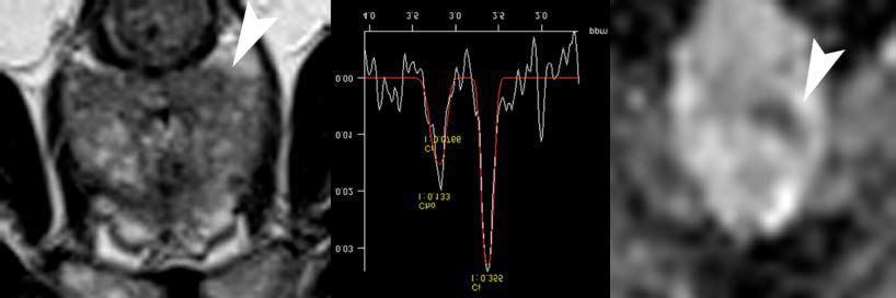
Figure 1. Transverse TSE T2 weighted sequence (left image; TR/TE, 3800/104 ms; flip angle, 180°)
showed a small ill defined nodular hypointense area (mMRI= 3) in the right peripheral midgland
(arrowhead). MRS was negative, while positive findings were evident on ADC maps (right image,
arrowhead). cMRI score was 4. Gleason score was 8.
Imaging Med. (2018) 10(1) 23RESEARCH PAPER Fusco, Petrillo, Setola, Granata , Raiano, et al.
Figure 2. On transverse T2 weighted sequence (left image; TR/TE, 3800/104 ms; flip angle, 180°) a
diffuse hypointense area (mMRI=2), particularly in the right peripheral midgland (arrowhead)
was evident. MRS was positive, and corresponding ADC map showed hypointense areas (right image,
arrowhead). cMRI score was 4. Gleason score was 8.
Figure 3. Transverse T2 weighted sequence (left image) shows an ill defined nodular hypointense
area (mMRI=3) in the left peripheral midgland (arrowhead). Positive findings were evident on MRS and
on ADC maps (right image, arrowhead). cMRI score was 5. Gleason score was 7.
Figure 4. On transverse T2 weighted sequence (left image) a diffuse hypointense area (mMRI=2) in
the left peripheral apex (arrowhead) is evident. MRS was negative while ADC map showed a hypointense
area (right image, arrowhead). cMRI score was 3. Gleason score was 7.
Figure 5. ROC curves for cMRI score in two coil setting.
Coefficients for each score using ERC and comparing the cMRI score to the Gleason score
PAC. A significant correlation was obtained for both coil conditions (FIGURE 5).
24 Imaging Med. (2018) 10(1)A multiparametric MRI score for prostate cancer detection: Performance in patients with
RESEARCH PAPER
and without endorectal coil
Table 1. Patients characteristics.
Variables ERC PAC
Patients, n 136 166
Age, years 66.35 (8.4) 64.23 (8.6)
PSA, ng/ml 6.8 (2.4) 6.6 (2.3)
f/t PSA, % 18.5 (7.3) 19.1 (6.9)
Values are expressed as a mean (SD)
Table 2. Diagnostic performance.
Variables Sensitivity (%) Specificity (%) PPV (%) NPV (%) AUC
cMRI with ERC 84 50 28 93 0.74
cMRI with PAC 87 50 49 87 0.72
Abbreviations: cMRI: combined MRI score; PPV: positive predictive value; NPV: negative predictive
value; AUC: area under ROC curve
Table 3. Rho Spearman’s coefficient for each score using ERC.
Variables mMRI Score dMRI Score sMRI Score cMRI Score Gleason Score
1.000 .304** .053 .901** .424**
mMRI Score
- .000 .502 .000 .000
.304** 1.000 -.262** .509** -.011
dMRI Score
.000 - .001 .000 .886
Rho
.053 -.262** 1.000 .311** .345**
Spearman's sMRI Score
.502 .001 - .000 .000
Coefficient
.901** .509** .311** 1.000 .668**
cMRI Score
.000 .000 .000 - .000
Gleason .424** -.011 .345** .6608* 1.000
Score .000 .886 .000 .000 -
** p value < 0.01
* p value < 0.05
Table 4. Rho Spearman’s coefficient for each score using PAC.
Variables mMRI Score dMRI Score sMRI Score cMRI Score Gleason Score
1.000 .195* .035 .847** .206*
mMRI Score
- .045 .719 .000 .034
.195* 1.000 -.071 .508** .283**
dMRI Score
.045 .471 .000 .003
Rho .035 -.071 1.000 .389** .283**
Spearman's sMRI Score
.719 .471 .000 .003
Coefficient
.847** .508** .389** 1.000 .650**
cMRI Score
.000 .000 .000 - .000
Gleason .206* .283** .283** .650** 1.000
Score .034 .003 .003 .000
** p-valueRESEARCH PAPER Fusco, Petrillo, Setola, Granata , Raiano, et al.
of MRI interpretation and reporting [24] using PAC of 0.95-0.99 and 0.93-0.97, respectively
a multi-parametric approach. A recent meta- (p=0.1395). They concluded that T2 weighted
analysis of 14 studies evaluating use of the imaging and DWI performed at 3T for prostate
PI-RADS scoring system for prostate cancer cancer lesion identification and evaluation did
detection on multi-parametric MRI showed not differ significantly with both coil setups and
good diagnostic accuracy [25]. that patients preferred MRI without an ERC.
Instead, our results are in contrast compared to
In our previous study [10] we demonstrated
the findings of Fütterer et al. [27] and Costa et al.
that the cMRI score had higher sensitivity
[30]. Futterer et al. [27] showed an accuracy and
and higher NPV than either single techniques
a specificity significantly better with endorectal-
(mMRI, DWI and MRS), or their combinations
pelvic phased-array coils (PA multiparametric MRI score for prostate cancer detection: Performance in patients with
RESEARCH PAPER
and without endorectal coil
Recent developments in sequence design MRI was compared to TRUS biopsy. Even if
with the introduction of turbo fast spin echo TRUS biopsy is reported to have a low diagnostic
sequences and in design of pelvic phased array accuracy, it remains the only current diagnostic
coils have undoubtedly improved image quality technique for the diagnosis of prostate cancer.
of prostate cancer imaging. Phased array coil The purpose of our study was to evaluate the
imaging has the advantage that the anterior diagnostic performance of the scoring system
pelvis, bladder and pelvic lymph nodes can be for patients work-up, where TRUS biopsy is the
evaluated with high resolution images and that gold standard. When surgery is used as standard
the technique is non-invasive. Although the reference in a study, a large selection bias is
endorectal coil can be combined with a PAC introduced because many patients can be selected
coil as part of a multicoil array [13], use of for non-surgical treatment or active surveillance
this facility has been slow to develop in clinical [34], as occurred in our population. According to
practice and hence experience remains limited. the literature, we adopted a consensus evaluation
method for scoring MRI findings in order to
Another limitation is that patients with
have more reliability in image interpretation
rectal stenosis or immediately after surgery
[25]. We evaluated the scoring system on a
or radiotherapy may not be good candidates
per-patient basis because a reasonably accurate
for the use of the endorectal coil during MR
evaluation on a per-site basis was not achievable
examination. When higher field strengths or
[34]. As reported [22], during needle biopsy the
phased array coil multichannel (8 channel or
path does not usually correspond to any MRI
more) and additional functional techniques
plane. Moreover, biopsy tumour localization is
were used, studies that used an ERC showed
affected by cores classification according to the
lower sensitivity and heterogeneous specificity
needle entry site, without considering the real
than studies without an ERC [8]. Lee et al.
needle path [35]. Furthermore, in our study, the
[26] reported that the use of ERC MRI did not
urologist was blinded to the MRI findings during
significantly improve the staging of prostate
the prostatic biopsy. Further investigation should
cancer (AUC 0.67 versus 0.66 respectively
be performed to obtain reslicing and registration
with and without ERC) and presented several
of MR images in order to reliably correlate MRI
complications in 11.4% of patients. Margolis et
findings to TRUS biopsy.
al. [17] reported that an endorectal coil is not
absolutely necessary and that the utility will Conclusion
depend on the performance of the scanner in In conclusion, the cMRI score showed high
question. Rectal distention with the associated accuracy both in term of sensitivity than in terms
susceptibility can markedly degrade DWI and of NPV independently if an endorectal coil is
potentially MRSI. An 8-channel external phased used or a phased array coil alone. Therefore,
array could replace the use of an endorectal coil the use of an ERC may be omitted in a prostate
and the use of additional functional imaging cancer detection setting and multi-channel
techniques seemed to improve the accuracy of phased-array coil MRI is a better alternative
local staging (17, 26). Also the ESUR prostate considering comorbidity.
MR guidelines 2012 reported in acquisition
protocols minimum requirements that imaging Funding
can adequately be performed at 1.5T using a This study was not funded.
good 8 to 16-channel pelvic phased array [33].
Considering our results the use of PAC Conflict of Interest
All Authors declare that have no conflict of
alone for morphological and functional MRI
interest.
acquisition did not limit overall images quality
and diagnostic accuracy and it should be prefer
Informed consent
for patient comfort.
Informed consent was obtained from all
A single limitation of the study is reported. individual participants included in the study.
Imaging Med. (2018) 10(1) 27RESEARCH PAPER Fusco, Petrillo, Setola, Granata , Raiano, et al.
Multiparametric MRI for prostate cancer negative biopsy and elevated PSA level-can it
REFERENCES detection: Preliminary results on quantitative rule out clinically significant prostate cancer?.
1. Cooperberg MR, Lubeck DP, Meng MV, et al. analysis of dynamic contrast enhanced imaging, Urol. Oncol. 32, 17-22 (2014).
The changing face of low-risk prostate cancer: diffusion-weighted imaging and spectroscopy
Trends in clinical presentation and primary imaging. Magn. Reson. Imaging. 34, 839-45 25. Hamoen EH, de Rooij M, Witjes JA, et al.
management. J. Clin. Oncol. 22, 2141-2149 (2016). Use of the prostate imaging reporting and data
(2004). system (PI-RADS) for prostate cancer detection
13. Hricak H, White S, Vigneron D, et al. with multiparametric magnetic resonance
2. Thompson IM, Pauler DK, Goodman PJ, et Carcinoma of the prostate gland: MR imaging imaging: A diagnostic meta-analysis. Eur. Urol.
al. Prevalence of prostate cancer among men with pelvic phased-array coils versus integrated 67, 1112-1121, (2005).
with a prostate specific antigen level ≤ 4.0 ng endorectal-pelvic phased-array coils. Radiology.
per millilitre. N. Engl .J. Med. 350, 2239-2246 193, 703-709 (1994). 26. Lee SH, Park KK, Choi KH, et al. Is endorectal
(2004). coil necessary for the staging of clinically
14. Futterer JJ, Engelbrecht MR, Jager GJ, et al. localized prostate cancer? Comparison of non-
3. Draisma G, Boer R, Otto SJ, et al. Lead times Prostate cancer: comparison of local staging endorectal versus endorectal MR imaging.
and overdetection due to prostate-specific accuracy of pelvic phased-array coil alone versus World. J. Urol. 28, 667-672 (2010).
antigen screening: Estimates from the European integrated endorectal-pelvic phased-array coils.
Randomized Study of Screening for Prostate Local staging accuracy of prostate cancer using 27. Fütterer JJ, Engelbrecht MR, Jager GJ, et al.
Cancer. J. Natl. Cancer. Inst. 95, 868-878 endorectal coil MR imaging. Eur. Radiol. 17, Prostate cancer: comparison of local staging
(2003). 1055-1065 (2007). accuracy of pelvic phased-array coil alone versus
integrated endorectal-pelvic phased-array coils.
4. Sanders A, Buchan N. Infection-related hospital 15. Heijmink SW, Futterer JJ, Hambrock T, et al. Local staging accuracy of prostate cancer using
admissions after transrectal biopsy of the Prostate cancer: body-array versus endorectal endorectal coil MR imaging. Eur. Radiol. 17,
prostate. ANZ. J. Surg. 83, 246-248 (2013) coil MR imaging at 3 T-comparison of image 1055-1065 (2007).
5. Baur AD, Daqqaq T, Wagner M, et al. T2- quality, localization, and staging performance.
Radiology. 244, 184-195 (2007). 28. Westphalen AC, Coakley FV, Qayyum A, et
and diffusion-weighted magnetic resonance al. Peripheral zone prostate cancer: accuracy of
imaging at 3T for the detection of prostate 16. Turkbey B, Albert PS, Kurdziel K, et al. Imaging different interpretative approaches with MR
cancer with and without endorectal coil: An localized prostate cancer: current approaches and MR spectroscopic imaging. Radiology. 246,
intraindividualcomparison of image quality and new developments. Am. J. Roentgenol. 192, 177-184 (2007).
and diagnostic performance. Eur. J. Radiol. 85, 1471–1480 (2009).
1075-1084 (2016). 29. Chen H, Sutedjo J, Wang L, et al. Prostate
17. Margolis DJA. Multiparametric MRI for Cancer Magnetic Resonance Spectroscopy
6. Javali TD, Dwivedi DK, Kumar R, et al. Localized Prostate Cancer: Lesion Detection Imaging at 1.5 and 3.0T: A Meta-Analysis.
Magnetic resonance spectroscopy imaging- and Staging. BioMed. Res. Intl. 14, 1-11 (2014). Technol. Cancer. Res. Treat. 15, 625-631 (2016).
directed transrectal ultrasound biopsy increases
prostate cancer detection in men with prostate- 18. Shukla-Dave A, Hricak H, Kattan MW, et al. 30. Costa DN, Yuan Q, Xi Y, Rofsky NM, et al.
specific antigen between 4-10 ng/mL and The utility of magnetic resonance imaging Comparison of prostate cancer detection at 3-T
normal digital rectal examination. Int. J. Urol. and spectroscopy for predicting insignificant MRI with and without an endorectal coil: A
21, 257-262 (2014). prostate cancer: an initial analysis. BJU. 99, prospective, paired-patient study. Urol. Oncol.
786-793 (2007). 34, 257-213 (2016).
7. Sankineni S, Osman M, Choyke PL. Functional
MRI in Prostate Cancer Detection. Biomed. Res. 19. Lim HK, Kim JK, Kim KA, et al. Prostate 31. Shah ZK, Elias SN, Abaza R, et al. Performance
Int. 14, 1-8, (2014). cancer: apparent diffusion coefficient map with comparison of 1.5T endorectal coil MRI with
T2-weighted images for detection-amultireader 3.0T nonendorectal coil MRI in patients with
8. Loffroy R, Chevallier O, Moulin M, et al. study. Radiology. 250, 145-151 (2009). prostate cancer. Acad. Radiol. 22, 467-474
Current role of multiparametric magnetic (2015).
resonance imaging for prostate cancer. Quant. 20. Akin O, Sala E, Moskowitz CS, et al. Transition
Imaging. Med. Surg. 5, 754-764 (2015). zone prostate cancers: features, detection, 32. Jager GJ, Ruijter ET, Van de KCA, et al. Local
localization, and staging at Endorectal MR staging of prostate cancer with endorectal MR
9. Hegde JV, Mulkern RV, Panych LP, et al. Imaging. Radiology. 239, 784-792 (2006). imaging: correlation with histopathology. Am. J.
Multiparametric MRI of Prostate Cancer: An Roentgenol. 166, 845-852 (1996).
Update on State-of-the-Art Techniques and 21. Rooij DM, Hamoen EH, Witjes JA, et al.
Their Performance in Detecting and Localizing Accuracy of Magnetic Resonance Imaging for 33. Barentsz JO, Richenberg J, Clements R, et al.
Prostate Cancer. J. Magn. Reson. Imaging. 37, Local Staging of Prostate Cancer: A Diagnostic European Society of Urogenital Radiology.
1035-1054 (2013). Meta-analysis. Eur. Urol. 70, 233-245 (2016). ESUR prostate MR guidelines 2012. Eur.
Radiol. 22, 746-757 (2012).
10. Petrillo A, Fusco R, Setola SV, et al. 22. Cirillo S, Petracchini M, Della Monica P, et al.
Multiparametric MRI for prostate cancer Value of endorectal MRI and MRS in patients 34. Weinreb JC, Blume JD, Coakley FV, et al.
detection: performance in patients with with elevated prostate-specific antigen levels and Prostate cancer: sextant localization at MR
prostate-specific antigen values between 2.5 and previous negative biopsies to localize peripheral imaging and MR spectroscopic imaging before
10ng/mL. J. Magn. Reson. Imaging. 39, 1206- zone tumours. Clin. Radiol. 63, 871-879 (2008). prostatectomy—results of ACRIN prospective
1212 (2014). multi-institutional clinicopathologic study.
23. Umbehr M, Bachmann LM, Held U. Combined Radiology. 251, 122-133 (2009).
11. Perdonà S, Di Lorenzo G, Autorino R, et al. magnetic resonance imaging and magnetic
Combined magnetic resonance spectroscopy resonance spectroscopy imaging in the diagnosis 35. Schulte RT, Wood DP, Daignault S, et al. Utility
and dynamic contrast-enhanced imaging for of prostate cancer: a systematic review and meta- of extended pattern prostate biopsies for tumor
prostate cancer detection. Urol. Oncol. 31, 761- analysis. Eur. Urol. 55, 575–590 (2009). localization: pathologic correlations following
765 (2013). radical prostatectomy. Cancer. 113, 1559-1565
24. Abd-Alazeez M, Ahmed HU, Arya M, et al. The (2008).
12. Fusco R, Sansone M, Petrillo M, et al. accuracy of multiparametric MRI in men with
28 Imaging Med. (2018) 10(1)You can also read