Development and growth of the pelvic fin in the extant coelacanth Latimeria chalumnae
←
→
Page content transcription
If your browser does not render page correctly, please read the page content below
Received: 23 January 2020    Revised: 12 March 2020    Accepted: 24 March 2020
DOI: 10.1002/ar.24452

FULL LENGTH ARTICLE

Development and growth of the pelvic fin in the extant
coelacanth Latimeria chalumnae

Rohan Mansuit1,2                     Gaël Clément1 | Anthony Herrel2
                                     |                                                                      |   Hugo Dutel3,4                    |
Paul Tafforeau5                   | Mathieu D. Santin6 | Marc Herbin2

1
 Département Origines and Evolution,
UMR 7207 Centre de Recherche en
                                                   Abstract
Paléontologie, Paris, MNHN - Sorbonne              The ontogeny of the paired appendages has been extensively studied in
Université - CNRS, Paris, France                   lungfishes and tetrapods, but remains poorly known in coelacanths. Recent
2
 Département Adaptations du Vivant,
                                                   work has shed light on the anatomy and development of the pectoral fin in
UMR 7179 MECADEV, MNHN - CNRS,
Paris, France                                      Latimeria chalumnae. Yet, information on the development of the pelvic fin
3
 School of Earth Sciences, University of           and girdle is still lacking. Here, we described the development of the pelvic fin
Bristol, Bristol, UK                               and girdle in Latimeria chalumnae based on 3D reconstructions generated
4
 School of Engineering and Computer                from conventional and X-ray synchrotron microtomography, as well as MRI
Science, University of Hull, Hull, UK
5                                                  acquisitions. As in other jawed vertebrates, the development of the pelvic fin
 European Synchrotron Radiation
Facility, Grenoble Cedex, France                   occurs later than that of the pectoral fin in Latimeria. Many elements of the
6
 Inserm U 1127, CNRS UMR 7225, Centre              endoskeleton are not yet formed at the earliest stage sampled. The four meso-
for NeuroImaging Research, ICM                     meres are already formed in the fetus, but only the most proximal radial ele-
(Brain & Spine Institute), Sorbonne
University, Paris, France
                                                   ments (preaxial radial 0–1) are formed and individualized at this stage. We
                                                   suggest that all the preaxial radial elements in the pelvic and pectoral fin of
Correspondence                                     Latimeria are formed through the fragmentation of the mesomeres. We docu-
Rohan Mansuit, Département
Adaptations du Vivant, UMR 7179                    ment the progressive ossification of the pelvic girdle, and the presence of a
MECADEV, MNHN - CNRS, 55 rue                       trabecular system in the adult. This trabecular system likely reinforces the car-
Buffon, 75005 Paris, France.
                                                   tilaginous girdle to resist the muscle forces exerted during locomotion. Finally,
Email: rohan.mansuit@mnhn.fr
                                                   the presence of a preaxial element in contact with the pelvic girdle from the
Funding information                                earliest stage of development onward questions the mono-basal condition of
Agence Nationale de la Recherche in the
LabEx ANR-10-LABX-0003-BCDiv, Grant/
                                                   the pelvic fin in Latimeria. However, the particular shape of the mesomeres
Award Number: ANR-11-IDEX-0004-02                  may explain the presence of this element in contact with the girdle.

                                                   KEYWORDS
                                                   Actinistia, appendages, endoskeleton, ontogeny, sarcopterygians

                                                                                   1 | INTRODUCTION

                                                                                   Among sarcopterygians, the living coelacanths Latimeria
                                                                                   chalumnae (Smith, 1939) and L. menadoensis (Pouyaud
Abbreviations: act., acetabulum; ant.p., anterior process; lat.p., lateral
process; med.pro., medial process; mes., mesomere; po.rad., postaxial
                                                                                   et al., 1999) are the only living representatives of the
radial; pos.p., posterior part; pr.acc., preaxial accessory elements; pr.          Actinistia, a clade closely related to lungfishes and tetra-
rad., preaxial radial; ps.pro., posterosuperior process.                           pods (Amemiya et al., 2013; Clack, 2012; Forey, 1998;

Anat Rec. 2020;1–18.                           wileyonlinelibrary.com/journal/ar                       © 2020 American Association for Anatomy       1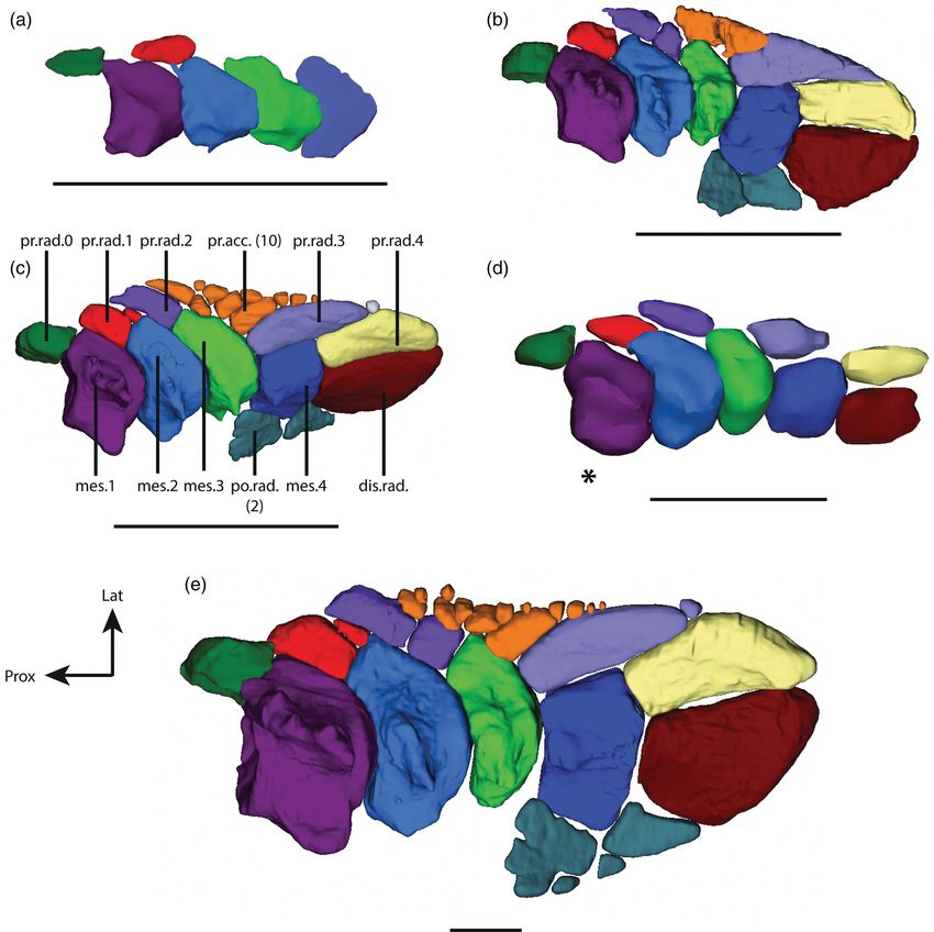
2                                                                                                                           MANSUIT ET AL.

Friedman, Coates, & Anderson, 2007). Although the anat-                2 | MATERIALS AND METHODS
omy of Latimeria chalumnae is well known (Millot &
Anthony, 1958, 1965; Millot, Anthony, & Robineau, 1978),               2.1 | Specimens
its development remains largely unknown. Indeed, only a
few embryos and one juvenile are present in collections                The developmental series includes five stages including
worldwide (Nulens, Scott, & Herbin, 2011). Due to the                  specimens from several Museum collections (Figure 1). The
improvement of noninvasive imaging techniques such as                  first stage is a fetus of 5 cm total length (TL) (international
conventional and synchrotron micro-tomography, as well                 number: CCC 202.1) found inside the female specimen
as magnetic resonance imaging, it is now possible to study             CCC 202 captured off the coast of Tanzania in 2005 and
the development of small and complex internal structures               conserved in the collection of the South African Institute for
of the coelacanth like the lung (Cupello et al., 2015), the            Aquatic Biodiversity (SAIAB) in Grahamstown (collection
neurocranium (Dutel et al., 2019) and the pectoral fin                 number: SAIAB 76199). The stage two is a pup with yolk
(Mansuit et al., 2019).                                                sac of 32.3 cm TL (CCC 29.5) found inside the female speci-
    The endoskeleton elements of the paired fins or                    men CCC 29 captured in the Comores in 1969 and con-
limbs are organized along a single metapterygial axis                  served in the collection of the Muséum national d'Histoire
in sarcopterygians (Millot & Anthony, 1958; Shubin &                   naturelle (MNHN), Paris (collection number: MNHN AC
Alberch, 1986). However, whereas the development                       2012-22). The stage three is a late pup without a yolk sac of
of paired appendages in lungfishes and tetrapods is                    34.8 cm TL (CCC 162.21) found inside the female CCC
relatively well known (Boisvert, Joss, & Ahlberg, 2013;                162 captured off the coast of Mozambique in 1991 and con-
Jude, Johanson, Kearsley, & Friedman, 2014; Shubin &                   served in the collections of the Zoologische Staatssammlung
Alberch, 1986), it remains poorly studied in coelacanths               of Munich (collection number: ZSM 28409). The stage four
(but see (Mansuit et al., 2019). Yet, the development of               is a juvenile of 42.5 cm TL (CCC 94) captured off the coast
the pelvic appendages of vertebrates occurs later than the             of Grande Comore Island in 1974 and conserved in the col-
pectoral appendages (Cubbage & Mabee, 1996; Mabee &                    lections of the MNHN, Paris (collection number: MNHN
Noordsy, 2004; Mabee & Trendler, 1996). Therefore,                     AC 2012-27). Imaging reconstructions and observations on
documenting the development of the pelvic fin of Lat-                  adult stage (Stage 5) were mainly based on a male specimen
imeria is important to fully understand the development                of 132 cm TL (CCC 27) captured off the coast of Grande
of the paired fins in this species. Following our recent               Comore Island in 1961 and conserved in the collections of
work on pectoral fin development (Mansuit et al., 2019),               the MNHN, Paris (collection number: MNHN AC 2012-21)
we here describe the anatomy and development of the                    (Nulens et al., 2011). Direct anatomical observations were
pelvic appendage of Latimeria based on an ontogenetic                  also made from on a dissected and prepared pelvic fin skele-
series of five different stages.                                       ton of adult specimen CCC 7 (collection number: MNHN

FIGURE 1         Latimeria chalumnae—Ontogenetic series in left lateral view. (a) Early embryo (CCC 202.1), (b) Embryo with yolk sac
(CCC 29.5), (c) Late embryo without yolk sac (CCC 162.21), (d) Juvenile (CCC 94), (e) Adult (CCC 27). Scale bar = 5 cm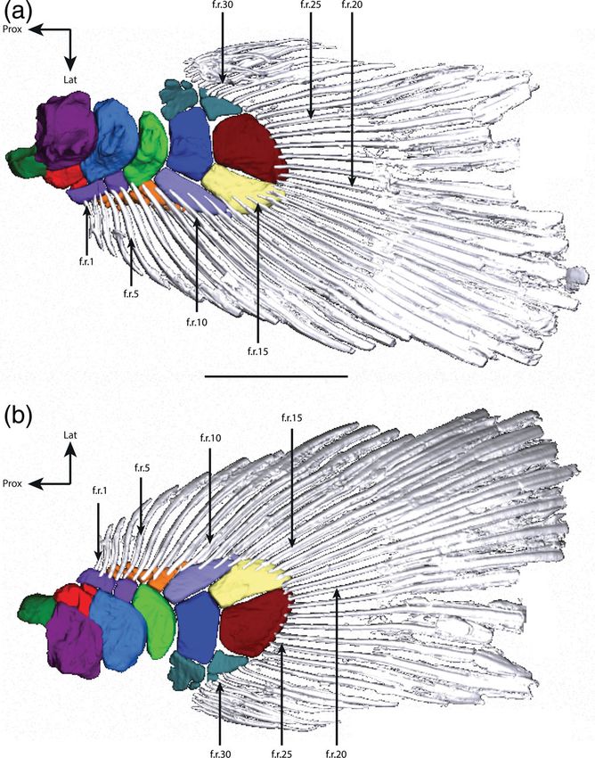
MANSUIT ET AL.                                                                                                                        3

AC 2012-5). All specimens of the MNHN, Paris are con-                    the ID19 beamline of the European Synchrotron Radiation
served in a 6–7% formaldehyde solution, while the others                 Facility (ESRF), Grenoble (France). It was imaged in a
are preserved in an aqueous ethanol solution (70%).                      glass cylinder filled with ethanol, at a voxel size of 6.5 μm,
                                                                         with a high-quality pink beam using the ID19 W150 wig-
                                                                         gler and a gap of 50 mm filtered by 2 mm of aluminum,
2.2 | Imaging                                                            0.25 mm of copper, and 0.2 mm of gold. The scintillator
                                                                         was a 250-μm-thick LuAG:Ce (lutetium–aluminum–
Figure 2 illustrates sections through the girdle and fins of             garnet) crystal. The resulting detected spectrum was then
the different specimens illustrating the quality of the raw              centered on 77 keV, with a bandwidth of 17 keV full width
data used in this article.                                               at half maximum (FWHM). The detector was a FreLoN 2K
                                                                         charge-coupled device (CCD) camera mounted on a lens
                                                                         system. To obtain a sufficient propagation phase-contrast
2.2.1     |      Stage 1: Fetus (CCC 202.1)                              effect, a distance of 3 m between the sample and the detec-
                                                                         tor was used. Synchrotron data were reconstructed
The specimen was scanned using long propagation                          using a filtered back-projection algorithm coupled with
phase-contrast synchrotron X-ray microtomography at                      a single distance phase-retrieval process (Paganin, Mayo,
                                                                         Gureyev, Miller, & Wilkins, 2002; Sanchez, Ahlberg,
                                                                         Trinajstic, Mirone, & Tafforeau, 2012). All the sub-scans
                                                                         were reconstructed separately, converted into 16-bit
                                                                         TIFF stacks and then concatenated to generate a single
                                                                         complete volume. The ring artifacts were corrected on the
                                                                         reconstructed slices using a specific tool developed at the
                                                                         ESRF (Lyckegaard, Johnson, & Tafforeau, 2011). The final
                                                                         volume used for the present study (13 μm voxel size) was
                                                                         obtained after isotropic 2-times binning with the software
                                                                         ImageJ.

                                                                         2.2.2 | Stage 2: Pup 1 (with yolk sac)
                                                                         (CCC 29.5)

                                                                         The specimen was scanned on the ID19 beamline on
                                                                         the ESRF in an empty plastic tube, at a voxel size of
                                                                         23.34 μm, using a propagation distance of 13 m to maxi-
                                                                         mize the phase-contrast effect. The beam produced by the
                                                                         W150 wiggler at a gap of 59 mm was filtered by 2.8 mm
                                                                         of aluminum and 1.4 mm of copper, resulting in an aver-
                                                                         age detected energy of 77.4 keV. The scintillator was a
                                                                         2000-μm-thick LuAG:Ce (lutetium–aluminum–garnet
                                                                         doped with cerium) crystal. The detector was a PCO edge
                                                                         4.2 sCMOS. Tomographic slices were reconstructed using
                                                                         the same protocol as the one described above. The final
                                                                         volume (46.68 μm voxel size) was obtained after isotropic
                                                                         2-times binning with the software ImageJ.

F I G U R E 2 Transverse section of the pelvic fin and girdle in
                                                                         2.2.3 | Stage 3: Pup 2 (with yolk sac
the fetus (a), pup 1 (b), pup 2 (c), and adult (d, e) showing the
quality of the scans and the contrast for the different stages, at
                                                                         resorbed) (CCC 162.21)
different section along the fin. Transverse section for the fetus,
pup 1, and 2 are zoomed on the pelvic fin (a, b, c). Concerning          The specimen was scanned in a similar manner to Stage 2,
the adult stage, the pelvic fins were isolated and the left fin was      at a voxel size of 23.34 μm. The scintillator, detector,
dissected (d, e). lat.pro., lateral process; med.pro., medial process;   and distance between the sample and the detector were
mes., mesomere; pr.rad., preaxial radial                                 the same as for the stage 2 embryo. The final volume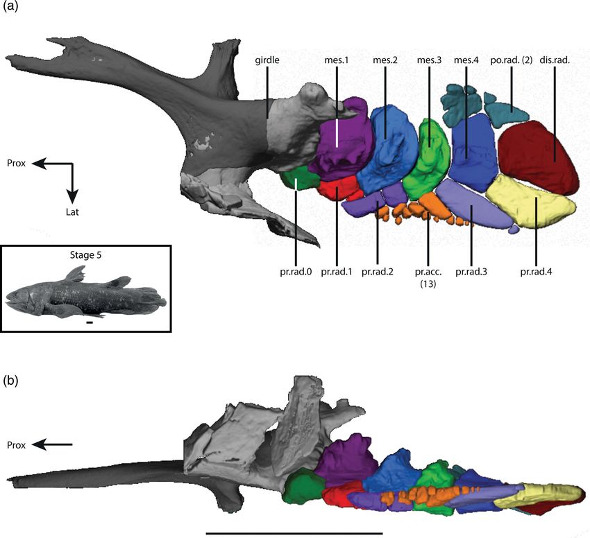
4                                                                                                               MANSUIT ET AL.

(46.68 μm voxel size) was obtained after isotropic 2-times     3 | RESULTS
binning in ImageJ.
                                                               The pelvic appendages are located on the ventral side of
                                                               the animal, in the middle of the body on both sides of the
2.2.4   |   Stage 4: Juvenile (CCC 94)                         cloaca (Millot et al., 1978).

The specimen was scanned twice, once at the ESRF on
the beamline ID19 and once using magnetic resonance            3.1 | The pelvic girdle
imaging (MRI). At the ESRF, the specimen was scanned
in a plastic tube filled with water, at a voxel size of        In the adult stage, the pelvic girdle is a massive element
28.43 μm, using a propagation distance of 13 m to maxi-        and supports several processes: the anterior process (ant.p)
mize the phase-contrast effect. The beam produced by           (“segment antérieur” according to Millot and Anthony),
the W150 wiggler at a gap of 30 mm was filtered by             the lateral process (lat.pro.) (“apophyse latérale externe”
2 mm of aluminum and 15 mm of copper, resulting in an          according to Millot and Anthony), the medial process
average detected energy of 170 keV with a bandwidth of         (med.pro) (“apophyse latérale interne” according to Millot
85 keV FWHM. The detector camera was a FreLoN 2K               and Anthony), and the posterosuperior process (ps.pro)
charge-coupled device mounted on a lens system coupled         (“apophyse postéro-supérieure” according to Millot and
to a 750-mm-thick LuAG:Ce scintillator. Tomographic            Anthony) (Figures 2e and 3). The anterior process is a long
slices were reconstructed using the same protocol as the       cartilaginous and rod-like structure that gently flares ante-
ones described above. The final volume (56.86 μm voxel         riorly to form a flattened blade. The lateral process is large
size) was obtained after isotropic 2-times binning with        and flat, concave on its dorsal side, with an anterior peak,
the software ImageJ.                                           whereas the medial process is a short and triangular expan-
    As the contrast was not sufficient due to the partial      sion. The posterosuperior process is robust and short, with
demineralization of the bones linked to the long preserva-     a flattened apex. The posterior part of the girdle presents a
tion in formalin solution, the specimen was re-scanned         convex articular head, called the acetabulum, which articu-
using MRI at a lower resolution. The MRI was performed         lates with the first axial element of the fin. The two girdles
at 3T with a Siemens Tim TRIO (Siemens, Germany) sys-          are not fused in Latimeria (Figure 4c) but joined by liga-
tem. Images were acquired with a 3D Flash sequence using       ments between the two medial processes and between
an isotropic resolution of 300 μm. Parameters were matrix      the distal edges of the two anterior processes. The micro-
size = 640 × 300 × 256, TR/TE (msec) = 18/4.73, flip           tomographic data allow, for the first time, the visualization
angle = 10 , spectral width = 100 kHz, number of aver-        of a partially denser region at the surface of the pelvic gir-
ages = 20, total acquisition time was 7 hr, and 41 min.        dle that could correspond to an endochondral ossification
                                                               of the girdle (Figures 2e–4). In the adult, this region covers
                                                               the anterior process, the medial process, and extends poste-
2.2.5   |   Stage 5: Adult (CCC 27)                            riorly until the base of the posterosuperior process and of
                                                               the lateral process with a convex expansion. This denser
The specimen CCC 27 was scanned at the AST-RX facil-           area is internally associated with a trabecular system
ity of the MNHN (Paris, France) using the stacking of          (Figures 2e–4b) that is well developed in the posterior part
multiple scans with the following scanning parameters:         and the medial process of the girdle. The anterior process
voltage 110 kV, current 950 μA, filter 0.2 mm Cu, voxel        and the base of the lateral process present a trabecular sys-
size 105 μm, and 3,258 views.                                  tem that appears to be less developed. This ossification is
                                                               also visible on the isolated pelvic girdle of the specimen
                                                               CCC 7.
2.3 | Segmentation and 3D-                                         In the fetus, the pelvic girdle is relatively smaller and
reconstruction method                                          shows a very different shape from what is observed in the
                                                               next stages (Figure 5a). The different processes have mor-
For all the specimens, segmentation and three-dimensional      phologies different from those observed in the adult stage.
rendering were done using the software MIMICS Innova-          The anterior process of the girdle is only represented by a
tion Suite 20.0 (Materialise) (Stages 1–4) and MIMICS          small pointed extension. The medial process is large with a
Innovation Suite 21.0 (Materialise) (Stage 5). The different   square shape and the lateral process forms only a short
objects were exported in STL format and transformed into       crest. The posterosuperior process is a reduced convex pro-
a 3D PDF with the software 3-matic 11.0 (Materialise)          cess. From pup 1 onward, the girdle becomes more com-
(Stages 1–4) and 3-matic 13.0 (Materialise) (Stage 5).         plete and all its processes are fully formed (Figure 5b–e).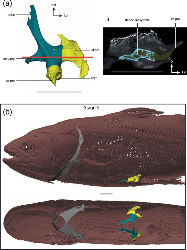
MANSUIT ET AL. 5
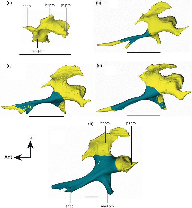
FIGURE 3 Latimeria chalumnae—Adult stage. Right pelvic girdle in dorsal view (a), ventral view (b), and lateral view (c). act.,
acetabulum; ant.p., anterior process; lat.p., lateral process; med.pro., medial process; pos.p., posterior part; ps.pro., posterosuperior process.
Blue = dense part of the girdle; yellow = endoskeletal pelvic girdle. Scale bar = 50 mm
In pup 1, the anterior process of the right girdle does not are five preaxial radial elements (Forey, 1998; Millot &
form a blade, but a bifid process. The dense superficial sur- Anthony, 1958), a variable number of preaxial accessory
face only partially covers the anterior process of the girdle elements, and postaxial radials (Millot & Anthony, 1958).
(Figure 5b). From pup 2 onward, the anterior process of the The fin rays insert on the preaxial and postaxial elements
girdle forms a blade. The dense superficial surface covers of the fin (see descriptions below). As described by Millot
almost the entire anterior process of the girdle and the most and Anthony (1958), the so-called “fifth article” of the
anterior part of the girdle (Figure 5c). In the juvenile, the metapterygial axis has a different shape compared to the
ossification expands to the posterior part of the girdle and previous elements of the metapterygial axis, and the fin
partially covers the medial process (Figure 5d). No trabecu- rays insert on it. Following Forey (1998), we, therefore,
lar system can be observed, in contrast to the adult stage. consider this element as a distal radial belonging to the
postaxial elements (Figure 6). In the adult stage, the organi-
zation of the endoskeleton of the pelvic fin is similar to that
3.2 | The pelvic fin observed for the pectoral fin (Mansuit et al., 2019; Millot &
Anthony, 1958) (Figure 7). However, unlike in the pelvic
The pelvic fin (Figure 6) is composed of several elements: girdle, there is no trabecular system in any element of the
mesomeres on the metapterygial axis, preaxial elements pelvic fin (as for the pectoral fin) (Figure 2).
(preaxial radials + preaxial accessory elements), and post-
axial elements (postaxial radials + distal radial). According
to Millot and Anthony (1958), the metapterygial axis of 3.2.1 | The metapterygial axis
the fin consists of five axial elements (named “articles”),
numbered from proximal to distal, while Forey (1998) iden- As for the pectoral fin of L. chalumnae (Mansuit
tified four mesomeres and one distal radial element. There et al., 2019), we use the term mesomere for subcylindrical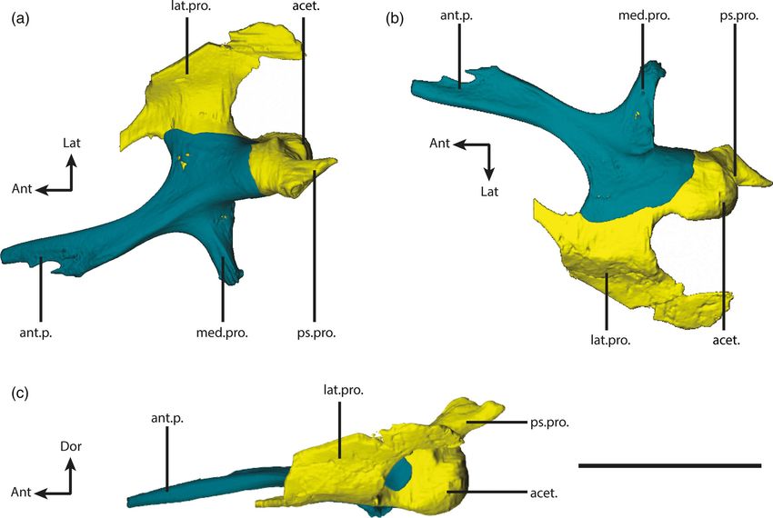
6                                                                                                                            MANSUIT ET AL.

                                                                         arc-shaped. It articulates with the acetabulum of the pel-
                                                                         vic girdle by its concave proximal edge and with the next
                                                                         mesomere by its convex distal edge (Figure 6). The angle
                                                                         between the distal edge and the medial edge forms a
                                                                         large process. The dorsal side presents a strong but irreg-
                                                                         ular oblique crest, directed from proximo-lateral to disto-
                                                                         medial. The ventral facet of the mesomere is flatter with
                                                                         only a small swelling on the medial part of its surface.
                                                                              In the fetus, the shape of the first mesomere is similar to
                                                                         that seen in the adult specimen: concave on the proximal
                                                                         edge and convex on the distal edge, with a ridge on its dorsal
                                                                         face (Figure 7a). There is a bilateral asymmetry to the mor-
                                                                         phology of the mesomere, since the ridge is oblique on the
                                                                         right fin whereas it is longitudinal on the left fin (Figure 10a).
                                                                         This first mesomere is squarer in the fetus than in the adult
                                                                         stage. There is no process on the medial side of the meso-
                                                                         mere. The ventral side of the mesomere does not have a
                                                                         swelling on the medial part of the surface, but a longitudinal
                                                                         ridge close to the medial edge. From pup 1 onward, the first
                                                                         mesomere has already its adult shape, arc-shaped and
                                                                         slightly wider than long (Figure 7b–e). The dorsal ridge is
                                                                         well developed, with an irregular surface. In pup 1, the ven-
                                                                         tral face of the first mesomere has an important swelling on
                                                                         the medial part of its surface. From pup 2 onward, the swell-
                                                                         ing is less important and the ventral side of the mesomere is
                                                                         flatter. The oblique crest on the ventral face of this mesomere
FIGURE 4          Latimeria chalumnae—Adult stage. Right pelvic          described by Millot and Anthony (1958) is not present from
girdle in dorsal view (a) and transverse section (b). Location of the    pup 1 onward, neither is it on the isolated pelvic fin.
pectoral and pelvic girdle inside the body in the left and ventral
view (c). The red line in A shows the location of the transverse
section. The trabecular system and the dense radio-opaque part of        3.2.3 |      The second mesomere (mes.2)
the girdle are clearly visible. The color code of the transverse
section and the 3D model is identical. act., acetabulum; ant.p.,
                                                                         In the adult stage, the second mesomere has a morphol-
anterior process; lat.p., lateral process; med.pro., medial process;
                                                                         ogy similar to that of the first mesomere, but more flat-
pos.p., posterior part; ps.pro., posterosuperior process. Blue = dense
part of the pelvic girdle; yellow = endoskeletal pelvic girdle;
                                                                         tened and slightly shorter (Figure 6). The medial process
grey = pectoral girdle. Scale bar = 50 mm                                is smaller and the dorsal ridge less developed. The latter
                                                                         is less oblique and more latero-medially oriented than on
radial segments of the principal axis in sarcopterygian                  the mesomere 1.
fins (Jarvik, 1980).                                                          In the fetus, the second mesomere is trapezoid in
    As described by Millot and Anthony (1958) for the                    shape. There is no process on its medial edge, and the
adult stage, the four mesomeres have a similar length, and               ridge is poorly developed (Figure 7a). Moreover, this
the three first mesomeres have a similar shape (Figure 6a).              ridge is longitudinal on the left fin whereas it is more
There is a progressive dorsoventral flattening of the meso-              oblique on the right fin (Figure 10a). From pup 1 onward,
meres from proximal to distal (Figure 6b). From proximal                 the second mesomere has its adult shape (Figure 7b–e).
to distal, the ridges on the dorsal side of the mesomeres are            As for the first mesomere, the swelling surface on the
also progressively less developed and have an increasingly               ventral face of the second mesomere is well marked in
medial position from the first to the third mesomere.                    pup 1, yet becomes flatter from pup 2 onward.

3.2.2     |   The first mesomere (mes.1)                                 3.2.4 |      The third mesomere (mes.3)

As described by Millot and Anthony (1958) in the adult                   In the adult stage, the length of this element is smaller
stage the first mesomere is wider than long and slightly                 than those of previous mesomeres, but the width is more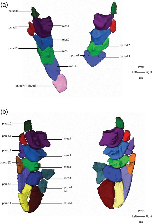
MANSUIT ET AL.                                                                                                                                      7

F I G U R E 5 Latimeria chalumnae. Right pelvic girdle in dorsal view at five different developmental stages. a: Stage 1, b: Stage 2, c: Stage 3, d:
Stage 4, and e: Stage 5. ant.p., anterior process; lat.p., lateral process; med.pro., medial process; pos.p., posterior part; ps.pro., posterosuperior
process. Blue = dense part of the girdle; yellow = endoskeletal pelvic girdle. a: Scale 2 mm; b–e: Scale bar = 10 mm

or less the same (Figure 6). The general morphology is                       extension could correspond to the second preaxial radial
boomerang-liked. Like for the previous mesomeres the                         in a fragmentation process of this early mesomere.
dorsal ridge is well developed, but less pronounced. How-                    The dorsal ridge is longitudinal and in medial position.
ever, the medial process is barely visible.                                  From pup 1 onward, the third mesomere has its boomer-
    In the fetus, the third mesomere is square in shape,                     ang shape and its well-developed ridge, as in the adult
with a proximo-lateral extension (Figure 7a). This                           (Figure 7b–e). As for the previous mesomeres, the ventral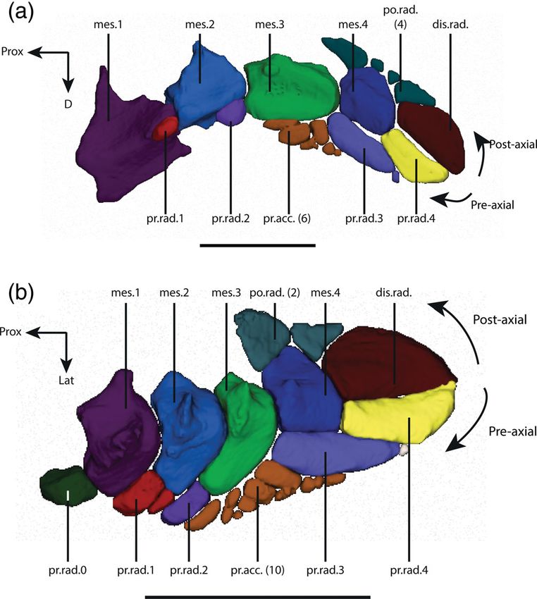
8                                                                                                                                MANSUIT ET AL.

FIGURE 6        Latimeria chalumnae—Adult stage. Left pelvic fin in dorsal (a) and lateral (b) view. The lateral process of the girdle is
broken. mes., mesomere; po.rad., postaxial radial; pr.acc., preaxial accessory elements; pr.rad., preaxial radial. Scale bar = 50 mm

side of the third mesomere has an important swelling                      previous mesomeres, there is a small ridge on the dorsal
that becomes flatter from pup 2 onward.                                   side of this mesomere. This ridge has a proximo-distal
                                                                          orientation and decreases in height from proximal to
                                                                          distal. This element is not aligned with the previous axial
3.2.5     |   The fourth mesomere (mes.4)                                 elements but is medially shifted. It is surrounded by the pre-
                                                                          axial radial 3, the preaxial radial 4, the distal radial, and the
In the adult stage, this mesomere is trapezoid in shape,                  postaxial radials from its preaxial side (corresponding to its
less wide than the previous mesomeres and without the                     lateral edge) to its postaxial side (corresponding to the
presence of a medial process (Figure 6). The proximal                     medial edge).
edge of the mesomere is slightly concave and the distal                       In the fetus, the fourth mesomere of the right fin
edge slightly convex, whereas the lateral and medial edges                presents an arrowhead-shape, wider than long with a
are straight. This mesomere is the only one in contact                    strongly concave proximal edge (Figure 7a). The shape of
with the postaxial elements. The medial edge of this                      the left mesomere is elongated, with a slightly concave
mesomere is thinner than the lateral edge. As for the                     proximal edge (Figure 10a). From pup 1 onward, the
MANSUIT ET AL. 9
mesomere. It is in contact with the ventral side of
the latero-distal part of the pelvic girdle (Figure 6). The
medial articular surface of the preaxial radial 0, in con-
tact with the pelvic girdle, is slightly concave.
In the fetus, this element is rectangular in dorsal view
with a triangular section (Figure 7a). From pup 1 onward,
this element is square in shape (Figure 7b–e).
3.2.8 | The preaxial radial 1 (pr.rad.1)
In the adult stage, this element forms the prolongation of
the lateral edge of the second mesomere and is in contact
with the lateral part of the distal edge of the first meso-
mere (Figure 6). The preaxial radial 1 is formed by two
parts, a large and rectangular proximal element and a
small distal element with a triangular shape.
In the fetus, this element is single and elongated
(Figure 7a). In pup 1, this element is also single and rect-
angular in shape (Figure 7b). In pup 2, the preaxial radial
1 is fully formed and composed of two elements
(Figure 7c–e). In the juvenile, only one element can be
FIGURE 7 Latimeria chalumnae—pup 2. Comparison reconstructed from the MRI scans but this may be due to
between the left pectoral (a) and pelvic fin (b). The pectoral fin is the lack of precision in the MRI data (Figure 7d).
reversed to have a better comparison with the pelvic fin. The
organization of the endoskeletal elements of the pectoral and pelvic
fins is the same, except for the presence of the preaxial radial 0 on
the pelvic fin. mes., mesomere; po.rad., postaxial radial; pr.acc.,
3.2.9 | The preaxial radial 2 (pr.rad.2)
preaxial accessory elements; pr.rad., preaxial radial. Scale
bar = 10 mm In the adult stage, the preaxial radial 2 forms the prolon-
gation of the lateral side of the third mesomere and lies in
contact with the lateral part of the distal edge of the sec-
mesomere has its trapezoidal shape, as in the adult ond mesomere (Figure 6). The preaxial radial 2 is formed
(Figure 7b–e). by two elements, a basal square element, adjacent to the
mesomere 3, and a rectangular element aligned with the
basal element (Figure 7b). This element carries fin rays
3.2.6 | The preaxial elements 1–6 on its dorsal side, whereas it only carries the fin rays
1–4 inserting more distally on its ventral side (Figure 9).
The preaxial elements are situated on the lateral edge of In the fetus, this element does not appear individual-
the pelvic fin. The organization of the radial elements ized from the mesomere 3. From pup 1 onward, the pre-
along the metapterygial axis is similar on the pelvic fin axial radial 2 has its adult shape, with its elongated
and on the pectoral fin, except for the presence of a pre- rectangular shape and the two aligned pieces (Figure 7b).
axial radial (called preaxial radial 0) on the pelvic fin only There is some variation in the morphology of this ele-
(Figure 7). As in the pectoral fin (Mansuit et al., 2019; ment as in pup 2 and the juvenile specimens preaxial
Millot & Anthony, 1958) there is an important difference radial 2 only consists of a single elongated element
in morphology of the preaxial radial elements between (Figure 7c,d), instead of two aligned elements (Figure 7b,e).
the most proximal elements (pr.rad. 0–2) and the distal However, these observations could be due to the low resolu-
elements (pr.rad. 3–4). tion of the MRI data of the juvenile specimen.
3.2.7 | The preaxial radial 0 (pr.rad.0) 3.2.10 | The preaxial radial 3 (pr.rad.3)
In the adult stage, this element has a square shape and In the adult stage, the preaxial radial 3 is a thin and
lies in the prolongation of the lateral edge of the first elongated element, longer than the fourth mesomere,10 MANSUIT ET AL.
F I G U R E 8 Latimeria chalumnae. Right (a–d) and left (e - mirror) pelvic fin of five different stages in dorsal view. a: fetus, b: pup 1, c:
pup 2, d: juvenile, e: adult. *: The juvenile was scanned using magnetic resonance imaging (MRI) at a low resolution, which did not allow
the segmentation of the smallest elements (po.rad., pr.acc.). Corresponding elements of the fin are indicated in the same color. mes.,
mesomere; po.rad., postaxial radial; pr.acc., preaxial accessory elements; pr.rad., preaxial radial. (a) Scale 2 mm; (b–e) Scale bar = 10 mm
with three straight edges and one distal convex edge latero-distal corner forms a concave angle to fit a preaxial
(Figure 6). The proximal edge of this element is straight accessory element. This angle could correspond to some
and articulates with the third mesomere. A very small elements of preaxial accessory elements that are seg-
globular element is present at its distal tip. This preaxial mented later in the pup 2 to form the three most distal ele-
radial carries the fin rays 10–13 (Figure 9). ments (Figure 7b,c). On the left fin, the distal part of the
In the fetus, this element is not separated from the radial is surrounded by the preaxial radial 4. From pup
fourth mesomere (Figures 7a and 10a). In pup 1, the 2 onward, this element has its adult shape, with an elon-
preaxial radial 3 is thin and elongated, without a sepa- gated trapezoidal shape and a separated distal small ele-
rated tip (Figure 7b). Its medial edge is straight and its ment (Figure 7c–e).MANSUIT ET AL. 11
FIGURE 9 Latimeria chalumnae—Adult stage. Insertion of
the fin rays on the endoskeletal elements of the left fin in dorsal
(a) and ventral view (b). The insertion of the fin rays on the
preaxial edge of the fin is more proximal on the dorsal edge
compared to the ventral edge. f.r., fin ray. Scale bar = 50 mm FIGURE 10 Latimeria chalumnae—Fetus (a) and pup
1 (b) stages. Right and left pelvic fins in dorsal. Dotted lines show
the supposed splitting zone between the mesomere and the preaxial
3.2.11 | The preaxial radial 4 (pr.rad.4) and postaxial elements. (b) There is an asymmetry between the
right and left fin in the pup 1 stage, on the distal part of the fin.
The preaxial radial 4, situated at the distal end of the fin mes., mesomere; po.rad., postaxial radial; pr.acc., preaxial accessory
endoskeleton, closely resembles the mirror image of the elements; pr.rad., preaxial radial. Not to scale
distal radial. As the preaxial radial 3, this element is
elongated with three straight edges and a convex distal 3.2.12 | The preaxial accessory elements
edge (Figure 6). This element carries the fin rays 14–18 (pr.acc.)
(Figure 9).
In the fetus, the data do not allow the separation of In the adult stage, these small elements (called “éléments
the preaxial radial 4 from the fin rays and muscles on the accessoires de la troisième pièce radiale préaxiale” by
right fin (Figure 7a), so this element was not segmented. Millot & Anthony, 1958) are positioned in a patch
However, it is assumed that the preaxial radial 4 is already between the distal edges of the third mesomere, the pre-
present on the right fin. On the left fin, this radial is axial radial 2, and the lateral edge of the preaxial radial
not separated from the distal radial (Figure 10a). In pup 3 (Figure 6). According to Millot and Anthony (1958), the
1, there is a bilateral asymmetry, since the medial edge of pelvic fin presents four of these elements at the adult
the preaxial radial 4 is straight on the right fin (Figure 7b), stage. However, both our segmentation of the scan of the
but concave on the left fin, forming a space between this pelvic fin and the observation of the isolated fin show a
element and the distal radial (Figure 10b). The lateral edge variation in the number of preaxial accessory elements.
of the preaxial radial 4 forms an angle that surrounds the Indeed, there are 13 elements in adult stage CCC 27
distal part of the preaxial radial 3. From pup 2 onward, (Figures 6 and 7e), whereas there are at least five acces-
the preaxial radial 4 has the same trapezoidal shape as sory elements in the isolated fin of CCC 7. The difference
described for the adult (Figure 7c–e). in numbers of elements observed in the different fins can12 MANSUIT ET AL.
be explained by the size of the smallest elements, not visi- proximal element on the right fin, as is observed for the
ble to the naked eyes and hidden by dermal fin rays cov- isolated prepared fin of CCC 7 and as described by Millot
ering them. The preaxial accessory elements carry the fin and Anthony (1958). The distal element is slightly smaller,
rays 7–9 on the dorsal side of the fin and the fin rays 5–9 with a quadrangular shape and located on the medial part
on the ventral side of the fin (Figure 9). of the distal edge of the fourth mesomere and on the
These elements are not present in the fetus and appear medial edge of the distal radial (Figure 6). The third ele-
at the pup 1 stage (Figure 7). In the pup 1 stage, there ment is very small compared to the other postaxial radials,
are only two elements, as large as the preaxial radials 2 triangular-shaped, and located between the proximal and
(Figure 7b). These two elements become fragmented dur- the distal one. The postaxial radials carry the fin rays 28–34
ing development since there are at least 10 small elements (Figure 9).
in pup 2 (Figure 7c) and 13 elements in the adult. In the In the fetus, these elements cannot be individually
juvenile, we assume that the preaxial accessory elements identified or separated from the mesomere 4 in the right
are present, but the MRI data did not allow the visualiza- fin (Figures 7a and 10). In the pup 1 and pup 2 the post-
tion of the preaxial accessory elements. axial radials are well developed, but with only two ele-
ments (Figure 7b,c).
3.2.13 | The postaxial elements
3.2.16 | The fin rays
3.2.14 | The distal radial (dis.rad.)
The pelvic fin in the adult stage presents 37 fin rays
In the adult stage, the distal radial is similar in shape to (Figure 9), which are numbered 1 from the preaxial (lat-
the preaxial radial 4 as a mirror image, although almost eral) to 37 at postaxial (medial) side of the fin. As for the
twice as wide (Figure 6). It has three more or less straight pectoral fin, the proximal part of each fin ray is bifurcated,
edges and a convex distal edge. It carries the fin rays one branch inserting on the dorsal face of the fin and one
19–27 (Figure 9). branch inserts on the ventral face of the fin. The first ray is
In the fetus, only the left distal radial could be seg- very small, and the next rays become longer along the pre-
mented (Figure 10a). We assumed that the right distal radial axial side of the fin, up to the axis of the fin, to progres-
is also present at this stage, but as for the preaxial radial sively decrease in size along the postaxial side of the fin. As
4, the data did not allow the separation of this element from for the pectoral fin, the rays of the preaxial side of the fin
the muscles and the fin web. This element is small and insert largely on the preaxial elements, whereas the rays on
square except for its proximal edge that is slightly concave. the postaxial side of the fin insert only on the medial edges
In pup 1, the left fin presents an ovoid space between the of the distal radial and the postaxial radials. The three last
distal radial and the preaxial radial 4 (Figure 10b). Such a fin rays 35–37 are free and do not insert on fin elements.
space is not present on the right fin (Figure 7b). Its medial The fins rays are inserted more proximally on the dorsal
edge forms an angle that surrounds the postaxial radials in than on the ventral side on the preaxial side of the fin.
the left fin. From pup 2 onward, the distal radial is fully
formed with the trapezoidal shape observed in the adult
stage (Figure 7c–e). According to Millot and Anthony (1958) 4 | DISCUSSION
there is a longitudinal ridge on the dorsal side of this ele-
ment, but such a ridge does not appear on the 3D modeling As discussed in our previous study (Mansuit et al., 2019),
or on the observed isolated pelvic fin. even if the gestation time for coelacanths is estimated at
more to 13 months (Hureau & Ozouf, 1977), it is not pos-
sible to precisely determine the time between the differ-
3.2.15 | The postaxial radials (po.rad.) ent stages.
The postaxial radials are located at the medial edge of the
pelvic fin in articulation with the fourth mesomere and the 4.1 | Morphology of the pelvic girdle
distal radial. In the adult stage, Millot and Anthony (1958)
described three elements among the postaxial radials. The Unlike the pectoral girdle, the pelvic girdle is only
proximal element presents a large triangular shape and is formed by a unique endoskeletal bone as described by
located on the medial edge of the fourth mesomere. On the Millot and Anthony (1958). Unlike in lungfishes as
left fin of CCC 27, this element is split into a large piece Neoceratodus (Boisvert et al., 2013) the two hemi-girdles
and a small one (Figure 6), whereas there is only one are not fused (Figure 4c) but only linked by ligamentsMANSUIT ET AL. 13
(Millot & Anthony, 1958). The general shape of the dipnomorphs as a derived character. Even if the precise
pelvic girdle of Latimeria chalumnae is similar to what interrelationships of sarcopterygians remain debated, the
is observed in fossil coelacanths with an anterior rod-like current consensus presents coelacanths as sister-taxa of
process, a lateral and a medial processes (Forey, 1998). tetrapods and lungfishes (e.g., Ahlberg, 1991; Amemiya
According to Forey (1998), the lateral process corresponds et al., 2013; Cloutier & Ahlberg, 1996; Friedman et al.,
to the ilium (iliac process of Coates & Ruta, 2007), the 2007). It thus appears that a convex acetabulum is the
medial process to the ischium (ischial process of Coates & primitive condition for sarcopterygians.
Ruta, 2007), and the anterior process to the pubis of the The micro-tomographic imaging highlights, for the first
pelvic girdle of tetrapods. However, the homology between time, the presence of a dense surface around part of the
the lateral and medial processes with the ilium and ischium pelvic girdle, and the presence of a dense trabecular system
remains uncertain (Ahlberg, 1989). Therefore, we preferred within the girdle (Figure 4b). The pelvic girdle of Latimeria
to use the neutral terminology proposed by Millot and is cartilaginous (Forey, 1998; Millot & Anthony, 1958), and
Anthony (1958) (i.e., medial and lateral processes, and this dense surface and the trabecular system might be an
anterior process) to avoid any ambiguities regarding the endochondral ossification of the girdle. The presence of a
homology of these structures. In extant tetrapods, the trabecular system in the pelvic girdle of Latimeria might
ilium, ischium, and pubis are formed through different reinforce its structural resistance when loaded by pelvic
ossification centers (Malashichev, 2001; Maxwell & muscle forces. Indeed, many levator and depressor muscles
Larsson, 2009; Rocková & Rocek, 2005). In the extant of the pelvic fin insert on the anterior part of the girdle and
coelacanth Latimeria, as in lungfish (Schultze, 1986) the on the medial process (Millot & Anthony, 1958) (personal
girdle is cartilaginous (Millot & Anthony, 1958) and there observations). In tetrapods, it has been shown that the
is only an ossification of the anterior part of the girdle ossification of the bones is influenced by muscular activity
and medial process, but not for the lateral process during the embryonic development (Boisvert et al., 2013;
(Figures 2e–4). Moreover, the ossification of the pelvic Hall, 1986; Newman & Müller, 2005). It is therefore likely
girdle begins at the anterior process of the girdle, subse- that the same process occurs in Latimeria where the young
quently extending to the medial process of the girdle develop in the mother and likely show muscular contrac-
(Figure 5). Consequently, only a single ossification center tions before birth. The ossification of the bone occurs only
appears to be present in the pelvic girdle of Latimeria. from the pup 1 stage. Forey (1998) noted that the pelvic gir-
Only based on anatomical observations, it is thus not pos- dle of extinct coelacanths often presents an open concave
sible to assign a homology to the different parts of the gir- posterior end, and that the acetabulum is missing. It could
dle in Latimeria relative to the different bones of the be assumed that only the ossified part of the pelvic girdle is
girdle of tetrapods. preserved in the fossil record, and that the cartilaginous
The anterior process/blade of the pelvic girdle seems to posterior part of the girdle does not fossilize. The trabecular
be common in actinopterygian fishes (e.g., Andrews & system highlighted in Latimeria has never been described
Westoll, 1970; Faustino & Power, 1999; Grandel & Schulte- in the fossil records for coelacanths and other osteichthyan
Merker, 1998; Yamanoue, Setiamarga, & Matsuura, 2010) fishes, and remains to be determined whether this feature
and sarcopterygian fishes (Andrews & Westoll, 1970; is specific to Latimeria or if other bony fishes possess a tra-
Boisvert et al., 2013; Forey, 1998). According to Andrews becular system in the pelvic girdle.
and Westoll (1970), this anterior process forms an insertion
area for abdominal muscles and ligaments. In Latimeria,
however, only the extremity of this process supports the 4.2 | Morphology of the pelvic fin
abdominal muscles. The main part of the blade forms
the attachment site for the pelvic fin muscles (Millot & In Latimeria, the general organization of the pelvic fin
Anthony, 1958) (personal observations). endoskeleton is similar to that of the pectoral fin (Millot &
As for the articular head of the pectoral girdle Anthony, 1958; Panchen & Smithson, 1990), particularly
(Millot & Anthony, 1958), the acetabulum of the pelvic the distal part of the fin (pr.rad. 3–4, pr.acc., dis.rad., po.
girdle of Latimeria is convex. This convex articular surface rad.) (Figure 7). This endoskeletal morphology of the pecto-
is a feature shared by coelacanths and dipnomorphs ral and pelvic fins/limbs is shared by all sarcopterygians
(Boisvert et al., 2013; Shubin, 1995). In tetrapodomorphs, (Rosen, Forey, Gardiner, & Patterson, 1981). In all crown-
the pelvic girdle shows a concave acetabulum, where the sarcopterygians, these paired appendages are connected
first mesomere fits (Ahlberg, 1989; Boisvert et al., 2013; to their respective girdle via a mono-basal articulation
Shubin, 1995). A concave acetabulum was considered (Clack, 2012; Janvier, 1996; Rosen et al., 1981), whereas
as the primitive condition for sarcopterygians (Ahlberg, stem-sarcopterygians are assumed to have a poly-basal fin
1989), and the convex acetabulum of coelacanths and articulation (Zhu & Yu, 2009). In Latimeria and in other14 MANSUIT ET AL.
sarcopterygian fishes, the pelvic fin is smaller than the pec- axis compared to the pectoral fin, that is, the organization
toral one (e.g., Ahlberg, 1989; Andrews & Westoll, 1970; and arrangement of the elements are different between
Boisvert, 2005; Forey, 1998; Jeffery, Storrs, Holland, the preaxial and the postaxial edge of the fin. As for the
Tabin, & Ahlberg, 2018). The major difference between the pectoral fin (Mansuit et al., 2019; Millot & Anthony,
pelvic and pectoral fins, in Latimeria, lies in the presence 1958), the arrangement of the pre-axial and postaxial ele-
of a preaxial radial 0, which articulates with the pelvic gir- ments along the metapterygial axis of the pelvic fin of
dle and the first mesomere (Figure 7b). This element is Latimeria is asymmetrical. The mesomeres of the pelvic
absent in the pectoral fin (Figure 7a). The preaxial radial fin are all associated with preaxial radials, whereas only
0 was previously described (Millot & Anthony, 1958) and the fourth mesomere is associated with postaxial elements
is also present in the pelvic fin of the Triassic coelacanth (postaxial radials and distal radial). Moreover, the pres-
Laugia groenlandica (Stensiö, 1932) (specimen NHMD ence of the preaxial radial 0 on the pelvic fin, not present
152716, personal observation), but never received devel- on the pectoral fin, increases the asymmetry to the central
opmental or phylogenetic consideration. Indeed, the pres- axis of the pelvic fin. However, as for the pectoral fin, the
ence of this small element questions the mono-basal most proximal preaxial radials (0–2) of the pelvic fin are
condition of the pelvic fin of Latimeria and coelacanths in small and globular or rectangular shape, whereas the dis-
general, and may question the synapomorphy of crown- tal preaxial radials (3–4) are thin and elongate (Millot &
sarcopterygians [a mono-basal articulation of paired fins Anthony, 1958) (Figure 6). This difference of shape is
(Janvier, 1996; Rosen et al., 1981; Zhu & Yu, 2009)]. The related to muscle insertions. Indeed, some muscles insert
development of the pelvic fin suggests that the presence on the preaxial radials 0–2, whereas for the preaxial radial
of this element in contact with the girdle is due to the par- 3–4, the muscles inserted at the base of the fin rays that
ticular morphology of the mesomeres. This element may are associated with the radials (Mansuit et al., in prep.).
correspond to the preaxial radial 1 of the pectoral girdle Considering the general endoskeletal arrangement, the
(see below). Thus, the pluri-basal condition of the pelvic pelvic fin of Latimeria shows a short lobe-shaped silhou-
fin of Latimeria would be a specificity of coelacanths. ette compared to that of the more elongate lobed pectoral
Although the general organization of the endoskeleton fin. The insertion of the fin rays on the fin is also more
of the pelvic and pectoral fins is similar, some differences asymmetrical on the pelvic fin compared to the pectoral
can be observed (Figure 6). The mesomeres of the pectoral fin (Friedman et al., 2007; Millot & Anthony, 1958). Fin
fin are longer than wide, with a quadrangular shape rays of the pelvic fin insert more proximally on the preax-
(Mansuit et al., 2019; Millot & Anthony, 1958). By con- ial side than on the postaxial side (Figure 9) compared to
trast, the mesomeres of the pelvic fin are shorter than the pectoral fin where the fin web shows a more symmet-
wide with an arc shape, dorsoventrally flattened, with a rical arrangement around the metapterygial axis. How-
ridge on their dorsal side. These differences between the ever, as for the pectoral fin, the most proximal preaxial
shape of the mesomeres of the pelvic and pectoral fins radials (preaxial radial 0–1) are not associated with the
appear to be also present in the Triassic coelacanth Laugia pelvic fin rays. An asymmetrical arrangement of the pel-
groenlandica (Stensiö, 1932) (personal observations). vic fin rays along the metapterygial axis has also been
There are marked differences in the size and shape of suggested in the Triassic coelacanth Laugia groenlandica
the most proximal radial elements between the pectoral (Forey, 1998; Stensiö, 1932). An asymmetry is also present
and pelvic fins. The preaxial radials 1–2 of the pectoral fin on the fin ray arrangements between the dorsal and ven-
are small compared to the mesomeres, ovoid in shape, and tral sides of the pelvic fin. Indeed, the fin rays insert
in contact with the distal part of the associated mesomere more proximally on the dorsal side of the fin than on the
(Figure 7a). By contrast, the preaxial radials 1–2 of the pel- ventral side (Figure 9). This asymmetrical coverage of
vic fin are proportionally larger and in contact with both the paired fin rays is also shared by the fossil coelacanths
the associated and the following mesomeres (Figures 6 and Laugia groenlandica (Forey, 1998; Stensiö, 1932), the
7b). In the fossil tetrapodomorph fishes Eusthenopteron extant lungfish Neoceratodus and fossil tetrapodomorph
and Panderichthys, the preaxial radials 1–2 of the pelvic fishes, such as Eusthenopteron and Tiktaalik (Stewart
fin have a similar shape, although slightly smaller, et al., 2019).
than those of the pectoral fin (Andrews & Westoll, 1970;
Boisvert, 2005). Their morphology is different from that of
Latimeria: they are elongated elements that articulate with 4.3 | Development of the pelvic
the distal edge of the associated mesomere, and more or appendage
less parallel to the long edge of the following mesomere.
According to Forey (1998), the pelvic fin of Latimeria The most pronounced changes in the pelvic girdle mor-
presents an important degree of asymmetry to the central phology occur between the fetus and pup 1, and notablyMANSUIT ET AL. 15
entail the elongation of the anterior part of the girdle is a progressive increase in the number of elements on
(Figure 5a,b). The elongation of the girdle during the the preaxial side of the fin. An additional preaxial radial
development seems to be common in osteichthyan fishes, and two preaxial accessory elements are observed in
since it is observed both in actinopterygians, for example pup 1, and the number of preaxial accessory elements
in Pagrus major (Matsuoka, 1985), Chanos chanos increases in pup 2 (10) and in the adult (13) (Figure 8).
(Taki, Kohno, & Hara, 1986), Danio rerio (Grandel & This pattern suggests that the preaxial radials are formed
Schulte-Merker, 1998), and Sparus aurata (Faustino & through the fragmentation of the radial extension of the
Power, 1999), and in the lungfish Neoceratodus forsteri adjacent mesomere, following a proximo-distal sequence
(Boisvert et al., 2013). However, the pelvic girdle mainly during the development (i.e., the first mesomere splits
grows in a posterior direction in the axolotl Ambystoma first). In this scenario, the extension of mesomere
mexicanum (Boisvert et al., 2013), whereas the pubis has 3 observed in the fetus gives rise to the preaxial radial
a small anterior growth. 2 in the pup. From pup 1 onward, a series of preaxial and
Although very similar, the development of the pelvic fin postaxial radials surrounds the mesomere 4. We suggest
appears to lag behind that of the pectoral fin in Latimeria. that these elements derive, respectively, from the lateral
The assumption of a delay in the development of the pelvic and medial extensions of the mesomere 4 observed in the
appendage is based on the morphology of the endoskeletal fetus. The distal end of the left fin is formed by a distal
elements observed in the fetus. At this stage, all mesomeres plate in the fetus, which might correspond to the distal
of the pectoral fin are formed, and the radial elements are radial and the preaxial radial 4 from pup 1 onward
present, at least as cartilaginous plates. However, most of (Figure 10). Yet, additional developmental stages are
the radial elements are not differentiated from the meso- needed to better understand the origin of the distal ele-
mere 4 and the distal radial in the pelvic fin (Figures 8 and ments of the pelvic fin, such as the preaxial radial 3, and
10a), which suggests that its development is delayed com- the potential contribution of the mesomere 4 and distal
pared to the pectoral fin. A delay in the development of plate in the formation of these elements.
the pectoral and pelvic appendages is also observed in The formation of the preaxial radials is not clearly
chondrichthyans (Ballard, Mellinger, & Lechenault, 1993; understood in the extant lungfish Neoceratodus. Previous
Didier, Leclair, & Vanbuskirk, 1998; Ziermann, Freitas, & studies suggest two different patterns of development to
Diogo, 2017), actinopterygians (Faustino & Power, 1999; explain their formation. In the first scenario, the radials
Grandel & Schulte-Merker, 1998), and sarcopterygians arise de novo process (Joss & Longhurst, 2001): they are
(Boisvert et al., 2013; Joss & Longhurst, 2001). Therefore, formed by a distinct mesenchymal condensation and
Latimeria likely follows the general gnathostome pattern of are unconnected to other elements (Johanson et al., 2007;
paired appendages development. In the Latimeria fetus, Shubin & Alberch, 1986). In the second scenario, the
only the most proximal preaxial radials (pr.rad. 0–1) are radials arise from the fragmentation of the associated
present in the pelvic fin (Figure 8). However, from pup mesomere (Joss & Longhurst, 2001), i.e., the element is
1 stage onward all the elements of the fin are present. Con- formed by a continuous plate of precartilage that subse-
trary to what is observed during the development of the quently breaks up into two separate elements (Shubin &
pectoral fin (Mansuit et al., 2019), there is no cartilaginous Alberch, 1986). This second scenario could corroborate
plate around the metapterygial axis of the pelvic fin. our hypothesis of the splitting of the mesomere. How-
Based on the comparison of the morphology of the ever, we suggested previously that the preaxial radials are
mesomeres during the development, we suggest a devel- formed by the fragmentation of the following mesomeres,
opmental mechanism for the endoskeletal elements of and not the associated mesomeres as in Neoceratodus.
the pelvic fin. Along the preaxial edge of the fin, the dis- Since the mesomeres of the pelvic fin of Latimeria have
tribution of the radials matches that of the mesomeres an arc-shape it is possible that the position of the preaxial
(e.g., mes.1 and pr.rad.0) in the pups and adult. In the radials is different compared to that observed for the pec-
fetus, only the radials 0 and 1 are present. In addition, toral fin. If so, this would suggest that the preaxial radial
the mesomere 3 presents a general shape that is different 0 of the pelvic fin corresponds to the preaxial radial 1 of
from that observed in later stages, with the presence of a the pectoral fin (and the pr.rad.1 of the pelvic fin to the
well-developed proximo-lateral extension (Figures 8 and pr.rad.2 of the pectoral fin). The preaxial radial 3 of the
10a). Similarly, the mesomere 4 (observed in the right pelvic fin would then not correspond to any preaxial
pelvic fin), which is the most distal endoskeletal element radial in the pectoral fin, and this element could have
in the fetus fin, has an arrowhead shape with lateral and been lost in the pectoral fin. If there is a correspondence
medial extensions around the distal end of the mesomere between pectoral and pelvic preaxial radials, the poly-
3. These lateral extensions of the mesomeres 3 and 4 are basal condition of the pelvic fin in Latimeria may be a
lost in later developmental stages (Figure 8), while there derived character in sarcopterygians due to the shape of16 MANSUIT ET AL.
the mesomeres. Concerning the formation of the postax- We thank F. Goussard (UMR 7207 CR2P MNHN-CNRS-
ial elements, these elements are supposed to arise de Sorbonne Université, Paris, France) for his assistance in the
novo in Neoceratodus (Johanson et al., 2007; Joss & 3D imaging work. We thank C. Bens and A. Verguin of the
Longhurst, 2001), but a fragmentation process cannot be Collections de Pièces anatomiques en Fluides of the MNHN
excluded in Latimeria, as explained above. de Paris. We thank B. Lindow (Natural History Museum of
Denmark) for the loan of fossils of Laugia groenlandica.
This work was supported by a grant from Agence Nationale
5 | C ON C L U S I ON de la Recherche in the LabEx ANR-10-LABX-0003-BCDiv,
program “Investissements d'avenir” No. ANR-11-IDEX-
As in other vertebrates, the development of the pelvic fin 0004-02.
occurs later than that of the pectoral fin in Latimeria:
many elements of the endoskeleton are not formed yet in AUTHOR CONTRIBUTIONS
the earliest stage sampled here. In the fetus, only the four Rohan Mansuit: Conceptualization; data curation;
mesomeres and the most proximal radial elements (pre- formal analysis; methodology; software; writing-original
axial radial 0–1) are formed. The mesomeres 3 and 4 show draft. Gaël Clément: Conceptualization; data curation;
prominent extensions in the fetus but not in later stages. formal analysis; funding acquisition; supervision; valida-
We suggest that the radial elements (preaxial radials 0–4, tion; writing-review and editing. Anthony Herrel: Con-
preaxial accessory elements, and postaxial radials) origi- ceptualization; data curation; formal analysis; funding
nate from the fragmentation of the mesomeres (e.g., pr. acquisition; supervision; validation; writing-review and
rad.0 fragments from mes. 1). Since the pectoral and pel- editing. Hugo Dutel: Formal analysis; methodology;
vic fins show a similar organization of their endoskeleton software; validation; writing-review and editing. Paul
and development, it is most probable that the same Tafforeau: Formal analysis; methodology; software;
mechanism underpins the formation of both paired fins. validation; writing-review and editing. Mathieu Santin:
The progressive ossification of the pelvic girdle and the Formal analysis; methodology; software; validation;
formation of a trabecular system in the adult stage writing-review and editing. Marc Herbin: Conceptuali-
are documented here. This trabecular system might rein- zation; data curation; formal analysis; funding acquisi-
force the cartilaginous girdle to withstand the muscle tion; supervision; validation; writing-review and editing.
forces exerted during locomotion. However, it remains
unknown whether this trabecular system is unique to DATA AVAILABILITY STATEMENT
Latimeria chalumnae or shared by other extinct coela- The synchrotron data will be available on the ESRF data-
canths or early sarcopterygians. Finally, the presence of a base, at the following address: http://paleo.esrf.fr/ (fetus,
preaxial element in contact with the pelvic girdle from pup1, pup2, juvenile). The CT-scan data are available at the
the earliest stage of development onward questions the following address: http://coldb.mnhn.fr/catalognumber/
mono-basal condition of the pelvic fin in Latimeria. The mnhn/za/ac-2012-21 (adult). The MRI data will be avail-
presence of this element raises questions a synapomor- able at the following address: https://www.morphosource.
phy of crown-sarcopterygians: the mono-basal articula- org/ (juvenile). All the data are also available by request
tion of paired fins. But the particular shape of the from the authors.
mesomeres of the pelvic fin and the process of develop-
ment of preaxial radials may explain the presence of this
ORCID
element in contact with the girdle. This element could be
Rohan Mansuit https://orcid.org/0000-0002-4727-3650
homologous to the preaxial radial 1 on the pectoral fin.
Anthony Herrel https://orcid.org/0000-0003-0991-4434
Hugo Dutel https://orcid.org/0000-0002-1908-5150
A C K N O WL E D G M E N T S
Paul Tafforeau https://orcid.org/0000-0002-5962-1683
We thank R. Bills and A. Paterson (South African Institute
Mathieu D. Santin https://orcid.org/0000-0003-4251-
for Aquatic Biodiversity, SAIAB) and D. Neumann
0538
(Zoologische Staatssammlung München, ZSM) for the loan
of the fetus and pup 2 specimens, respectively. We are grate-
RE FER EN CES
ful to the European Synchrotron Radiation Facility (ESRF,
Ahlberg, P. E. (1989). Paired fin skeletons and relationships of the
Grenoble, France) for granting beam time and providing
fossil group Porolepiformes (Osteichthyes: Sarcopterygii). Zoo-
assistance in using beamline ID19 (Proposal EC-1023), and logical Journal of the Linnean Society, 96, 119–166.
M. Garcia and M. Bellato at AST-RX, plate-forme d'accès Ahlberg, P. E. (1991). A re-examination of sarcopterygian interrela-
scientifique à la tomographie à rayons X (UMS 2700, tionships, with special reference to the Porolepiformes. Zoologi-
MNHN, Paris, France) for the X-ray tomography scans. cal Journal of the Linnean Society, 103, 241–287.You can also read