Stereological Quantification of Extraocular Muscles in Humans
←
→
Page content transcription
If your browser does not render page correctly, please read the page content below
Int. J. Morphol.,
39(2):506-511, 2021.

    Stereological Quantification of Extraocular Muscles in Humans

                       Cuantificación Estereológica de Músculos Extraoculares en Humanos

                San Martín, J.1; Luna, C.1; Garretón, R.1; Araneda, S.1; Salgado, C.1; Rodríguez, A.2 & Salgado, G.3

SAN MARTÍN, J.; LUNA, C.; GARRETÓN, R.; ARANEDA , S.; SALGADO, C.; RODRÍGUEZ, A. & SALGADO, G. Stereological
quantification of extraocular muscles in humans. Int. J. Morphol., 39(2):506-511, 2021.

         SUMMARY: The aim of this study is to quantify muscular and connective tissue volumes of extraocular muscles (EOM) in
humans with no ophthalmological disease using stereology. EOM from five cadaveric non-strabismic humans were obtained. The number
of muscle fibers in 5,000 µm2 and volume density (Vv) of muscle and collagen were measured using stereology. Comparisons between
antagonist EOM were conducted using Wilcoxon signed rank test for paired samples. A secondary analysis examining differences between
pairs of EOM was also conducted. Bilateral tests were performed, and significance was set at 0.05. The horizontal rectus muscles (medial
and lateral rectus) had the highest Vv of muscle and the lowest Vv of collagen. The inferior rectus muscle tended to have a fewer number of
fibers per 5,000 µm2 than the rest of the EOM. However, these differences did not reach statistical significance. This is the first published
study describing the normal histology of human EOM using stereology. Our investigation, through the quantification of the proportion of
muscle and collagen tissue, as well as the number of muscle fibers in 5,000 µ2, establishes normal stereological parameters for EOM of
humans without ophthalmological disease.

        KEY WORDS: Oculomotor muscles; Humans; Histology; Anatomy.

INTRODUCTION

        The oculomotor system has the role of providing a                 the sclera (Büttner-Ennever, 2007). In relation to motor units
stable image in the retina and allows searching visual targets            (MU), the EOM MU sizes are one order of magnitude smaller
in space, with both rapid-saccadic and slow-pursuit                       than those in limbs muscles, and also have about 4 times higher
movements. The ocular alignment and a steady fixation are                 firing frequencies, and faster contractile properties (Yu Wai
key factors in order to achieve high grade visual acuity and              Man et al., 2005). These characteristics allow higher precision
stereopsis. The extreme precision of the ocular movements                 and velocity in movements. EOM fibers express all known
relies not only on the complex supranuclear and nuclear neural            heavy-chain myosin isotypes (Davis et al., 1986), while limbs
control, but also on the complex biological organization of               muscles express only 4 of them (Briggs & Schachat, 2002;
the extraocular muscles (EOM) (Porter et al., 1995; Spencer               Schachat & Briggs, 2002). This in part explains why the
& Porter, 2006).                                                          contractile properties of both muscle groups are not similar.
                                                                          EOM have also higher mitochondrial content, metabolic rate
       The EOM are a group of seven skeletal muscles:                     and a more developed microvascular network, features that
medial rectus (MR), lateral rectus (LR), inferior rectus (IR),            make them particularly fatigue resistant (Wooten & Reis,
superior rectus (SR), inferior oblique (IO), superior oblique             1972; Fuchs & Binder, 1983; Carry et al., 1986). EOM fibers
(SO) and levator palpebrae (LP). These muscles show                       do not fit in the classical 4-type classification of skeletal
numerous differences when compared to other skeletal                      muscle fibers. They are actually divided in 6 types of EOM
muscles, such as masticatory or limbs muscles. For this reason,           fibers, regarding its location (orbital/global), color (white,
they are actually defined as a different allotype (Hoh et al.,            intermediate or red) and innervation patterns (single or
1989). In contrast to limbs muscles, EOM are divided in two               multiple). The multiple innervated fibers, also known as tonic-
main compartments, an orbital, more external compartment                  type fibers, are exclusively present in EOM. These fibers are
which inserts in the orbital pulleys, approximately at globe              smaller, located more superficially in the muscle, contract
equator, and a global, more internal compartment that reaches             slower, and they are involved in slow pursuit movements.

1
  Departamento de Oftalmología, Facultad de Medicina, Pontificia Universidad Católica de Chile, Chile.
2
  Departamento de Ciencias Morfológicas, Escuela de Medicina, Universidad Autónoma de Barcelona, España.
3
  Estudiante de Doctorado, Departamento de Ciencias Morfológicas, Escuela de Medicina, Universidad Autónoma de Barcelona, España.

506
SAN MARTÍN, J.; LUNA, C.; GARRETÓN, R.; ARANEDA , S.; SALGADO, C.; RODRÍGUEZ, A. & SALGADO, G. Stereological quantification of extraocular muscles in humans.
Int. J. Morphol., 39(2):506-511, 2021.
The single innervated fibers, or twitch-type fibers, are seen muscles and two oblique muscles were isolated from each
in all types of skeletal muscle. They are bigger, located deeper orbit. The muscles were fixated in a tamponade solution of
in the muscle and involved in rapid-saccadic movements. formaldehyde 10 % (pH = 7.4) for 10 days and then embedded
EOM have even a distinct immune response, having less in paraffin. Following the stereological procedure described
capacity to control complement reaction (Yu Wai Man et al.; by Mandarim-de-Lacerda & delSol (Mandarim-de-Lacerda
Spencer & Porter). All these differences make EOM prone to & del Sol, 2017) multiple sections of 5-µm thickness were
certain diseases, being selectively spared in Duchenne mus- taken. The sections were stained, obtaining five hematoxylin-
cular dystrophy and targeted in thyroid eye disease or ocular eosin, five periodic acid-Schiff (PAS) and five Van Gieson
myasthenia gravis. stained sections from each EOM. Five different random fields
were evaluated from each stained section. So finally, for each
The EOM are particularly relevant in strabismus, being muscle group (for example medial rectus muscles), we
the main therapeutic target in procedures that modify their analyzed 125 hematoxylin-eosin fields (estimate number of
function (i.e. botulinum toxin injection) and anatomy (i.e. muscle fibers), 125 PAS fields (estimate volume density of
strabismus surgery) in order to correct ocular misalignment vessels) and 125 Van Gieson (estimate volume density of
(Davis et al.; McNeer et al., 2000). EOM histology also muscle and connective tissue).
undergoes changes in strabismic individuals. Semi-
quantitative studies have described certain changes in EOM For the stereological analysis, every field was digitized
samples of humans with horizontal or vertical strabismus, using a Nikon® microscope, model Eclise 80-i, with 4x, 20x
showing an increased amount of elastin and collagen, and an and 40x magnification. Volume Density (Vv) is defined as
augmented expression of connective tissue related genes the volume of a component (in this case muscle, vessels or
compared to EOM of non-strabismic human controls (Stager connective tissue) per unit volume of the containing reference
Jr. et al., 2013; Agarwal et al., 2016). Nevertheless, space (the field analyzed) and is expressed as a percentage.
histopathology of EOM in strabismus is far to be fully In relation to data analysis, pairwise comparisons of Vv
understood (Schiavi, 2016). In fact, there are no histological Muscle, Vv Collagen and number of fibers per 5,000 µm2
studies published that quantitatively describe muscle volume, between antagonist EOM were conducted using Wilcoxon
and connective and vascular tissue of EOM, in both healthy signed rank test for paired samples. A secondary analysis
and strabismic humans. examining differences between pairs of EOM (vertical, hori-
zontal and oblique) was also conducted. Significance values
The stereological method quantitatively analyses were based on two-tailed tests, and p-values 0.05). However, the Vv of muscle of the
orbits from five male adults without known ophthalmic medial and lateral rectus tended to be higher when compared
disease. The samples were donated to the Pontificia Univer- with the rest of the EOM (Fig. 1). This was more evident
sidad Católica de Chile, and the study was conducted with when comparing each horizontal muscle, with the superior
prior approval of the ethic committee of our institution (project and inferior rectus, and the superior oblique (p-value 0.06
number 12-074). each pairwise comparison).
The orbits were dissected within the first 24 hours post- In terms of Vv of collagen, the lateral rectus had the
mortem and were preserved in cold between 0-4º Celsius. lowest mean of 26.6 followed by the medial rectus with a
The orbital content was removed in toto within the periorbita, mean of 28.4 (Table I). The vertical (superior and inferior)
through the superior and medial orbital walls. Four rectus rectus and superior oblique muscles showed a similar mean
507SAN MARTÍN, J.; LUNA, C.; GARRETÓN, R.; ARANEDA , S.; SALGADO, C.; RODRÍGUEZ, A. & SALGADO, G. Stereological quantification of extraocular muscles in humans.
Int. J. Morphol., 39(2):506-511, 2021.
Table I. Summary of characteristics by extraocular muscle.
Mean ± SD
Muscle Number of Volume density of Volume density Number of fibers in
samples muscle of collagen 5,000um2
Superior rectus 5 54.9 ± 2.7 32.3 ± 3.1 17.4 ± 1.3
Inferior rectus 5 55.0 ± 1.9 32.4 ± 3.0 16.8 ± 0.9
Medial rectus 5 62.4 ± 4.7 28.4 ± 3.0 18.4 ± 3.5
Lateral rectus 5 61.4 ± 4.8 26.6 ± 3.8 18.8 ± 2.0
Superior oblique 5 51.5 ± 3.4 32.6 ± 2.4 18.2 ± 0.8
Inferior oblique 5 57.3 ± 1.9 30.8 ± 3.4 19.4 ± 2.2
of Vv of collagen, higher than the ho-
rizontal rectus. However, when
comparing each horizontal rectus with
the superior rectus (p-value 0.1), the
inferior rectus (p-value 0.06), and the
superior oblique (p-value 0.06)
muscles, this difference was not
statistically significant. The Vv of
collagen by extraocular muscle is
shown in Figure 2.
Concerning the number of
muscle fibers per 5,000 µm2, greater
dispersion was seen in the horizontal
rectus and inferior oblique muscles
Fig. 1. Boxplot of volume density of muscle by extraocular muscle. The medial and lateral compared to the vertical rectus and su-
rectus tended to have higher volume density of muscle. There was no statistically significant perior oblique (Fig. 3). No statistical
difference between extraocular muscles (p-value > 0.05). differences in pairwise comparison (p-
value > 0.05) were found. However,
the inferior rectus tended to have a
fewer number of fibers than the rest of
the EOM. The number of fibers per
5,000 µm2 by extraocular muscle is
shown in Figure 3.
Examples of Van Gieson
(collagen and muscle volume
densities) and Hematoxylin-Eosin
(number of fibers per 5,000 µm 2)
stains, for each muscle group, are
shown in Figures 4 and 5.
Fig. 2. Boxplot of volume density of collagen by extraocular muscle. The medial and lateral
rectus tended to have lower volume density of collagen. There was no statistically significant
difference between extraocular muscles (p-value > 0.05).
508SAN MARTÍN, J.; LUNA, C.; GARRETÓN, R.; ARANEDA , S.; SALGADO, C.; RODRÍGUEZ, A. & SALGADO, G. Stereological quantification of extraocular muscles in humans.
Int. J. Morphol., 39(2):506-511, 2021.
Fig. 3. Boxplot of number of muscular
fibers per 5,000 µm2 by extraocular
muscle. The horizontal rectus and infe-
rior oblique had greater dispersion
compared to the vertical rectus and su-
perior oblique. There was no
statistically significant difference
between extraocular muscles (p-value
> 0.05).
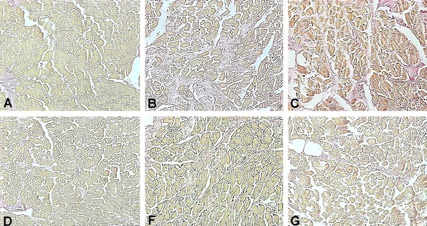
Fig. 4. Optical microscopy, Hematoxylin-Eosin (40X): Each plaque corresponding to a different extraocular muscle: inferior oblique (a),
superior oblique (b), inferior rectus (c), lateral rectus (d), medial rectus (e) and superior rectus (f).
DISCUSSION
The stereological method allows the study of three- et al., 2010; Mühlfeld & Ochs, 2013; Mandarim-de-Lacerda
dimensional biological structures by analyzing two-dimen- & del Sol, 2017). The present study describes the normal
sional histological sections, giving as result reliable numeric histology of the extraocular muscles using stereology and
parameters. This technique is being used more in a wide range specifically determines the proportion of muscle and collagen
of biomedical fields and is considered the gold standard in tissue and quantifies the number of muscle fibers in 5,000
quantitative morphometry (Howard & Reed, 2005; Mühlfeld µm2. To our best knowledge, there are no previously published
509SAN MARTÍN, J.; LUNA, C.; GARRETÓN, R.; ARANEDA , S.; SALGADO, C.; RODRÍGUEZ, A. & SALGADO, G. Stereological quantification of extraocular muscles in humans.
Int. J. Morphol., 39(2):506-511, 2021.
Fig. 5. Optical microscopy, Van Gieson (40X): Each plaque corresponding to a different extraocular muscle: inferior oblique (a), superior
oblique (b), inferior rectus (c), lateral rectus (d), medial rectus (e) and superior rectus (f).
studies in which these variables are described in humans, using The present study has certain limitations among which
stereology. are the limited number of individuals analyzed, representing
only a specific age range and ethnicity. Additionally, we only
Regarding our results, it is interesting to mention how described three histological parameters and we do not
the histological parameters we studied have a morphological distinguish between muscle fiber types and collagen subtypes.
– functional correlation. The horizontal rectus (medial and
lateral) showed higher proportion of muscular volume density
(Vv) when compared to cyclovertical muscles (vertical rectus CONCLUSION
and oblique muscles). This is somewhat predictable because
lateral versions are the most frequently executed eye
movements. The medial rectus showed the highest muscle This study establishes normal stereological parameters
Vv, a result that is also expected because it is involved in in extraocular muscles of humans without ophthalmological
both convergence and lateral movements. Nonetheless, those disease. This data will allow future comparisons if similar
differences did not achieve statistical significance. Certain studies are performed. It would be particularly interesting to
level of similarity is expected between the extraocular muscles see if there is any difference in these stereological parameters
considering that the oculomotor system is highly balanced, in the extraocular muscles of strabismic humans.
in which yoke (synergistic) and antagonist muscles have been
clinically described (Hering et al., 1977; Davis et al.;
Bruenech & Haugen, 2015). SAN MARTÍN, J.; LUNA, C.; GARRETÓN, R.; ARANEDA,
S.; SALGADO, C.; RODRÍGUEZ, A. & SALGADO, G.
Unfortunately, there are no published stereological Cuantificación estereológica de músculos extraoculares en huma-
studies regarding extraocular muscles histology, in both nos. Int. J. Morphol., 39(2):506-511, 2021.
animals and humans. Several studies have employed
RESUMEN: El objetivo de este estudio es cuantificar el
stereology for the description of limbs muscle fibers in volumen de tejido muscular y conectivo de los músculos
humans, but they all analyze ultrastructural features, such as extraoculares en humanos sin enfermedad oftalmológica conocida
mitochondrial volume density (Broskey et al., 2013; Picard utilizando estereología. Los músculos extraoculares fueron obteni-
et al., 2013). Thus, there is no available data for our results to dos de cinco cadáveres humanos sin estrabismo. El número de fi-
be compared. bras musculares en 5.000 µm2 y la densidad de volumen (Vv) de
510SAN MARTÍN, J.; LUNA, C.; GARRETÓN, R.; ARANEDA , S.; SALGADO, C.; RODRÍGUEZ, A. & SALGADO, G. Stereological quantification of extraocular muscles in humans.
Int. J. Morphol., 39(2):506-511, 2021.
músculo y colágeno fueron medidas usando estereología. Las com- Mühlfeld, C. & Ochs, M. Quantitative microscopy of the lung: a problem-
paraciones entre músculos extraoculares antagonistas se realizaron based approach. Part 2: stereological parameters and study designs in
a través de la prueba de los rangos con signo de Wilcoxon para various diseases of the respiratory tract. Am. J. Physiol. Lung Cell. Mol.
Physiol., 305(3):L205-21, 2013.
muestras pareadas. Un análisis secundario examinando diferencias
Mühlfeld, C.; Nyengaard, J. R. & Mayhew, T. M. A review of state-of-the-
entre pares de músculos extraoculares también fue llevado a cabo. art stereology for better quantitative 3D morphology in cardiac research.
Se realizaron pruebas bilaterales y la significancia fue fijada en 0,05. Cardiovasc. Pathol., 19(2):65-82, 2010.
Los músculos rectos horizontales (recto medial y lateral) tuvieron Picard, M.; White, K. & Turnbull, D. M. Mitochondrial morphology, topology,
el mayor Vv de músculo y el menor Vv de colágeno. El músculo and membrane interactions in skeletal muscle: a quantitative three-di-
recto inferior tuvo la tendencia a poseer menos número de fibras por mensional electron microscopy study. J. Appl. Physiol. (1985),
5.000 µm2 que el resto de los músculos extraoculares. No obstante, 114(2):161-71, 2013.
estas diferencias no llegaron a ser estadísticamente significativas. Porter, J. D.; Baker, R. S.; Ragusa, R. J. & Brueckner, J. K. Extraocular
muscles: basic and clinical aspects of structure and function. Surv.
Este es el primer estudio publicado describiendo la histología nor-
Ophthalmol., 39(6):451-84, 1995.
mal de los músculos extraoculares usando estereología. Nuestra in- Schachat, F. & Briggs, M. M. Phylogenetic implications of the superfast
vestigación, a través de la cuantificación de la proporción de tejido myosin in extraocular muscles. J. Exp. Biol., 205(Pt. 15):2189-201, 2002.
de músculo y colágeno, así como el número de fibras musculares en Schiavi, C. Extraocular muscles tension, tonus, and proprioception in infantile
5.000 µm2, establece parámetros estereológicos normales para mús- strabismus: role of the oculomotor system in the pathogenesis of infantile
culos extraoculares en humanos sin enfermedad oftalmológica. strabismus-Review of the literature. Scientifica (Cairo), 2016:5790981,
2016.
PALABRAS CLAVE: Músculos oculomotores; Huma- Spencer, R. F. & Porter, J. D. Biological organization of the extraocular
muscles. Prog. Brain Res., 151:43-80, 2006.
nos; Histología; Anatomía. Stager Jr., D.; McLoon, L. K. & Felius, J. Postulating a role for connective
tissue elements in inferior oblique muscle overaction (an American
Ophthalmological Society thesis). Trans. Am. Ophthalmol. Soc., 111:119-
REFERENCES 32, 2013.
Wooten, G. F. & Reis, D. J. Blood flow in extraocular muscle of cat. Arch.
Neurol., 26(4):350-2, 1972.
Yu Wai Man, C. Y.; Chinnery, P. F. & Griffiths, P. G. Extraocular muscles
Agarwal, A. B.; Feng, C. Y.; Altick, A. L.; Quilici, D. R.; Wen, D.; Alan, L. have fundamentally distinct properties that make them selectively vul-
A. & von Bartheld, C. S. Altered protein composition and gene expression nerable to certain disorders. Neuromuscul. Disord., 15(1):17-23, 2005.
in strabismic human extraocular muscles and tendons. Investig.
Ophthalmol. Vis. Sci., 57(13):5576-85, 2016.
Briggs, M. M. & Schachat, F. The superfast extraocular myosin (MYH13) is
localized to the innervation zone in both the global and orbital layers of Corresponding author:
rabbit extraocular muscle. J. Exp. Biol., 205(Pt. 20):3133-42, 2002. Dr. Guillermo Salgado
Broskey, N. T.; Daraspe, J.; Humbel, B. M. & Amati, F. Skeletal muscle Estudiante de Doctorado
mitochondrial and lipid droplet content assessed with standardized grid Departamento de Ciencias Morfológicas
sizes for stereology. J. Appl. Physiol. (1985), 115(5):765-70, 2013.
Escuela de Medicina
Bruenech, J. R. & Haugen, I. B. K. How does the structure of extraocular
muscles and their nerves affect their function? Eye (Lond.), 29(2):177-
Universidad Autónoma de Barcelona
83, 2015. Los Militares 4777
Büttner-Ennever, J. A. Anatomy of the oculomotor system. Dev. Ophthalmol., Torre 1, Piso 8
40:1-14, 2007. Las Condes, Santiago
Carry, M. R.; Ringel, S. P. & Starcevich, J. M. Mitochondrial morphometrics CHILE
of histochemically identified human extraocular muscle fibers. Anat. Rec.,
214(1):8-16, 1986.
Davis, G.; McNeer, K. W. & Spencer, R. F. Myectomy of the inferior oblique
E-mail: gsalgadoalarcon@gmail.com
muscle. Arch. Ophthalmol., 104(6):855-8, 1986.
Fuchs, A. F. & Binder, M. D. Fatigue resistance of human extraocular muscles.
J. Neurophysiol., 49(1):28-34, 1983.
Hering, E.; Bridgeman, B. & Stark, L. The Theory of Binocular Vision. New Received: 18-12-2020
York, Plenumm Press, 1977. Accepted: 13-01-2021
Hoh, J. F. Y.; Hughes, S.; Hugh, G. & Pozgaj, I. Three Hierarchies in Skeletal
Muscle Fiber Classification: Allotype, Isotype and Phenotype. In Kedes,
L. H. & Stockdale, F. E. (Eds.). Cellular and Molecular Biology of Muscle
Development. New York, Alan R. Liss, 1989.
Howard, C. V. & Reed, M. G. Unbiased Stereology: Three-Dimensional
Measurement in Microscopy. 2nd ed. New York, BIOS Scientific
Publishers, 2005.
Mandarim-de-Lacerda, C. A. & del Sol, M. Tips for studies with quantitative
morphology (morphometry and stereology). Int. J. Morphol., 35(4):1482-
94, 2017.
McNeer, K. W.; Tucker, M. G. & Spencer, R. F. Management of essential
infantile esotropia with botulinum toxin A: review and recommendations.
J. Pediatr. Ophthalmol. Strabismus, 37(2):63-7, 2000.
511You can also read