Applied anatomy and clinical significance of the maxillofacial and mandibular regions of the barking deer (Muntiacus muntjak) and sambar deer ...
←
→
Page content transcription
If your browser does not render page correctly, please read the page content below
Folia Morphol.
                                                                                                                  Vol. 80, No. 1, pp. 170–176
                                                                                                                DOI: 10.5603/FM.a2020.0061
                                                    ORIGINAL              ARTICLE                               Copyright © 2021 Via Medica
                                                                                                                             ISSN 0015–5659
                                                                                                                            eISSN 1644–3284
                                                                                                                         journals.viamedica.pl

Applied anatomy and clinical significance
of the maxillofacial and mandibular regions
of the barking deer (Muntiacus muntjak)
and sambar deer (Rusa unicolor)
K. Keneisenuo1, O.P. Choudhary1 , P. Priyanka2 , P.C. Kalita1, A. Kalita1, P.J. Doley1,
J.K. Chaudhary3
1
 Department of Veterinary Anatomy and Histology, College of Veterinary Sciences and Animal Husbandry,
Central Agricultural University (I), Selesih, Aizawl, Mizoram, India
2
 Department of Veterinary Microbiology, College of Veterinary Sciences and Animal Husbandry,
Central Agricultural University (I), Jalukie, Peren, Nagaland, India
3
 Department of Animal Genetics and Breeding, College of Veterinary Sciences and Animal Husbandry,
Central Agricultural University (I), Selesih, Aizawl, Mizoram, India

[Received: 15 January 2020; Accepted: 20 February 2020]

                                           Background: There is no previously reported information on the applied anatomy
                                           and clinical significance of the maxillofacial and mandibular regions of the barking
                                           deer and sambar deer.
                                           Materials and methods: Therefore, the present study was designed to provide some
                                           important clinical landmarks related to tracking of the infraorbital, mental and man-
                                           dibular nerves with its clinical implications in regional anaesthesia in both the species.
                                           Results: In the present study, the distance between the most lateral bulging of the
                                           facial tuberosity to the infraorbital foramen and from the latter to the root of the
                                           alveolar tooth directly ventral to it was found to be 2.65 ± 0.01 cm and 0.90 ±
                                           ± 0.02 cm in males; 2.75 ± 0.01 cm, 1.11 ± 0.01 cm in females of barking deer and
                                           4.57 ± 0.01 cm and 1.83 ± 0.02 cm in males; 4.52 ± 0.02 cm and 1.76 ± 0.02 cm in
                                           females of sambar deer. The infraorbital foramen was small, elliptical and was located
                                           at the level of first superior premolar teeth in barking deer and sambar deer. The
                                           facial tuberosity was located above the third superior premolar teeth in the barking
                                           deer but was located at the level of the first superior molar teeth in sambar deer.
                                           The distance between the lateral alveolar root of the third inferior incisor tooth to
                                           the mental foramen was 2.84 ± 0.01 cm in males, 2.78 ± 0.01 cm in females of
                                           barking deer and 3.04 ± 0.02 cm in males, 2.96 ± 0.01 cm in females of sambar
                                           deer which is an important landmark for achieving the location of the mental foramen
                                           nerve for the regional nerve block in both the species. The mandible of both the
                                           species showed oval-shaped mental foramen with unossified mandibular symphysis.
                                           Conclusions: The present study revealed that most of the parameters showed
                                           a statistically significant difference between the sexes in barking deer and sambar
                                           deer; however, from the practical point of view, these differences were meager.
                                           The results were discussed with regard to their clinical applications in various
                                           regional anaesthesia performed in maxillofacial and mandibular regions of both
                                           the species. (Folia Morphol 2021; 80, 1: 170–176)
                                           Key words: barking deer, sambar deer, infraorbital, mental, mandibular,
                                           nerve, regional anaesthesia

Address for correspondence: Dr. O.P. Choudhary, Department of Veterinary Anatomy and Histology, College of Veterinary Sciences and Animal
Husbandry, Central Agricultural University (I), Selesih, Aizawl-796015, Mizoram, India, tel: +919928099090, e-mail: dr.om.choudhary@gmail.com

                                                                    170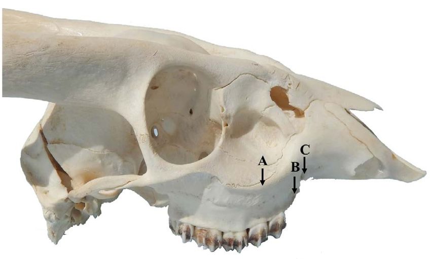
K. Keneisenuo et al., Applied anatomy of the head region of barking deer and sambar deer
INTRODUCTION to block the infraorbital and mental nerve. Knowing
The barking deer (Muntiacus muntjak) is a cervid the topographic and morphometric anatomy of the
species deer native to South and Southeast Asia that infraorbital and mental foramen provides simplicity
has been listed as ‘least concern’ on the International in veterinary surgery during an emergency [12, 27].
Union for the Conservation of Nature (IUCN) red list There is no previously reported information on
[32]. They are relatively small tropical deer that have the applied anatomy and clinical significance of the
a solitary lifestyle [21] and wide natural distribution, maxillofacial and mandibular region of barking deer
ranging throughout the large parts of South-east Asia and sambar deer. Therefore, the present study has been
[24]. The males have short antlers protruding from carried out to provide information on some clinically
long body hair-covered pedicles above the eyes, while important parameters and landmarks on the maxillo-
females have small bony knobs. Males have preorbital facial and mandibular region in both the species. Thus,
glands which are larger than in females [2] and they the results shown in this study will aid the clinicians
use these glands to mark the ground [1, 3]. in the implication of regional anaesthesia in the max-
The sambar deer (Rusa unicolor) is a large deer of illofacial and mandibular regions in both the species.
cervid species native to the Indian subcontinent, South-
ern China and Southeast Asia, that has been listed as MATERIALS AND METHODS
‘vulnerable’ by the IUCN red list since 2008 due to The study was conducted on the maxillofacial and
decrease in their population year by year [20, 33]. The mandibular regions of eight adult barking deer and
males have rugged antlers having simple brow tines sambar deer of either sex. The skull samples were
along with forked beams at its tip and are dropped an- collected from the Aizawl Zoological park, Aizawl, Mi-
nually. The barking and sambar deer are protected under zoram after taking official permission from the Prin-
Schedule III of the Indian Wildlife Protection Act, 1972. cipal Chief Conservator of Forest and Chief Wildlife
The regional anatomy is the important branch of Warden, Government of Mizoram, Aizawl vide letter
anatomy that deals with the form and relationships no. A. 33011/5/2017-CWLW/91 dated 15.03.2019.
of various anatomic structures present in a specific After collection, the samples were processed by the
area. It is one of the major foundations of clinical and hot water maceration technique. The present study
surgical practice as it enables the clinician/surgeon was carried out in the Department of Veterinary Anat-
to visualise the details of the structures relevant to omy and Histology, College of Veterinary Sciences
the case at hand [12]. The knowledge of the regional and Animal Husbandry, Selesih, Aizawl, Mizoram and
anatomy of the head is crucial due to the presence Interpretation Centre, Aizawl Zoological Park, Aizawl,
of the vital organs and structures such as the brain, Mizoram. The radiograph of the mandible was car-
tongue, eye, ears, nose, lips, horn and skull. Due to ried out by the Siemens X-Ray machine (500 mA) at
the presence of these structures, the function of the Diagnostic Division Radiology and Imaging, Mizoram
head is to coordinate the body, deglutition, olfaction Health Care, Aizawl, Mizoram.
and defence [12]. Numerous investigations have been Altogether a total of eleven measurements were
done on the regional anatomy of the head region of taken in the upper jaw and mandibles of both the
the domestic and wild animals including ox, horse, species by using digital vernier calliper (Resolution
sheep, goat, dog, pig and camel [4–6, 12, 15, 26, 28]. 0.01 mm or 0.0005 inches: Accuracy ± 0.03 mm)
The relationship and forms of all organs that are and the results were expressed as mean ± standard
located in a particular area are directly concerned deviation (SD). The parameters taken in maxillofacial
with regional anatomy helping the clinician as well as and mandibular regions are delineated below and
surgeon to visualise details of the structures relevant revealed in Figures 1–3.
to the case at hand and form one of the important A. Facial tuberosity to the infraorbital foramen:
foundations for clinical and surgical practice [12]. It It was measured from the level of most lateral
has been reported previously that the infraorbital bulging of facial tuberosity to mid-level of the
nerve and mental nerve pass from the infraorbital infraorbital foramen.
foramen and mental foramen, respectively [13, 14]. B. Infraorbital foramen to root of alveolar tooth: It
In an emergency situation that requires surgical inter- was measured from the mid-level of the infraor-
vention, it is very easy to locate this region as a topo bital foramen to the alveolar root of the superior
graphical landmark for quick and easy anaesthesia first premolar tooth.
171
Folia Morphol., 2021, Vol. 80, No. 1
C. Lateral alveolar root to mental foramen: It was
measured from the lateral extent of the alveolar
root of the third inferior incisor tooth to the
mental foramen.
D. Mental foramen to the caudal mandibular border:
It was measured from the level of the mental
foramen to caudal border of the ramus of the
mandible.
E. Mandibular length: It was measured from the
level of the rostral extremity of the alveolar root
of the central inferior incisor tooth to the trans-
Figure 1. Lateral view of the skull of sambar deer showing facial versal plane at the level of the caudal border of
tuberosity to infraorbital foramen (A) and infraorbital foramen to
root of the superior first premolar tooth (B). the mandible.
F. Mandibular foramen to the horizontal plane at
the level of the ventral margin of the mandible:
It was measured from the ventral limit of the
mandibular foramen to the horizontal plane at
the level of the ventral margin of the mandible.
G. Caudal border of the mandible to below mandib-
ular foramen: It was measured from the caudal
most border of the mandible to the vertical line
produced by a description of measurement of
mandibular foramen to the ventral margin of
the mandible.
H. Condylar process to the height of mandible: It
was measured from the condylar process to the
maximum height of the mandible.
I. Maximum mandibular height: It was measured
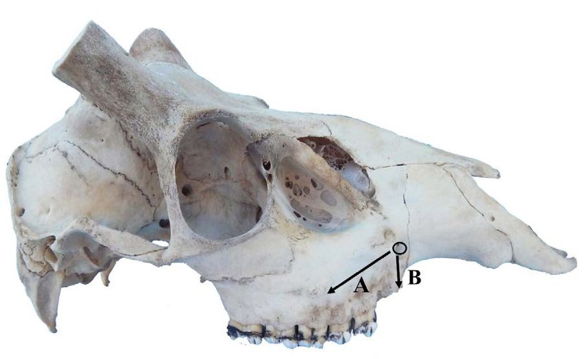
Figure 2. Lateral view of the mandible of sambar deer showing
from the highest level of the coronoid process
measurements; lateral alveolar root of inferior third incisor tooth
to mental foramen (C), mental foramen to the caudal mandibular perpendicularly to the ventral mandibular margin
border (D) and mandibular length (E). of the mandible.
J. Condylar process to the ventral margin of the
mandible.
K. Mandibular angle to mandibular foramen: It was
measured from the extreme caudal border of
angle of the mandible to mandibular foramen.
Statistical analysis
All the above parameters of the maxillofacial
and mandibular regions of barking deer and sambar
deer were obtained. All the measurements obtained
were analysed by routine statistical analysis [30]
and Student “t” test by the Statistical Package
for the Social Sciences (IBM, SPSS, 20.0 version)
programme.
Figure 3. Medial view of mandible of sambar deer showing meas-
urements; mandibular foramen to the horizontal plane at the level of
the ventral margin of the mandible (F), below mandibular foramen RESULTS
to caudal mandibular border (G), maximum height of mandible to The infraorbital foramen, mandibular foramen and
the condylar process (H), maximum mandibular height (I), condylar
process to the ventral margin of the mandible (J) and mandibular mental foramen on the medial and lateral surface of
foramen to the border of mandibular angle (K). the maxillofacial and mandibular regions are shown
172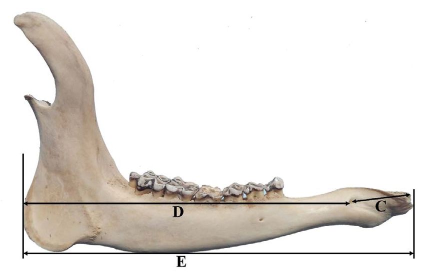
K. Keneisenuo et al., Applied anatomy of the head region of barking deer and sambar deer

Figure 4. Lateral view of the skull of barking deer showing facial                                Figure 5. Mediolateral radiographical view of the mandible of bark-
tuberosity (A); root of the superior first premolar tooth (B) and                                 ing deer showing mandibular foramen (A), mandibular foramen (B),
infraorbital foramen (C).                                                                         mental foramen (C) and root of the lateral alveolar tooth (D).

Table 1. The measurements of the maxillofacial and mandibular region in barking deer and sambar deer in centimetres
(mean ± standard deviation)
 Parameters#                                      Barking deer                                   P                               Sambar deer                      P
                                        Male                        Female                                             Male                           Female
 A                                  2.65 ± 0.01                  2.75 ± 0.01                < 0.01**               4.57 ± 0.01                  4.52 ± 0.02    < 0.05*
 B                                  0.90 ± 0.02                  1.11 ± 0.01                < 0.01**               1.83 ± 0.02                  1.76 ± 0.02    < 0.01**
 C                                  2.84 ± 0.01                  2.78 ± 0.01                < 0.01**               3.04 ± 0.02                  2.96 ± 0.01    < 0.01**
 D                                 12.87 ± 0.01                 14.46 ± 0.02                < 0.01**              23.66 ± 0.01                  23.59 ± 0.01   < 0.01**
 E                                 15.77 ± 0.01                 17.27 ± 0.02                < 0.01**              28.10 ± 0.01                  27.96 ± 0.01   < 0.01**
 F                                  2.49 ± 0.01                  2.39 ± 0.02                < 0.01**               5.17 ± 0.01                  5.10 ± 0.02    < 0.01**
 G                                  2.15 ± 0.01                  2.09 ± 0.01                < 0.01**               3.80 ± 0.01                  3.71 ± 0.01    < 0.01**
 H                                  2.89 ± 0.01                  2.79 ± 0.01                < 0.01**               5.29 ± 0.01                  5.21 ± 0.01    < 0.01**
 I                                  7.88 ± 0.01                  7.78 ± 0.02                < 0.01**              15.96 ± 0.01                  15.78 ± 0.02   < 0.01**
 J                                  4.89 ± 0.01                  4.39 ± 0.01                < 0.01**              10.60 ± 0.01                  10.50 ± 0.01   < 0.01**
 K                                  2.49 ± 0.03                  2.39 ± 0.02                < 0.01**               5.16 ± 0.02                  5.11 ± 0.01    < 0.01**

#As mentioned in alphabetical order in materials and method. Level of significance at 5%; *Significant (p < 0.05); **Highly significant (p < 0.01).

in Figures 4, 5. The measurement points taken to de-                                                 The present study revealed that all the obtained
termine the topographic and morphometric location                                                 parameters differed statistically significantly (p < 0.01
of the mental foramen are shown in Figures 2, 3.                                                  and p < 0.05) between the males and females of
In the present study, the infraorbital foramen was                                                barking deer and sambar deer respectively; however,
small, elliptical and was located at the level of first                                           from the practical point of view, these differences
superior premolar tooth in barking deer and sambar                                                were meager.
deer (Figs. 1, 4). The alveolar border of mandible
presented 6 alveoli for the 3 superior premolar and                                                                              DISCUSSION
3 superior molar teeth. The facial tuberosity was                                                     In the present study, facial tuberosity of barking
located dorsally to the third superior premolar tooth                                             deer was located above the third superior premolar
in barking deer but was at the first superior molar                                               tooth which has also been reported in ox [13], chital
tooth in sambar deer. The preorbital fossa was wide                                               [18] and blackbuck [9], while it was located at the
and deep in both the species with upper canine teeth                                              level of first superior molar tooth in sambar deer.
on the maxilla. The mandible of both the species                                                  However, the facial tuberosity of the Madras Red
showed oval mental foramen with unossified man-                                                   sheep was prominent and placed at the level of 5th
dibular symphysis. The results of the measurements                                                cheek tooth (second superior molar tooth) [31]. The
are listed in Table 1.                                                                            distance between the most lateral bulging of the

                                                                                          173
Folia Morphol., 2021, Vol. 80, No. 1
facial tuberosity to the infraorbital foramen and from foramen. The mental nerve block is useful for desen-
the latter to the root of the alveolar tooth directly ven- sitizing the lower lip during its surgical interventions.
tral to it was 2.65 ± 0.01 cm and 0.90 ± 0.02 cm in The distance from the mental foramen to caudal
males; 2.75 ± 0.01 cm, 1.11 ± 0.01 cm in females of border of the ramus of the mandible was 12.87 ±
barking deer and 4.57 ± 0.01 cm and 1.83 ± 0.02 cm ± 0.01 cm in males, 14.46 ± 0.02 cm in females of
in males; 4.52 ± 0.02 cm and 1.76 ± 0.02 cm in fe- barking deer and 23.66 ± 0.01 cm in males, 23.59 ±
males of sambar deer (Table 1). However, the same pa- ± 0.01 cm in females of sambar deer, while the same
rameter was reported as 1.6–1.8 cm and 1.3–1.6 cm parameters were 13.43 ± 0.08 cm in blackbuck [8];
in West African Dwarf goats [26]; 2.06 ± 0.14 cm 11.69 ± 0.40 cm in black Bengal goat [34]; 13.74 ±
and 1.13 ± 0.11 cm in Gwembe Valley dwarf goat ± 0.18 cm in Mehraban sheep [16]; 9.26 ± 0.49 cm
[17]; 2.8 cm and 2.5 cm in Iranian native cattle [23]; in Gwembe Valley Dwarf goat [17]; 32.12 ± 0.16 in
1.85 ± 0.14 cm and 1.75 ± 0.19 cm in black Bengal dromedary camel [5]; 15.23 ± 1.46 cm in Barbados
goat [34] and 2.37 ± 0.00 cm and 0.72 ± 0.00 cm black belly sheep [22]; 11.8 ± 0.89 cm in black Bengal
in blackbuck [10]. The infraorbital nerve block can be goat [29], 12.38 ± 1.52 cm in Abaza goats [11] and
achieved extraorally by injecting anaesthetic drugs 18.47 ± 0.01 cm in local pig of Mizoram [6].
approximately 1 cm in barking deer and 1.8 cm in The length and height of the mandible was
sambar deer above the root of the first superior pre- 15.77 ± 0.01 cm, 7.88 ± 0.01 cm in males and 17.27 ±
molar tooth in the infraorbital foramen. The infraor- ± 0.02 cm, 7.78 ± 0.02 cm in females of barking
bital nerve block is used in the surgical interventions deer; and 28.10 ± 0.01, 15.96 ± 0.01 cm in males and
related to the upper lip, nose and skin supplied by 27.96 ± 0.01 cm, 15.78 ± 0.02 cm in females of sambar
the infraorbital nerve. deer, respectively. The same mandibular parameters
The above-recorded parameters were of clinical were 12.00 ± 0.89 cm, 6.90 ± 1.09 cm for West Afri-
importance because the facial tuberosity is remark- can Dwarf goats [26]; 11.24 ± 0.52 cm, 6.64 ± 0.44 cm
able even in live animals providing a clear guide for in Gwembe Valley Dwarf goat [17]; 16.53 ± 0.12 cm,
tracking the infraorbital nerve and its desensitisation 10.69 ± 0.02 cm in blackbuck [7]; 42.98 ± 0.62 cm,
during the manipulations in the skin of the upper lip, 22.58 ± 0.28 cm in dromedary camel [5] and 25.02 ±
nostril and face at the level of the foramen. The in- ± 0.09, 10.54 ± 0.07 cm in local pig of Mizoram [6].
fraorbital foramen was small, elliptical and located at The distance between the condylar process to
the level of the first superior premolar tooth in bark- the height of the mandible, condylar process to the
ing deer and sambar deer which was also reported ventral margin of the mandible was 2.89 ± 0.01 cm,
in chital [18] and blackbuck [9]. However, the same 4.89 ± 0.01 cm in males and 2.79 ± 0.01 cm, 4.39 ±
foramen was located dorsal to the second premolar ± 0.01 cm in females of barking deer; and 5.29 ±
in red Sokoto (Maradi) goats [25]. ± 0.01 cm, 10.60 ± 0.01 cm in males and 5.21 ±
The distance between the lateral alveolar root of ± 0.01 cm, 10.50 ± 0.01 cm in females of sambar deer.
the third inferior incisor tooth to the mental foramen However, the same parameter has been reported to
was 2.84 ± 0.01 cm in males, 2.78 ± 0.01 cm in fe- be 3.09 ± 0.00 cm, 7.57 ± 0.02 cm in blackbuck [7].
males of barking deer and 3.04 ± 0.02 cm in males, The distance between the vertical line drawn
2.96 ± 0.01 cm in females of sambar deer (Fig. 2, downward from the caudal border of the mandible
Table 1) which is an important landmark for achiev- (I) and the vertical line drawn from the mandibular
ing the location of the mental foramen nerve for the foramen downwards (F) was (G) 2.15 ± 0.01 cm in
regional nerve block in both the species, whereas it males, 2.09 ± 0.01 cm in females of barking deer and
was 1.6 ± 0.22 cm in West African Dwarf goat [26]; 3.80 ± 0.01 cm in males, 3.71 ± 0.01 cm in females
2.0 ± 0.3 cm in red Sokoto (Maradi) goats [25]; of sambar deer (Fig. 3). However, the same parameter
2.45 ± 0.00 cm in blackbuck [8]; 9.22 ± 0.05 cm was observed as 1.85 ± 0.01 cm in blackbuck [7].
in dromedary camel [5] and 3.57 ± 0.04 cm in local The mandibular nerve block is used to anesthetise
pig of Mizoram [6]. The mental nerve block can be the mandibular nerve during the clinical examinations
achieved extraorally by injecting anaesthetic drugs and surgical procedures involving the alveoli and
approximately 2.8 cm in barking deer and 3 cm in of teeth of the lower jaw in animals [19]. The distances
sambar deer from the lateral extent of the alveolar from the mandibular foramen to the ventral margin
root of inferior third incisor tooth into the mental of the mandible, caudal border of mandible to the
174K. Keneisenuo et al., Applied anatomy of the head region of barking deer and sambar deer
level of mandibular foramen, mandibular foramen to REFERENCES
the border of mandibular angle were 2.49 ± 0.01 cm, 1. Adnyane IKM, Zuki ABZ, Noordin MM, et al. Morpholog-
2.15 ± 0.01 cm, 2.49 ± 0.03 cm in males; 2.39 ± ical study of the infraorbital gland of the male barking
deer, muntiacus muntjak. Afr J Biotech. 2011; 10(77), doi:
± 0.02 cm, 2.09 ± 0.01 cm, 2.39 ± 0.02 cm in females
10.5897/ajb10.2634.
of barking deer and 5.17 ± 0.01 cm, 3.80 ± 0.01 cm, 2. Barrette C. Musculature of facial scent glands in the
5.16 ± 0.02 cm in males; 5.10 ± 0.02 cm, 3.71 ± 0.0 cm, muntjac. J Anat. 1976; 122(Pt 1): 61–66, indexed in Pu-
5.11 ± 0.01 cm in females of sambar deer (Fig. 3). bmed: 977477.
3. Barrette C. Social behavior of muntjac. Ph.D. Thesis
Whereas, the same parameters were recorded as
submitted to the University of Calgary, Calgary, Alberta,
4.18 ± 0.01 cm, 1.36 ± 0.01 cm, 3.07 ± 0.00 cm Canada. 1975.
in blackbuck [8]; 8.84 ± 0.08 cm, 5.88 ± 0.05 cm, 4. Choudhary O, Kalita P, Doley P, et al. Applied anatomy of
8.29 ± 0.07 cm in dromedary camel [5] and 4.56 ± the head region of the indian wild pig (sus scrofa) and
± 0.01 cm, 3.81 ± 0.00 cm, 4.84 ± 0.00 cm in local its clinical value during regional anesthesia. J Anim Res.
2017; 7(2): 339, doi: 10.5958/2277-940x.2017.00049.3.
pig of Mizoram [6]. Equivalent figures for West Af-
5. Choudhary OP, Kalita PC, Kalita A, et al. Applied anatomy
rican dwarf goats of Nigeria were 1.57 ± 0.44 cm, of the maxillofacial and mandibular regions of the drom-
2.58 ± 0.34 cm for the caudal border of the mandible edary camel (Camelus dromedarius). J Camel Prac Res.
to below mandibular foramen and the mandibular 2016; 23(1): 127, doi: 10.5958/2277-8934.2016.00021.7.
6. Choudhary O, Kalita P, Konwar B, et al. Morphological and
foramen to the ventral margin of the mandible [26].
Applied Anatomical Studies on the Head Region of Local
In horse and dogs, the distance between the mandib- Mizo Pig (Zovawk) of Mizoram. Int J Morphol. 2019; 37(1):
ular foramen and the ventral margin of the mandible 196–204, doi: 10.4067/s0717-95022019000100196.
was 3 cm and 1.5 to 2 cm, respectively [15]. The 7. Choudhary O, Singh I, Bharti S, et al. Gross and Morpho-
mandibular nerve is useful during the treatment of metrical Studies on Mandible of Blackbuck (Antelope
cervicapra). Int J Morphol. 2015; 33(2): 428–432, doi:
the injuries related to the lower incisors and premolar
10.4067/s0717-95022015000200003.
tooth, i.e. dental extraction, tumours etc. An extraoral 8. Choudhary O, Singh I. Applied Anatomy of the Maxillo-
mandibular nerve block can be achieved by injecting facial and Mandibular Regions of the Indian Blackbuck
anaesthetic drugs approximately 2.5 cm in barking (Antilope cervicapra). J Anim Res. 2015; 5(3): 497, doi:
10.5958/2277-940x.2015.00085.6.
deer and 5.0 cm in sambar deer from the horizontal
9. Choudhary O, Singh I. Morphological and Radiographic
plane at the level of the ventral margin of the man- Studies on the Skull of Indian Blackbuck (Antilope cervi-
dible to the ventral limit of the mandibular foramen. capra). Int J Morphol. 2016; 34(2): 775–783, doi: 10.4067/
s0717-95022016000200055.
CONCLUSIONS 10. Choudhary O, Singh I. Morphometrical Studies on the
Skull of Indian Blackbuck (Antelope cervicapra). Int
It is concluded that the measurements obtained J Morphol. 2015; 33(3): 868–876, doi: 10.4067/s0717-
from the present study will be useful for the surgeons 95022015000300011.
to locate the site for infiltration of the anaesthetic 11. Dalga S. Topographic and morphometric study of the mental
drugs for the nerves of maxillofacial and mandibular foramina of Abaza goats with its clinical implication for re-
gional anesthesia. Folia Morphol. 2019 [Epub ahead of print],
regions and can aid the veterinary practitioners in
doi: 10.5603/FM.a2019.0122, indexed in Pubmed: 31750539.
treating the head injuries related to both the species. 12. Dyce KM, Sack WO, Wensing CJG. Textbook of Veterinary
Further, it will be very useful in the future endeavours Anatomy. 2nd edn. Elsevier, Philadelphia 1996.
involving applied research works leading towards the 13. Getty R. Sisson and Grossman’s The Anatomy of the
Domestic Animals, 2nd edn. Vol. I. W.B. Saunders Co.,
massive improvement in the livestock sector of the in-
Philadelphia 1975.
ternational economy as well as conservation of wildlife. 14. Ghosh RK. Primary Veterinary Anatomy, 5th edn. Current
books international, Kolkata, West Bengal, India 2012.
Acknowledgements 15. Hall LW, Clarke KW, Trim CM. Wright’s Veterinary Anesthe-
sia. 10th edn. ELBS and Baillierre Tindall, London 2000.
The authors are thankful to the Dean, College of
16. Karimi I, Onar V, Pazvant G, et al. The cranial morphometric
Veterinary Sciences and Animal Husbandry, Central and morphologic characteristics of Mehraban sheep in
Agricultural University (I), Aizawl, Mizoram; Principal western Iran. Global Vet. 2011; 6(2): 111–117.
Chief Conservator of Forest and Chief Wildlife Warden 17. Kataba A, Mwaanga ES, Simukoko H, et al. Clinical anat-
(PCCF and CWW), Government of Mizoram, Aizawl omy of the head Region of Gwembe Valley dwarf goat in
Zambia. Int J Vet Sci. 2014; 3(3): 142–146.
and Director, Aizawl Zoological Park, Aizawl, Mizoram
18. Kumawat R, Joshi S, Mathur R, et al. Gross morphological
for providing all the necessary facilities to carry out studies on mandible of Indian spotted deer (Axis axis).
the research work. Indian Vet J. 2014; 91(9): 105–107.
175Folia Morphol., 2021, Vol. 80, No. 1
19. Lahunta ADE, Habel RE. Applied veterinary anatomy. W.B. 27. Ommer PA, Harshan KR. Applied Anatomy of Domestic
Saunders Co., Philadelphia 1986. Animals, 1st edn. Jaypee brother’s medical publisher, New
20. Leslie D. Rusa unicolor (Artiodactyla: Cervidae). Mamma- Delhi, India 1995.
lian Species. 2011; 43: 1–30, doi: 10.1644/871.1. 28. Onar V, Ozcan S, Pazvant G. Skull typology of adult male
21. Long JL. Introduced mammals of the world: their history, Kangal dogs. Anat Histol Embryol. 2001; 30(1): 41–48,
distribution and influence. CSIRO Publishing, Collingwood doi: 10.1046/j.1439-0264.2001.00292.x, indexed in Pu-
Victoria, Australia 2003. bmed: 11284162.
22. Mohamed R, Drisco M, Mootoo N. Clinical anatomy of 29. Poddar S, Faruq AA, Dey T, et al. Topographic and morpho-
the skull of the Barbados black belly sheep in Trinidad. Int metr ic anatomy of mental foramen of black Bengal goat
J Curr Res Med Sci. 2016; 2(8): 8–19. (Capra hircus) in Bangladesh with its clinical implication
23. Monfared AL. Gross anatomical measurements of for regional anesthesia. Int J Zoo Anim Biol. 2018; 1(1),
the head region of the Iranian native cattle (Bos tau- doi: 10.23880/izab-16000102.
rus) and their clinical value for regional anesthesia. 30. Snedecor GW, Cochran WG. Statistical Methods. 8th edn.
Global Vet. 2013; 10(2): 219–222, doi: 10.5829/idosi. Iowa State University Press, Ames, Iowa, USA 1994.
gv.2013.10.2.724. 31. Sundaram V, Dharani P, Gnanadevi R, et al. Studies on clini-
24. Ohtaishi N, Gao Y. A review of the distribution of all cal anatomy of the maxillofacial and mandibular regions of
species of deer (Tragulidae, Moschidae and Cervidae) in the Madras Red sheep (Ovis aries) in India. Folia Morphol.
China. Mammal Review. 1990; 20(2-3): 125–144, doi: 2019; 78(2): 389–393, doi: 10.5603/FM.a2018.0098,
10.1111/j.1365-2907.1990.tb00108.x. indexed in Pubmed: 30371934.
25. Olopade JO, Onwuka SK. Osteometric studies of 32. Timmins RJ, Duckworth JW, Hedges S. Muntiacus muntjak.
the red Sokoto (Maradi) goats (Capra hircus): Im- The IUCN Red List of Threatened Species. International Union
plication for regional anaesthesia of the head. Int for Conservation of Nature and Natural Resources, IUCN 2016.
J Morphol. 2007; 25(2): 407–410, doi: 10.4067/S0717- 33. Timmins RJ, Kawanishi K, Giman B, et al. Rusa unicolor.
95022007000200027. The red list of threatened species. IUCN 2015.
26. Olopade J, Onwuka S. Some Aspects of the Clinical Anat- 34. Uddin M, Ahmed S, Islam K, et al. Clinical anatomy of
omy of the Mandibular and Maxillofacial Regions of the the head region of the black bengal goat in bangla-
West African Dwarf Goat in Nigeria. Int J Morphol. 2005; desh. Int J Morphol. 2009; 27(4), doi: 10.4067/s0717-
23(1), doi: 10.4067/s0717-95022005000100006. 95022009000400048.
176You can also read