Collagen Quantification in Peri-implant Soft Tissues in Human Peri-Implantitis Lesions
←
→
Page content transcription
If your browser does not render page correctly, please read the page content below
Int. J. Morphol.,
39(3):683-687, 2021.

                       Collagen Quantification in Peri-implant Soft
                       Tissues in Human Peri-Implantitis Lesions

                           Cuantificación de Colágeno en Tejidos Blandos Periimplantarios
                                     en Lesiones de Periimplantitis en Humanos

                                Valentina Flores1,6; Bernardo Venegas2; Wendy Donoso2; Camilo Ulloa3;
                                        Alejandra Chaparro4; Vanessa Sousa5 & Víctor Beltrán6

FLORES, V.; VENEGAS, B.; DONOSO, W.; ULLOA, C.; CHAPARRO, A.; SOUSA, V. & BELTRÁN, V. Collagen quantification
in peri-implant soft tissues in human peri-implantitis lesions. Int. J. Morphol., 39(3):683-687, 2021.

        SUMMARY: Peri-implantitis is an inflammatory lesion of bacterial etiology characterized by inflammation of the mucosa and
bone loss. Chronic inflammation is characterized by neovascularization and collagen neoformation. Mast cells have been shown to
participate in the inflammatory process by releasing mediators and proteases such as chymase and tryptase, important in the collagen
neoformation process. Although a higher percentage of collagen has been described in periodontal disease, the literature is scarce about
the percentage of collagen in peri-implantitis. The aim of this study was to quantify the percentage of collagen fibers present in the peri-
implant soft tissue of patients with peri-implantitis lesions. A descriptive observational cross-sectional study was performed. Samples of
peri-implant soft tissue were collected from eleven patients with peri-implantitis and then processed by Masson's Trichrome Technique.
In microscopic analysis, collagen fibers were observed in all samples, with an average percentage of 39.89 %, standard deviation of
17.02 %, with a minimum value of 8.99 % and a maximum value of 75.65 % density. From these results, it can be concluded that in
human peri-implantitis lesions with bone loss greater than 50 %, there is an important percentage of collagen fibers, which is interpreted
as connective tissue in a permanent process of reparative response, in the presence of inflammatory infiltrate.

        KEY WORDS: Peri-implantitis; Inflammation; Collagen; Masson's Trichrome Technique.

INTRODUCTION

        Peri-implantitis (PI) is defined as the pathology                   studies that peri-implantitis lesions contain significantly
associated with bacterial plaque, in the tissues around den-                higher proportions of B cells and elastase positive cells
tal implants, characterized by inflammation in the peri-                    than mucositis lesions, which suggests that they exhibit
implant mucosa and the progressive loss of supporting bone                  properties different from mucositis but similar to
(Berglundh et al., 2018). Peri-implantitis is one of the                    periodontitis lesions (Gualini & Berglundh, 2003).
leading causes of failure and loss of dental implants. Its
prevalence at the subject level is 20 % and at the implant                          Mast cells have been shown to participate in the
level is 9.25 % (Lee et al., 2017).                                         inflammatory process by releasing mediators and proteases
                                                                            such as chymase and tryptase, which are important in the
       Peri-implantitis, as a chronic inflammatory lesion,                  collagen neoformation process (Zizzi et al., 2011).
is characterized by neovascularization and neoformation                     However, although a higher percentage of collagen has
of collagen. Lymphocytes and plasma cells are the most                      been described in periodontal disease, there is scarce
frequent inflammatory cells in peri-implantitis lesions. It                 literature that describes the percentage of collagen in peri-
has been possible to show through immunohistochemical                       implantitis.

1
  Programa de Magíster en Odontología, Universidad de La Frontera, Temuco, Chile.
2
  Department of Stomatology, Faculty of Health Sciences, Universidad de Talca, Talca, Chile.
3
  Department of Surgical Stomatology, Postgraduate Program in Periodontology, School of Dentistry, Universidad de Concepción, Concepción, Chile.
4
  Department of Periodontology, Faculty of Dentistry, Universidad de los Andes, Santiago, Chile.
5
  Centre for Clinical Oral Research, Centre for Oral Immunobiology and Regenerative Medicine, Barts and The London School of Medicine and Dentistry,
  Queen Mary University of London, London, UK.
6
  Clinical Investigation and Dental Innovation Center (CIDIC), Dental School and Center for Translational Medicine (CEMT - BIOREN), Universidad de
  La Frontera, Temuco, Chile.

                                                                                                                                             683
FLORES, V.; VENEGAS, B.; DONOSO, W.; ULLOA, C.; CHAPARRO, A.; SOUSA, V. & BELTRÁN, V. Collagen quantification in peri-implant soft tissues in human peri-implantitis lesions.
Int. J. Morphol., 39(3):683-687, 2021.
Knowing the histopathology of the peri-implant soft patients with peri-implantitis (six men and five women, 33
tissues in peri-implantitis lesions helps to understand the to 79 years old, function time of implants from five to sixty
pathogenesis of the inflammatory process and its consecutive months). The participants signed an informed consent during
fibrous reaction, in order to contribute to new therapies that the visit to the implant clinic of the Faculty of Dentistry,
prevent the early loss of the dental implant and its associated Universidad de La Frontera, Temuco, Chile and Universi-
consequences. dad de Concepción, Chile. The study was conducted in
accordance with the principles outlined in the Declaration
The aim of this study is to quantify the percentage of of Helsinky on experimentation with human participants.
collagen fibers present in the peri-implant soft tissue in Ethical approval for the study was granted by the Ethics
human peri-implantitis lesions. Committee of the Universidad de La Frontera, Report N °
024_2018. After written informed consent, the biopsies were
submitted to histopathological analysis at the Oral Pathology
MATERIAL AND METHOD Laboratory of Universidad de Talca. The parameters to de-
termine the diagnosis of peri-implantitis were based on the
classification proposed by Working Group 4 of the 2017
A descriptive study was conducted. Eleven peri-implant Global Workshop on the Classification of Peri-implant
soft tissue samples were obtained from the same number of Diseases and Conditions (Berglundh et al., 2018).
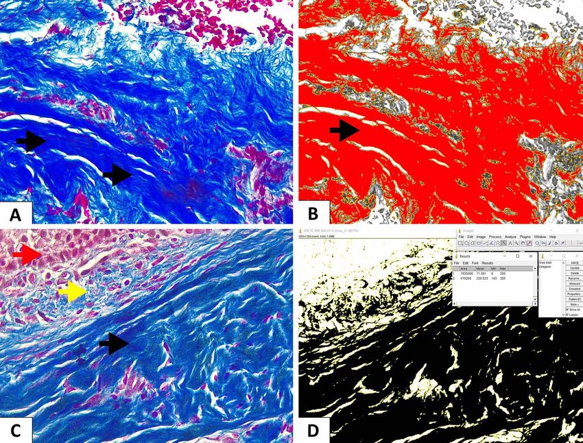
Fig. 1. A. Shows connective tissue in blue (Masson's trichrome technique 40x). Collagen fibers are marked with black arrows. B. Shows
the application of color deconvolution in Image J software to visualize selected collagen fibers (Red Option).C. Red arrow: Squamos
epithelium; Yellow arrow: scarce inflammatory cells in loose fibrous connective tissue; Black arrow: dense fibrous connective tissue
(Masson's trichrome technique 40x). D. Shows the selected areas to be quantified throw application of color deconvolution in Image J
software (B&W option) and summary table with the area results in square pixels.
684FLORES, V.; VENEGAS, B.; DONOSO, W.; ULLOA, C.; CHAPARRO, A.; SOUSA, V. & BELTRÁN, V. Collagen quantification in peri-implant soft tissues in human peri-implantitis lesions.
Int. J. Morphol., 39(3):683-687, 2021.
Inclusion criteria considered patients with RESULTS
osseointegrated implants with a function time equal to or
greater than five months, with a diagnosis of peri-
implantitis with bone loss greater than 50 % and significant Clinical Characteristics. Eleven peri-implant soft tissue
aesthetic compromise. The exclusion criteria were as samples from eleven patients diagnosed with peri-implantitis
follows: Children, adolescents, patients with autoimmune were analyzed. The mean age of the patients was 59 years,
diseases, and pregnant or lactating women. with a range of 33 to 79 years. Five patients were women
(55 %) and six men (45 %). Only two patients were smokers
Surgical Procedure. The soft tissue biopsies were obtained (less than 10 cigarettes a day) and five suffered from related
during the surgical peri-implantitis treatment. The biopsies chronic diseases, four were undergoing treatment for arterial
dimensions were approximately 2x3 mm. Each biopsy was hypertension (HA) and one patient for HA and Diabetes
immediately placed in 10 % buffered formalin for Mellitus (MD). Six patients did not report any chronic
histological analysis. disease. The functional time of implant (FTI) ranged from
five to sixty months.
Histological processing and analysis. All samples were
processed by Masson's trichromic technique (Fig. 1). From Histological Results. The lesions that were included in
the samples, 4 µm histological sections were obtained biopsies had similar histopathological characteristics to each
which were deparaffinized and hydrated in distilled water. other. In all peri-implant soft tissue samples with peri-
First, the samples were fixed in formaldehyde solution and implantitis lesions, collagen fibers were observed. The
then an assembly was carried out with Bouin's liquid for composition of the samples was not uniform with respect to
15 minutes at 56-60 ºC, cooled and washed in distilled collagen quantity. The values were measured in percentage
water to remove the yellow color. Weigert´s Iron with a minimum value of 8.99 %, and a maximum value of
hematoxylin staining was performed for 10 minutes and 75.65 % of the total area evaluated, with an average of 39.89
washed in distilled water for 5 minutes. Dyeing with acid % and a standard deviation of 17.02 % (Tables I and II).
scarlet fuchsin solution was carried out for 5 minutes and
washed with distilled water. A phosphomolybdic- Table I. Percentage value of collagen fibers per sample.
phosphotungstic acid solution treatment followed for 5 n= Minimum Maximum Average
minutes and then staining was carried out with aniline blue Value ( %) Value ( %) (%)
solution for 5 minutes. The slides were then treated with 1 1 19,57 49,24 33,53
% acetic acid for 2 minutes. After that, sections were 2 26,96 33,45 32,43
washed in distilled water, dehydrated, rinsed and mounted 3 11,31 35,99 30,38
on a slide. 4 10,49 65,67 53,82
5 11,67 27,18 23,07
The reading, interpretation of the results, and the 6 33,29 69,91 56,91
capture of images were carried out under optical 7 28,75 63,90 41,78
8 24,62 59,81 41,63
microscopy at 40X magnification with a camera (Canon
9 31,99 75,65 59,15
EOS Rebel XSI, Tokyo, Japan).
10 16,15 51,22 39,27
11 8,99 34,46 26,78
Quantification of collagen fibers. Five microscopic fields
per each sample were analyzed. The quantification of
Table II. Percentage value of collagen fibers of the total samples.
collagen fibers was carried out using the Image J software
(version 1.46j; National Institute of Health, USA) and the Mínimum Maximum Total Standard
Value (%) Value (%) Average (%) Deviation (%)
color deconvolution application. The image was
decomposed into 3 colors and a representative color area 8,99 75,65 39,89 17,02
was selected to be quantified. Once the imagen values are
adjusted, the “Red” option is changed to “B&W” option, DISCUSSION
and areas of interest areselected. Finally, the system
delivers a summary table with the area results in square
pixels, which will later allow to calculate the percentage Connective tissue constitutes a fundamental part in
of collagen. Collagen quantification was expressed as a every inflammatory disease. It is the place where cellular
percentage of the total area evaluated. The data were and vascular structures interact between them and induce
grouped to represent a mean value and standard deviation, the healing process, which is the final purpose of any
determining minimum and maximum values. inflammatory course. Therefore, determination of the amount
685FLORES, V.; VENEGAS, B.; DONOSO, W.; ULLOA, C.; CHAPARRO, A.; SOUSA, V. & BELTRÁN, V. Collagen quantification in peri-implant soft tissues in human peri-implantitis lesions.
Int. J. Morphol., 39(3):683-687, 2021.
of this tissue becomes of great importance in terms of According to a study carried out by Berglundh et al.
associating the histological structure with the dynamics of (2004), in which soft tissue biopsies from six patients with
the pathogenesis of disease. peri-implantitis were analyzed, it was shown that the
composition of the connective tissue was not uniform with
In chronic peri-implantitis, few reports on human respect to the densities of collagen, vascular and
studies have been found in the literature. Most of the research inflammatory structures in different compartments of the
is performed in animal studies using models with ligation- injury. The proportions of collagen and vascularization were
induced peri-implantitis which represents an acute process 3.6 % and 3.5 %, respectively.
(Lang et al., 1993; Comut et al., 2001; Martins et al., 2005;
Berglundh et al., 2007). Considering that peri-implantitis is In an immunohistochemical study that shows the
a chronic inflammatory disease, these studies could not be pattern of collagen distribution in healthy and peri-implant
comparable due to the differences in the pathogenesis mucosa, an important increase in collagen V was shown in
between an acute and a chronic process. Although peri-implantitis (Borsani et al., 2005). The authors propose
inflammatory process involves similar mechanisms, type of that this marked increase in collagen V could influence the
cells in one or another are not the same, and different homeostasis of gingival stroma and could permit greater
molecular interactions occur. Finally, considering a clinical bacterial penetration. Some authors refer to other
point of view, conclusions obtained in an experimental model investigation in which it is mentioned that Collagen type V
cannot always be extrapolated to a human process. Our study is resistant to collagenase digestion (Liotta et al., 1979) and
was based on the histological analysis of the amount of is found localized in inflamed gingival tissues. In our study,
collagen tissue directly on human soft tissue from peri- the collagen found could be type V collagen, which might
implant lesions, which allows a possible application in the be associated to the inflammatory process found, but more
clinical field. investigation needs to be performed.
There is scarce literature that evidences the It has been reported that when there is a permanent
percentage of collagen fibers in peri-implant soft tissue in association between pathogenic molecules, host tissue
human peri-implantitis. The composition of peri-implant soft destruction, mediators, and receptors in the context of a
tissue in healthy implants has been described, comparing chronic inflammatory process develops in peri-implant
the orientation of the collagen fibers with the natural tissues, as well as in periodontal disease. It has been reported
dentition, analyzing the adaptive process in relation to the that the extent of inflammation in the cellular infiltrate
implant (Sculean et al., 2014; Ivanovski & Lee, 2018). In seemed to be more pronounced in peri-implantitis, where it
the present study, it was possible to demonstrate that the can spread to the bone marrow (Heitz-Mayfield & Lang,
average quantity of collagen fibers in peri-implantitis lesions 2010), compared to that reported in periodontitis where the
is around 40 % of the total area evaluated, which is disease was well contained within the compartment. This
considered as an important percentage. When comparing the can be attributed to structural differences between the
quantification of collagen fibers in other similar studies, some periodontal mucosa and the peri-implant soft tissue.
differences in this figure were found.
The stain used in this study was Masson’s Trichrome,
In a descriptive observational study, eighteen implants which is a common technique that distinguishes 3 colors
were analyzed histologically; nine patients in the group with and allows collagen fibers to be clearly visible (Pujari et al.
peri-implantitis (PP) and nine in the group with healthy peri- 2013). Numerous studies have used Masson’s Trichrome
implant tissues (PH). This study shows that the PH group stain to identify the collagen fibers in different diseases
had a higher percentage of collagen fibers (28 %) compared (Cáceres et al., 2017; Ying et al. 2017). However, since this
to the PP group (20 %), with a significant difference. There procedure could cause a considerable number of errors, it
was also a significant negative correlation between the has been complemented with the use of Image J software
density of IL-17 and the percentage of collagen, indicating method to quantify collagen fibers using ImageJ with its
that a higher secretion of this interleukin decreases the color deconvolution plugin for a more exact quantification
percentage of collagen fibers (de Araújo et al., 2017). The of collagen fibers.
amount of collagen tissue described in this study differ from
results found in our investigation, in which we registered a This investigation is a descriptive design, therefore
mean total percentage of 39,89 %. A possible explanation it only refers to the amount of collagen in peri-implantitis
for this difference could be a greater healing reaction, in disease as a preliminary study.The main limitation of this
which the inflammatory cells are a stimuli for fibroblasts to research is the number of samples and the lack of a control
secrete more extracellular matrix components. group. Increasing the number of samples and comparing the
686FLORES, V.; VENEGAS, B.; DONOSO, W.; ULLOA, C.; CHAPARRO, A.; SOUSA, V. & BELTRÁN, V. Collagen quantification in peri-implant soft tissues in human peri-implantitis lesions.
Int. J. Morphol., 39(3):683-687, 2021.
quantification of collagen fibers with a control group would Berglundh, T.; Armitage, G.; Araujo, M. G.; Avila-Ortiz, G.; Blanco, J.;
Camargo, P. M.; Chen, S.; Cochran, D.; Derks, J.; Figuero, E.; et al. Peri-
be recommended in order to obtain more significant implant diseases and conditions: Consensus report of workgroup 4 of the
information about the pathogenesis of peri-implantitis. In the 2017 World Workshop on the Classification of Periodontal and Peri-
present study, an important percentage of collagen fibers is Implant Diseases and Conditions. J. Periodontol., 45 Suppl. 20:S286-S291,
reported; this is interpreted as connective tissue in a permanent 2018.
Berglundh, T.; Gislason, Ö.; Lekholm, U.; Sennerby, L. & Lindhe, J.
process of reparative response, in the presence of an Histopathological observations of human periimplantitis lesions. J. Clin.
inflammatory infiltrate. This phenomenon should be Periodontol., 31(5):341-7, 2004.
investigated in depth in future studies with a larger sample Borsani, E.; Salgarello, S.; Mensi, M.; Boninsegna, R.; Stacchiotti, A.; Rezzani,
R.; Sapelli, P.; Bianchi, R. & Rodella, L. F. Histochemical and
size, however, it serves as a preliminary result for future immunohistochemical evaluation of gingival collagen and
research. metalloproteinases in peri-implantitis. Acta Histochem., 107(3):231-40,
2005.
Cáceres, F.; Herrera, G.; Fernández, A.; Fernández, J.; Martínez, R.; Carvajal,
D. & Haidar, Z. S. Utility of Masson’s trichrome stain in the quantification
ACKNOWLEDGMENTS
of mean vascular density in normal oral mucosa, epithelial dysplasia and
oral squamous cell carcinoma. Int. J. Morphol., 35(4):1576-81, 2017.
Comut, A. A.; Weber, H. P.; Shortkroff, S.; Cui, F. Z. & Spector, M. Connective
Project REDI170658, funded by ANID (Agencia tissue orientation around dental implants in a canine model. Clin. Oral
Nacional de Investigación y Desarrollo), located at Moneda Implants Res., 12(5):433-40, 2001.
de Araújo, M. F.; Etchebehere, R. M.; Ribeiro de Melo, M. L.; Beghini, M.;
1375, Santiago, Chile. Severino, V. O.; de Castro Côbo, E.; Rodrigues, D. B. R. & de Lima Pereira,
S. A. Analysis of CD15, CD57 and HIF-1a in biopsies of patients with
peri-implantitis. Pathol. Res. Pract., 213(9):1097-101, 2017.
FLORES, V.; VENEGAS, B.; DONOSO, W.; ULLOA, C.; CHA- Gualini, F. & Berglundh, T. Immunohistochemical characteristics of
PARRO, A.; SOUSA, V. & BELTRÁN, V. Cuantificación de inflammatory lesions at implants. J. Clin. Periodontol., 30(1):14-8, 2003.
colágeno en tejidos blandos periimplantarios en lesiones de Heitz-Mayfield, L. J. A. & Lang, N. P. Comparative biology of chronic and
periimplantitis en humanos. Int. J. Morphol., 39(3):683-687, 2021. aggressive periodontitis vs. peri-implantitis. Periodontol. 2000, 53:167-
81, 2010.
Ivanovski, S. & Lee, R. Comparison of peri-implant and periodontal marginal
RESUMEN: La periimplantitis es una lesión inflamatoria de soft tissues in health and disease. Periodontol. 2000, 76(1):116-30, 2018.
etiología bacteriana caracterizada por inflamación de la mucosa y Lang, N. P.; Brägger, U.; Walther, D.; Beamer, B. & Kornman, K. S. Ligature-
pérdida ósea. La inflamación crónica se caracteriza por induced peri-implant infection in cynomolgus monkeys. I. Clinical and
neovascularización y neoformación de colágeno. Se ha demostrado radiographic findings. Clin. Oral Implants Res., 4(1):2-11, 1993.
que los mastocitos participan en el proceso inflamatorio liberando Lee, C. T.; Huang, Y. W.; Zhu, L. & Weltman, R. Prevalences of peri-implantitis
mediadores y proteasas como quimasa y triptasa, importantes en el and peri-implant mucositis: systematic review and meta-analysis. J. Dent.,
proceso de neoformación del colágeno. Aunque se ha descrito un ma- 62:1-12, 2017.
yor porcentaje de colágeno en la enfermedad periodontal, la literatura Liotta, L. A.; Abe, S.; Robey, P. G. & Martin, G. R. Preferential digestion of
basement membrane collagen by an enzyme derived from a metastatic
sobre el porcentaje de colágeno en la periimplantitis es escasa. El
murine tumor. Proc. Natl. Acad. Sci. U. S. A., 76(5):2268-72, 1979.
objetivo de este estudio fue cuantificar el porcentaje de fibras de Martins, M. C.; Shibli, J. A.; Abi-Rached, R. S. G. & Marcantonio Jr., E.
colágeno presentes en el tejido blando periimplantario de pacientes Progression of experimental chronic peri-implantitis in dogs: clinical and
con lesiones de periimplantitis. Se realizó un estudio observacional radiographic evaluation. J. Periodontol., 76(8):1367-73, 2005.
descriptivo transversal. Se recogieron muestras de tejido blando Pujari, R. K. V.; Vanaki, S. S.; Puranik, R. S.; Desai, R. S.; Motupalli, N. &
periimplantario de once pacientes con periimplantitis y luego se pro- Halawar, S. Histomorphometric analysis of vascularity in normal buccal
cesaron mediante la técnica tricrómica de Masson. En el análisis mi- mucosa, leukoplakia, and squamous cell carcinoma of buccal mucosa. J.
croscópico, se observaron fibras de colágeno en todas las muestras, Oral Maxillofac. Pathol., 17(3):334-9, 2013.
Sculean, A.; Gruber, R. & Bosshardt, D. D. Soft tissue wound healing around
con un porcentaje promedio de 39,89 %, desviación estándar de 17,02
teeth and dental implants. J. Clin. Periodontol., 41 Suppl. 15:S6-22, 2014.
%, con un valor mínimo de 8,99 % y un valor máximo de 75,65 % de Zizzi, A.; Aspriello, S. D.; Rubini, C. & Goteri, G. Peri-implant diseases and
densidad. De estos resultados se puede concluir que en las lesiones de host inflammatory response involving mast cells: a review. Int. J.
periimplantitis humana con pérdida ósea superior al 50 %, existe un Immunopathol. Pharmacol., 24(3):557-66, 2011.
porcentaje importante de fibras de colágeno, que se interpreta como
tejido conectivo en un proceso permanente de respuesta reparadora,
en presencia de infiltrado inflamatorio.
Corresponding author:
Prof. Víctor Beltrán
PALABRAS CLAVE: Periimplantitis; Inflamación; Dental School
Colágeno; Tricrómico de Masson. Universidad de La Frontera
Manuel Montt Nº 112
Temuco - CHILE
REFERENCES
E-mail: victor.beltran@ufrontera.cl
Berglundh, T.; Abrahamsson, I.; Welander, M.; Lang, N. P. & Lindhe, J.
Morphogenesis of the peri-implant mucosa: an experimental study in dogs. Received: 29-01-2021
Clin. Oral Implants Res., 18(1):1-8, 2007. Accepted: 03-04-2021
687You can also read