Percutaneous cartilage injection: A prospective animal study on a rabbit model
←
→
Page content transcription
If your browser does not render page correctly, please read the page content below
Beaudoin et al. Journal of Otolaryngology - Head and Neck Surgery 2013, 42:7
http://www.journalotohns.com/content/42/1/7

 ORIGINAL RESEARCH ARTICLE                                                                                                                    Open Access

Percutaneous cartilage injection: A prospective
animal study on a rabbit model
Olivier X Beaudoin1, Andrew Mitchell2 and Akram Rahal1*

 Abstract
 Background: Cartilage grafting is a useful technique in nasal reconstruction. Implantation of a whole graft is usually
 done through an incision. Crushed cartilage can also be used. Injection of cartilage could be an alternative to
 implantation. The objective of this study is to compare the long-term viability of percutaneously injected crushed
 auricular cartilage to surgically implanted cartilage in the rabbit.
 Methods: Auricular cartilage was harvested bilaterally in 10 New Zealand white rabbits. A 1 cm2 cartilage graft was
 implanted surgically on the upper nasal dorsum. The remaining cartilage was crushed and percutaneously injected
 on the lower nasal dorsum. Volume and mass of each graft were compared between pre-implantation and after
 3 months of observation. A histological study was conducted to evaluate chondrocyte viability and degree of
 fibrosis on pre and post-implantation cartilage.
 Results: Mass and volume remained similar for surgically implanted cartilage grafts. Mass and volume diminished
 by an average of 47% and 40% respectively after 3 months for the injected crushed cartilage grafts. Chondrocyte
 viability was an average of 25% lower in the injected grafts.
 Conclusions: Cartilage injection is a promising technique that must be refined to increase chondrocyte viability.
 Developing an appropriate injection apparatus would improve this technique.
 Keywords: Cartilage injection, Nasal dorsum, Rabbit

Background                                                                         after 120 days of implantation [4]. Noncrushed specimens
Nasal dorsum irregularities can result from multiple                               also demonstrated superior chondrocyte viability than
causes: trauma, surgical complications from previous                               crushed grafts with statistical significance [4]. Cakmak
rhinoplasty, cancer resection and congenital malformations                         et al. had obtained similar results in their own study,
[1]. Surgical repair of these defaults often requires the use                      finding decreasing proportions of chondrocyte viability
of cartilage grafting. There exist many donor sites for the                        with increased degree of cartilage crushing [5]. Despite
graft, such as the nasal septum, the conchal cartilage or                          these findings, the increased malleability of crushed cartil-
costal cartilage [2]. Once harvested, the cartilage graft may                      age is advantageous when molding nasal dorsum defects
be implanted whole or undergo different degrees of                                 or when the graft is delivered through injection.
crushing to render it more malleable [3]. Positioning of the                          Cartilage injection onto the nasal dorsum has first
graft onto the nasal dorsum can be accomplished by open                            been studied by Limberg in 1957 [6]. He reported the
rhinoplasty or closed rhinoplasty.                                                 existing technique which used large 1.55 mm diameter
  Crushing of the cartilage prior to grafting has been                             cannulas without an incision or dissection of a subcuta-
found to diminish the proportion of viable chondrocytes.                           neous tunnel [6]. He introduced a “revolver-syringe”
Ale de Souza et al. compared different degrees of cartilage                        which allowed high pressure injection [6]. However, no
crushing and found that non-crushed grafts maintained a                            other study was done until Noordzij et al. evaluated
higher area of recovered graft than crushed specimens                              preparation techniques to produce a cartilage of an easily
                                                                                   injectable consistency [7]. The otologic burr yielded the
* Correspondence: akramrahal@gmail.com                                             finest injectable slurry compared with mincing using a
1
 Division of Otolaryngology-Head and Neck Surgery, Maisonneuve-Rosemont
Hospital, University of Montreal, Montreal, Canada                                 scalpel, the Cottle cartilage crusher or the cartilage
Full list of author information is available at the end of the article

                                     © 2013 Beaudoin et al.; licensee BioMed Central Ltd. This is an Open Access article distributed under the terms of the Creative
                                     Commons Attribution License (http://creativecommons.org/licenses/by/2.0), which permits unrestricted use, distribution, and
                                     reproduction in any medium, provided the original work is properly cited.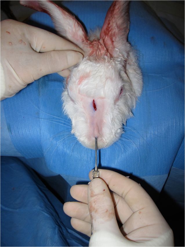
Beaudoin et al. Journal of Otolaryngology - Head and Neck Surgery 2013, 42:7 Page 2 of 6
http://www.journalotohns.com/content/42/1/7
morselizer [7]. The paste produced could easily be
injected through a 16 gauge needle [7]. However, in vivo
injection was not undertaken. No study evaluating the
viability of injected cartilage grafts on the nasal dorsum
has been found in the literature.
The aim of this study is to evaluate the long-term
viability of injected auricular cartilage in comparison
to surgically implanted cartilage on the nasal dorsum
of the rabbit.
Methods
The New Zealand white rabbit is a well-validated model
for the study of cartilage grafting, especially when involving
the nasal dorsum [4]. This study used 10 New Zealand
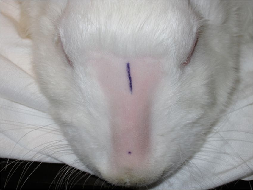
white male rabbits aged approximately 3 months and Figure 1 Incision for whole cartilage graft implantation and
weighing from 3.3 to 3.7 kg (average 3.5 kg). The research location for cartilage injection.
protocol was reviewed by a veterinarian and approved by
the University of Montreal Deontological Committee on
Animal Experimentation. Institutional guidelines regarding A 1 cm x 1 cm whole cartilage graft was harvested from
animal experimentation were followed. the rectangle for implantation. The graft was weighed and
For one week prior to surgery, the animals acclimatized its volume was determined by water displacement in a
to the elizabethan collars they had to wear for 10 post- 3 ml syringe. A 1.5 cm incision was made on the superior
operative days in prevention of self-trauma. General portion of each rabbit’s nasal dorsum followed by minimal
anesthesia was performed using Ketamine (35 mg\kg) and dissection to create a subcutaneous pocket. The graft was
Xylazine (5 mg/kg). Ocular protection was provided with then implanted and the wound was closed with plain 4–0
ointment. Breathing and heart rate were monitored by a interrupted sutures (Figure 4).
licensed animal technician.
The nasal dorsum and ears were shaved using an elec-
tric clipper. Infiltration of the ears and nasal dorsum with
xylocain 1% solution containing epinephrine 1:100,000
aided with pain control and hemostasis. Only the right ear
was used for the first three rabbits, but the amount of
cartilage yielded was insufficient, so both ears were
used for rabbits four through ten. Surgical disinfection
was done with four consecutive chlorhexidine sponges
and sterile techniques were used throughout. The injec-
tion site on the lower nasal dorsum and the implant-
ation site on the upper dorsum were marked (Figure 1).
Each ear was vertically incised on its posterior aspect
using a scalpel, with care to avoid the main auricular
vein (Figure 2). Hemostasis was done with a disposable
electrical cautery. The subperichondral plane was found
with a small scissor and the incision was extended in
that plane along its entire length. Blunt dissection of the
perichondrium to expose the cartilage was done with a
hemostatic clamp (Figure 2). A rectangular cartilage
window was cut with the scalpel. Care was taken to leave
sufficient cartilage architecture to maintain ear support.
The anterior aspect of the cartilage rectangle was then
carefully dissected to free it from the overlying perichon-
drium without puncturing the anterior skin flap. Once
the cartilage rectangle was obtained (Figure 3), the
Figure 2 Posterior auricular incision and dissection of
wound was closed with a plain 4–0 continuous running
cartilage specimen.
suture.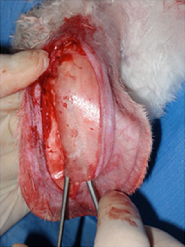
Beaudoin et al. Journal of Otolaryngology - Head and Neck Surgery 2013, 42:7                                                       Page 3 of 6
http://www.journalotohns.com/content/42/1/7

 Figure 3 Injection cannula, cartilage morselizer, cartilage              Figure 4 Injection of crushed cartilage graft in the inferior
 specimens from both ears and ruler (from left to right).                 portion of the nasal dorsum.

  The critical step was crushing the remaining cartilage                    Two injection cannulas were used: a large bore cannula
to produce an injectable slurry. Preliminary trials were                 (2.5 mm) for the first three rabbits and a small bore
conducted prior to surgery to determine the best cartil-                 (2 mm) cannula for rabbits four through ten. Injecting
age crushing technique. The otologic burr did not yield                  through the small bore cannula required more aggressive
a satisfactory volume of cartilage and was troublesome                   cartilage crushing than through the larger bore cannula
to use. The Cottle cartilage crusher is a hinged anvil.                     Injection resulted in a readily visible subcutaneous car-
The cartilage sample is placed between the closed moving                 tilage mound. A single interrupted plain 4–0 suture was
pieces and struck with a mallet. It did not break the cartil-            placed to prevent graft extrusion. A sample of crushed
age fibers sufficiently to render the graft injectable.                  cartilage was taken for the pre-implantation histological
  The optimal method was dicing the cartilage with a                     analysis. It should be noted again that only the right ear
scalpel before crushing it with the cartilage morselizer.                was used in the three first rabbits. In the remaining
The morselizer uses serrated jaws that tear through                      rabbits, we harvested cartilage from both ears. This
cartilage fibers. This process yielded a cartilage paste that            provided sufficient volumes for effective injection.
could pass through the injection cannula (Figure 3).                        Post-operative care consisted of antibiotic ointment
  The slurry was passed successively through 1 ml                        applied onto the nasal dorsum and the ears. Supportive
syringes. Once satisfactory consistency was obtained, the                compressive dressings were done on the ears. Analgesia
apparatus was weighted with the cartilage and cannula                    was provided by a dose of subcutaneous non-steroidal
on an analytical scale accurate to seven-decimal points.                 anti-inflammatories (NSAIDs) and was repeated if
The volume of cartilage was also noted in the syringe.                   needed. The elizabethan collars were kept for ten days
The skin on the inferior part of the nasal dorsum was                    postoperatively to prevent traumatic graft extrusion or
pinched upwards before the cartilage was injected                        wound dehiscence. Sutures were removed prior to removal
through the cannula (Figure 4). Sufficient pressure had                  of the collars. Some post-operative complications occurred,
to be applied to the syringe piston to inject the cartilage.             such as spontaneous partial graft extrusion of the injected
Insufficiently crushed cartilage obstructed the cannula.                 cartilage in one rabbit, auricular hematoma in two rabbits
This resulted in loss of cartilage volume and necessitated               and cellulitis in one rabbit. The partially extruded graft was
further crushing.                                                        removed without further complications. The hematomas
Beaudoin et al. Journal of Otolaryngology - Head and Neck Surgery 2013, 42:7                                                    Page 4 of 6
http://www.journalotohns.com/content/42/1/7

Table 1 Mass and volume measurements for whole cartilage grafts
             Pre-implantation                                Post-implantation                                 Difference
    Mass (mg)               Volume (ml)              Mass (mg)                 Volume (ml)        Mass (%mg)            Volume (%ml)
Average     Median      Average      Median      Average     Median      Average      Median   Average   Median       Average       Median
 77.4         66          0.09         0.1         109         110         0.11         0.1     +41       +67           +22            0

were drained daily until resolution and the cellulitis was               grafts (Table 1). Mass was decreased by an average of
treated with topical antibiotic cream.                                   47% (median 24%) and volume was decreased by an
  Britt and Park compared histological analyses of                       average of 40% (median 38%) for injected cartilage grafts
implanted cartilage grafts after four weeks, eight weeks, six            (Table 2).
months and twelve months [8]. They found superior tensile                  Histological analysis of pre-implantation whole cartil-
strength at four and eight weeks but stabilization of the                age grafts, pre-implantation injected cartilage grafts
chondrocyte viability between six and twelve months [8].                 (Figure 5) and post-implantation whole cartilage grafts
Another study compared crushed cartilage viability after                 were all similar and showed complete chondrocyte via-
two, five and ten months and did not find significant vari-              bility and absence of fibrosis in all samples.
ation despite degree of crushing [5]. Based on these stud-                 Histological analysis led to the exclusion of the
ies, we chose a three month implantation period, which is                crushed cartilage grafts from rabbits 5 and 10. Rabbit 5
equivalent to twelve human months [9].                                   demonstrated an abscess in lieu of viable cartilage and
  During this period, no complications were encountered                  rabbit 10 showed a granulomatous foreign body reaction.
and all the animals thrived. The animals were then put                   The remaining grafts were separated according to the
under general anesthesia with Ketamine (35 mg/kg) and                    diameter of the injection cannulas.
Xylazine (5 mg/kg) before they were euthanized with                        The first three crushed cartilage grafts were injected
pentobarbital. Shaving of the nasal dorsum was followed                  through a 2.5 mm large bore cannula and demonstrated
by a vertical incision on the nasal dorsum. Both grafts and              an average of 93% chondrocyte viability (median 90%)
their capsules, if present, were retrieved. Post-implantation            with an average and median limited degree of fibrosis
volume and mass were measured. The samples were then                     (Table 3). The following five crushed cartilage grafts
stored in formaldehyde to undergo post-implantation                      were injected through the 2 mm small bore cannula and
histological analysis.                                                   showed an average of 64% chondrocyte viability (median
  The histological analysis was performed by an experienced              75%) with an average and median limited degree of fibro-
pathologist. The stains used were hematoxylin-phloxin                    sis. The average chondrocyte viability was 75% (median
for chondrocyte viability and chondroid tissue appreci-                  82.5%) when all injected cartilage grafts were pooled
ation and alcian blue for glycosaminoglycan content.                     regardless of cannula diameter (Figure 6).

Results                                                                  Discussion
The non-crushed cartilage grafts were almost macro-                      The results obtained in our study compare favorably with
scopically identical to their pre-implantation state. The                similarly designed studies in the literature. Ale de Souza
surface area was conserved and they appeared viable. A                   et al. demonstrated superior surface area recovered with
fibrous capsule was formed around these grafts. Neo-                     non-crushed cartilage grafts when compared with crushed
vascularisation could be seen on some of the grafts. The                 cartilage grafts [4]. The percentage of viable chondrocytes
injected cartilage grafts were also surrounded by a fibrous              was also greater in non-crushed cartilage grafts [4].
capsule. The cartilaginous contents of this capsule was                    Cakmak et al. also found decreasing chondrocyte via-
similar to the pre-implantation state in all but one sample,             bility with increased degree of graft crushing [5]. The
which demonstrated necrosis of the cartilage (rabbit 5).                 moderately, significantly and severely crushed grafts
   Mass was increased by an average of 41% (median                       demonstrated 50%, 30% and 10% cartilage viability re-
67%) and volume was increased by an average of 22%                       spectively [5]. According to the Cakmak classification
(median 0%) during implantation for whole cartilage                      system for degrees of crushed cartilage, the injected

Table 2 Mass and volume measurements for injected crushed cartilage
             Pre-implantation                                Post-implantation                                 Difference
    Mass (mg)               Volume (ml)              Mass (mg)                 Volume (ml)        Mass (%mg)            Volume (%ml)
Average     Median      Average      Median      Average     Median      Average      Median   Average   Median       Average       Median
  335         310         0.35        0.32         179         235         0.21         0.2     −47       −24           −40          −38
Beaudoin et al. Journal of Otolaryngology - Head and Neck Surgery 2013, 42:7 Page 5 of 6
http://www.journalotohns.com/content/42/1/7
Figure 5 Injected cartilage graft (pre-implantation; Figure 6 Injected cartilage graft (post-implantation,
hematoxylin-eosin, 20 X). hematoxylin-eosin, 20 X).
crushed cartilage in our study is severely crushed, mean- In comparison to the other studies mentioned, the
ing its integrity is totally destroyed [10]. The injected injected crushed cartilage grafts offer similar or superior
cartilage in our study remained 64% viable on average, chondrocyte viability to surgically implanted crushed
with a median of 75%. grafts [4,6]. This indicates that the injection of cartilage
The crushed grafts injected through the 2.5 mm large is not detrimental to survival of chondrocytes in the
bore cannula also demonstrated greater chondrocyte via- same way that crushing is. Degree of graft crushing
bility than the crushed grafts injected through the 2 mm therefore seems to be the critical attribute predicting
small bore cannula. The latter necessitated more long-term chondrocyte viability. Long-term viability of
vigourous crushing to allow delivery through the smaller crushed cartilage grafts seems to be independent from
diameter. the delivery method.
Pre and post implantation histological studies used each
non-crushed cartilage graft as the control for the injected Conclusions
crushed graft. This provides greater confidence in attribut- Cartilage injection is a promising technique that delivers
ing volume, mass, change in chondrocyte viability and crushed cartilage without subcutaneous dissection.
degree of fibrosis to the grafting procedure itself. However, Chondrocyte viability and degree of fibrosis in the graft
the cartilage crushing technique and the injection are related to the degree of cartilage crushing. Delivery of
methods used were difficult to reproduce from graft to the cartilage graft through injection does not seem to
graft. This is due mostly to the severe degree of crushing influence chondrocyte viability. The major caveat with this
necessary to produce an easily injectable slurry which did method is the poor reliability of the injection apparatus
not obstruct the cannula. Loss of mass and volume were used. Development of a cartilage injection system would
mostly attributable to repeated filling and emptying of the increase efficiency of delivery and increase reproducibility
syringe following additional crushing maneuvers. of the results.
Table 3 Histological analysis for percutaneously injected cartilage
Cannula Rabbit Proportion of viable Degree of Mean/median chondrocyte Mean/median chondrocyte
chondrocytes (%) fibrosis viability (%) viability (%)
Large bore 1 100 None 93/90 75/82.5
2 90 Moderate
3 90 Slight
Small bore 4 50 Slight 64/75
6 90 Moderate
7 75 None
8 30 Modrate
9 75 NoneBeaudoin et al. Journal of Otolaryngology - Head and Neck Surgery 2013, 42:7 Page 6 of 6
http://www.journalotohns.com/content/42/1/7
Competing interests
This research was funded by a 5000$ grant from Stryker as well as a 2500$
donation from the Maisonneuve-Rosemont hospital Otolaryngology
foundation. These organisations have nothing to gain or lose from the
publication of this article.
Authors’ contributions
OXB has participated in the design, performed surgical manipulations,
analysed the data and drafted the manuscript. AM performed the
histological analysis. AR designed the study, performed surgical
manipulations and helped draft the manuscript. All authors read and
approved the final manuscript.
Acknowledgements
Thank you to the animal care team at Pavillion Liliane de Stewart and
especially Vanessa Lapointe Sasseville for their expertise in animal care.
Author details
1
Division of Otolaryngology-Head and Neck Surgery, Maisonneuve-Rosemont
Hospital, University of Montreal, Montreal, Canada. 2Department of
Pathology, Maisonneuve-Rosemont Hospital, University of Montreal,
Montreal, Canada.
Received: 29 November 2012 Accepted: 6 January 2013
Published: 31 January 2013
References
1. Ge NN, Rabinov CR, Crumley RL: Nasal reconstruction. In Cummings
otolaryngology head & neck surgery, Volume 1. 5th edition. Edited by
Thomas JR. Philadelphia: Mosby Elsevier; 2010:364–372.
2. Murrell GL: Auricular cartilage grafts and nasal surgery. Laryngoscope 2004,
114(12):2092–2102.
3. Cakmak O, Buyuklu F: Crushed cartilage grafts for concealing irregularities
in rhinoplasty. Arch Facial Plast Surg 2007, 9(5):352–357.
4. de Souza MM A, et al: Study of rabbit septal cartilage grafts placed on
the nasal dorsum. Arch Facial Plast Surg 2008, 10(4):250–254.
5. Cakmak O, et al: Viability of crushed and diced cartilage grafts: a study in
rabbits. Arch Facial Plast Surg 2005, 7(1):21–26.
6. Limberg AA: Plastie de soutien et de contour par injection de cartilage
broyé. La presse médicale 1957, 65(86):1961–1962.
7. Noordzij JP, Cates JM, Cohen SM, et al: Preparation techniques for the
injection of human autologous cartilage: an Ex vivo feasibility study.
Laryngoscope 2008, 118:185–188.
8. Britt JC, Park SS: Autogenous tissue-engineered cartilage: evaluation as an
implant material. Arch Otolaryngol Head Neck Surg 1998, 124(6):671–677.
9. Rudderman RH, Guyuron B, Mendelsohn G: The fate of fresh and
preserved, noncrushed and crushed autogenous cartilage in the rabbit
model. Ann Plast Surg 1994, 32:250–254.
10. Cakmak O, Altindas H: A classification for degree of crushed cartilage.
Arch Facial Plast Surg 2010, 12(No 6):435–436.
doi:10.1186/1916-0216-42-7
Cite this article as: Beaudoin et al.: Percutaneous cartilage injection: A
prospective animal study on a rabbit model. Journal of Otolaryngology -
Head and Neck Surgery 2013 42:7.
Submit your next manuscript to BioMed Central
and take full advantage of:
• Convenient online submission
• Thorough peer review
• No space constraints or color figure charges
• Immediate publication on acceptance
• Inclusion in PubMed, CAS, Scopus and Google Scholar
• Research which is freely available for redistribution
Submit your manuscript at
www.biomedcentral.com/submitYou can also read