Diagnosis and Therapy of Microscopic Colitis with Presence of Foamy Macrophages in Children
←
→
Page content transcription
If your browser does not render page correctly, please read the page content below
International Scholarly Research Network
ISRN Gastroenterology
Volume 2011, Article ID 756292, 9 pages
doi:10.5402/2011/756292

Research Article
Diagnosis and Therapy of Microscopic Colitis with Presence of
Foamy Macrophages in Children

          Jan Józefczuk1 and Bogdan Marian Woźniewicz2
          1 Pediatric   Ward, Hospital in Sandomierz, Schinzla 13, 27-600 Sandomierz, Poland
          2 Department    of Pathology, The Children’s Memorial Health Institute, Aleja Dzieci Polskich 20, 04-730 Warsaw, Poland

          Correspondence should be addressed to Bogdan Marian Woźniewicz, b.wozniewicz@czd.waw.pl

          Received 12 April 2011; Accepted 4 May 2011

          Academic Editor: C.-Y. Chen

          Copyright © 2011 J. Józefczuk and B. M. Woźniewicz. This is an open access article distributed under the Creative Commons
          Attribution License, which permits unrestricted use, distribution, and reproduction in any medium, provided the original work is
          properly cited.

          We discuss the diagnosis of and efficacy 5-amino-2-hydroxybenzoic acid (5-ASA), Saccharomyces boulardii, or magnesium in
          therapy of microscopic colitis with presence of foamy macrophages. A basis for diagnosis and inclusion to the analysed group
          was presence of characteristic foamy macrophages in histopathological examination of hematoxylin and eosin-stained specimens
          collected from the large intestine, reviewed under ×200 or ×320 magnification. No statistically significant improvement was found
          following the use of 5-amino-2-dihydroxybenzoic acid in therapy of the disease. The use of Saccharomyces boulardii was associated
          with statistically significant improvement in clinical, endoscopic, and histopathological condition. Use of magnesium caused a
          histological, statistically significant improvement but failed to have any effect on the clinical and endoscopic presentation. In the
          group of children in whom no therapeutic intervention was provided, a statistically significant spontaneous clinical improvement
          was observed, but no statistically significant changes in endoscopic and microscopic condition were found.

1. Introduction                                                                Evaluation of clinical, endoscopic, and microscopic ef-
                                                                           fects of therapy with 5-amino-2-dihydroxybenzoic acid, Sac-
Typical inflammatory bowel disease (IBD), such as ulcerative               charomyces boulardii, or magnesium of children with micro-
colitis or Crohn’s disease, is only a part of cases of persistent          scopic colitis with presence of foamy macrophages and
enteritis. Nonspecific IBDs are most often superficial pro-                evaluation of a group of children with no therapeutic inter-
cesses, nondamaging to the structure of intestinal mucosa                  vention.
and submucosa. In the medical literature, they are usually
referred to as microscopic enteritis [1–4].
                                                                           2. Material and Methods
     Foamy colitis with presence of foamy macrophages
within the intestinal lamina propria belongs to a group of                 A basis for diagnosis of the disease was presence of macro-
microscopic inflammations in children. That recently sepa-                 phages that stained with hematoxylin and eosin formed a
rated disease requires determination of effective therapeutic               characteristic image of foamy (clear cell) macrophages, in the
methods [5, 6].                                                            lamina propria of the large intestine.
     Observation of that type of inflammation, indicating a                    Additional examinations using electron microscopy Jeol
rather mild clinical course of the disease and no cases of                 100CX were performed in several cases. Sections of mucosa
its transformation into a typical inflammatory bowel disease               were fixed in cacodylate buffered 4% glutaraldehyde and
(IBD) justified undertaking a prospective observation of                   then in osmium tetroxide (OsO4 ) and analysed acc. to a
children treated with 5-amino-2-hydroxybenzoic acid, with a                typical procedure for Epon. Resulting ultrathin sections were
probiotic (Saccharomyces boulardii), or with magnesium, and                additionally stained with lead citrate and uranyl acetate.
of a group of children who were not treated pharmacologi-                      Uneven distribution and quantity of foamy macrophages
cally.                                                                     constituted a basis for development of our own classification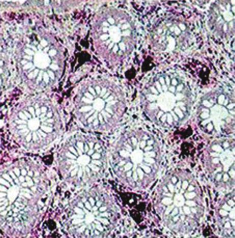
2                                                                                                        ISRN Gastroenterology

of microscopic changes, defining three levels of intensity:           The level of endoscopic lesions was determined by
                                                                  evaluation of vascular pattern, granulation, and mucosa
     (i) single cells on various levels of the lamina propria,    proneness for bleeding, as well as presence of mucus, pus,
    (ii) presence of aggregates composed of 3–10 cells,           erosions, and ulcerations on the surface of colic mucosa. The
    (iii) foamy cells covering evenly the whole lamina propria.   clinical activity index (CAI) acc. to Rachmilewitz was then
                                                                  applied [7].
    That type of cells was absent in other layers of the large        Evaluation of clinical and endoscopic changes was per-
intestine, and their presence was not found in other sections     formed at diagnosis and following 12 months of therapy and
of the alimentary tract.                                          observation of each analysed child.
    In the process of identification of foamy macrophages,            Due to nonparametric distribution of sets, a sign test was
except for hematoxylin and eosin staining of serial spec-         used for statistical evaluation of results (Statistica software).
imens, also a PAS reaction was used, with and without
diastase digestion.                                               3. Results and Discussion
    Additionally, immunocytochemical tests using mono-
clonal sera from DAKO: anti-CD3(T cell), CD4(T cell),             At diagnosis, histopathological examination of bioptates
CD22 (B cell), CD31 (Endothelia), CD34 (precursor cell),          collected from the large intestine revealed presence of large,
CD 68 (Macrophages), allowing identification of cells occur-      clear, foamy cells in the lamina propria of the mucosa. Those
ring in an inflammatory process, were completed on speci-         cells appeared single or in agglomerates, covering the whole
mens placed on hyalinized slides. Tests were also completed       width of the lamina propria—from the covering epithelium
to detect the PCNA-proliferation antigen, Ki67-mitotic            to the base of intestinal crypts and the level of muscularis
activity, chromogranin, synaptophysin, neurofilament, actin,      mucosa. (Figure 1)
P53 mutations, GFAP glial marker, TNF alpha, IL2, and                 Immunocytochemical tests indicated a strong positive
mucicarmin.                                                       reaction with the CD68 antibody, typical for activated
    Tests were competed to detect intestinal pathogens (Sal-      macrophages. Foamy macrophages showed a positive PAS
monella, Shigella, Camphylobacter, Escherichia coli, Yersnia,     reaction with periodic acid and Shiff ’s reagent. However,
Clostridium difficile) and Giardia infection.                       the reaction was negative following digestion with dias-
    Tests for enteroviruses were performed on cell lines (Hep     tase. Immunocytochemical reaction to neurofilament was
2 and RD) in the Department of Virusology of the National         negative. Using the marker PCNA-proliferation antigen
Health Institute, using WHO-recommended methods. Pres-            an increased activity was demonstrated only in cryptic
ence of rotaviruses was tested with the latex reaction, using     epithelium. No increased activity was found in epithelium
the Slidex Rota-Kit, and completing serological tests with        surrounding crypts (Figure 2).
ELISA, using Rabbit Anti-Rotavirus Human reagents from                Other numerous, mentioned in the Methods section,
DAKO.                                                             immunocytochemical markers gave a negative result in tests
    Children with presence of foamy macrophages confirmed         with foamy macrophages (Table 1).
in histopathological specimens and who had a clinical and             Before statistical analysis of results of clinical, endo-
endoscopic changes characteristic for colitis were random-        scopic, and microscopic evaluation results for three study
ized to individual therapeutic groups of a prospective, open      groups, they were evaluated at the time of intervention.
trial realised from 1999 to 2009.                                 Hypothesis of equality of those three groups (A, B, C, D) in
    In the analysis, there were included 144 children aged        clinical, endoscopic, and microscopic evaluation cannot be
from 3 to 18 years. For 12 months, children received              dismissed (Whitney-Mann test) (Figure 3).
5-amino-2-hydroxybenzoic acid (5-ASA) at dose of 20–                  In the group A, for 12 months, children received
25 mg/kg b.w./24 h (38 children-group A), Saccharomyces           Masalazine (5 amino-2-hydroxybenzoic acid). Clinical, endo-
boulardii at dose of 15–20 mg/kg b.w./24 h (35 children-group     scopic, and microscopic evaluation of the group A, treated
B), or magnesium 5–10 mg/kg b.w./24 h (35 children-group          with 5-amino-2-hydroxybenzoic acid, showed no significant
D).                                                               improvement, neither in clinical, endoscopic, nor in micro-
    Children who received no treatment constituted a control      scopic presentation (Figure 4).
group (36 children-group C).                                          In the group B, Saccharomyces boulardii was used for 12
    Clinical and endoscopic examinations were performed at        months. In the group B of children treated with Saccha-
the Pediatric Ward, and microscopic and immunocytochem-           romyces boulardii the 12-month period of therapy caused a
ical examinations—in the Department of Pathology in the           clinical, endoscopic, and microscopic, statistically significant
Children’s Memorial Health Institute in Warsaw.                   improvement (Figure 5).
    Clinical evaluation included general feeling, presence of         In the group C of children with microscopic colitis with
abdominal pain and its intensity, number of bowel move-           presence of foamy macrophages, no therapy was applied.
ments per week, presence of blood in stool, fever, presence           In the group C of untreated children the clinical
of extraenteral symptoms, ESR, haemoglobin concentration,         evaluation showed statistically significant improvement, and
and nutritional status expressed as Cole and Stanfield index.     the endoscopic and microscopic evaluation showed no
    Clinical activity index (CAI) acc. to Rachmilewitz was        regression of pathological lesions (Figure 6).
used for evaluation of clinical presentation of analysed              Children in the group D had magnesium administered
children [7].                                                     for 12 months. Statistical analysis of 12-month magnesium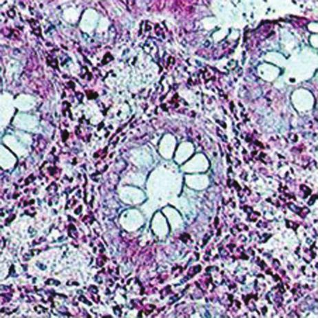
ISRN Gastroenterology 3
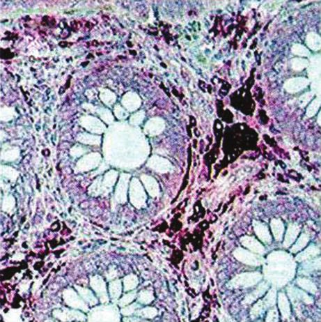
(a) (b) (c) (d)
Figure 1: (a–d) Microscopic features of colitis with presence of foamy macrophages—characteristic large, clear cells with a small nucleus,
single or aggregated within the lamina propria. HE staining. Magnification ×200.
(a) (b) (c) (d)
Figure 2: (a–d) Immunochemistry. (a-b) positive with a serum marking macrophages. C-negative to limfocytes T. D-neurofilament’s
negative reaction. Magnification ×200 (a) ×320 (b, c, d).
Table 1: Expression of immunocytochemical markers in the micro- Presence of characteristic foamy macrophages observed
scopic colitis with presence of foamy macrophages. at ×200 and ×320 magnification in hematoxylin and eosin-
stained specimens is a crucial diagnostic factor [5, 6]. The
Macrophages foamy Marker
disease is not associated with destruction of glands or their
Negative CD3 (Cell T) restructure. The lamina propria is not destroyed by lympho-
Negative CD4 (Cell T) cytic infiltration, characteristic for inflammations belonging
Negative CD22 (Cell B) to the group of IBD. Presence of foamy macrophages in
Negative CD34 (Cell precursor) examined histopathological specimens excludes diagnosis of
Negative CD31 (Entothelial) Colitis ulcerosa or Crohn’s disease.
Positive CD68 (Macrophages) In IBD-type inflammations, proliferation of the mono-
Negative Ki67 mitotic activity nuclear-phagocytic line is characterised by a positive expres-
Negative Chromogranin sion of TNF-alpha and IL-beta, absent in our examinations.
Negative Synaptophysin Microscopic evaluation of properly collected and prepared
Negative Neurofilament specimens, but observed under a routinely used low magni-
Negative Actin
fication, is a reason for underdiagnosis of the disease in other
centres. Observation of the whole group of children confirms
Negative P53 mutations
previous findings on mild and moderate course of that type
Negative GFAP glial marker
of inflammation [4, 6].
Negative TNF alpha
Children left without any therapeutic intervention show
Positive PAS no endoscopic or microscopic progression of lesions, and an
Positive Osmium tetraoxide observed surprising clinical improvement indicates an ability
Negative IL2 for independent regeneration of the intestine in that disease.
Negative Mucicarmin Existing reports indicate efficacy of therapy of mild colitis
with probiotics. Successful attempts were made in therapy
of ulcerative colitis and even of Crohn’s disease [8–12].
therapy shows microscopic and clinical improvement, but Part of favourable therapeutic effects of monotherapy with
without any endoscopic improvement. Microscopic diagno- probiotics could be a result of erroneous qualification of
sis of that unspecific inflammatory disease is relatively easy microscopic colitis to a group of mild IBD. It seems that
(Figure 7). probiotics may be useful as supplementary therapy, but their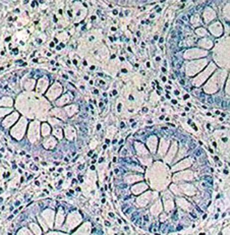
4 ISRN Gastroenterology
Clinical presentation of groups at diagnosis Endoscopic presentation of groups at diagnosis
9 25
8
7 20
Number of cases
Number of cases
6
15
5
4
10
3
2
5
1
0 0 Mg
1 1 N
2 3 4 2 3 E
5 6 7 8 4 5 M
Own classifica9 10 11 12 13 6 7 8
tion 14 Own classi 9
fication
M N M N
E Mg E Mg
Statistica U Mann-Whitney test: Statistica U Mann-Whitney test:
hypothesis of equality of distribution cannot be dismissed hypothesis of equality of distribution cannot be dismissed
(a) (b)
Microscopic presentation of groups at diagnosis
35
30
25
Number of cases
20
15
10
5 Mg
N
0 E
1 M
2
3
Own classi 4
fi cation
M N
E Mg
Statistica U Mann-Whitney test:
hypothesis of equality of distribution cannot be dismissed
(c)
Figure 3: Clinical, endoscopic, and microscopic evaluation at diagnosis (Whitney-Mann test).
use as a monotherapy is at least risky, particularly in the B12 ), folic acid, and pantoteic acid contained in that probiotic
Crohn’s disease. on enterocytes [8].
A favourable clinical effect of Saccharomyces boulardii Nicotinamide included in Saccharomyces boulardii may
demonstrated in this trial, and confirmed in control endo- reduce free oxygen radicals causing intensification of the
scopic and microscopic examinations, may be a result of a inflammatory process, and numerous enzymes contained
trophic effect of carbohydrates, B group vitamins (B1 , B2 , B6 , there; for example, proteases and disaccharidases may cause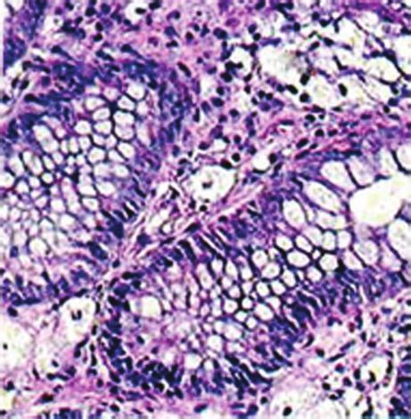
ISRN Gastroenterology 5
Results-clinical presentation Results-endoscopic evaluation
10 25
9
8 20
Number of cases
Number of cases
7
6 15
5
4 10
3
2 5
1
0 0
1 2 1 2
3 4 5 3 4
6 7 8 9 10 5 6
11 12 13 7 8
Own classifica 14 Own classifica 9
tion tion
Before treatment Before treatment
After treatment After treatment
Statistica ranked sign test: (Z = 2.085144, P = 0.037056) Statistica ranked sign test: (Z = 1.835326, P = 0.066457)
hypothesis of equality of distributions cannot be dismissed hypothesis of equality of distributions cannot be dismissed
(a) (b)
Results-microscopic presentation
35
30
25
Number of cases
20
15
10
5
0
0
1
2
Own classifica 3
tion
Before treatment
After treatment
Statistica ranked sign test: (Z = 0.948683, P = 0.342782)
hypothesis of equality of distributions cannot be dismissed
(c)
Figure 4: Clinical, endoscopic, and microscopic evaluation of children treated with 5-amino-2-hydroxybenzoic acid (group A).
digestion of protein content of foamy macrophages and be No favourable therapeutic effect of 5-amino-2-dihydrox-
responsible for reduction of their count, demonstrated in ybenzoic acid and relatively mild course of the disease jus-
control microscopic examinations [8, 13]. tify resignation from therapy with Mesalazine (5-amino-2-
A favourable therapeutic effect of Saccharomyces bou- dihydroxybenzoic acid) in case of that type of colitis.
lardii may be also a result of its confirmed immunostimula- A favourable therapeutic effect of magnesium, mani-
tory and anti-inflammatory effect, significant for limitation fested by regression of microscopic lesions, is most probably
and reduction of the inflammatory process in the intestine a result of supplementation of magnesium deficiency, result-
[13–15]. ing from erosion of soil and low content of the element in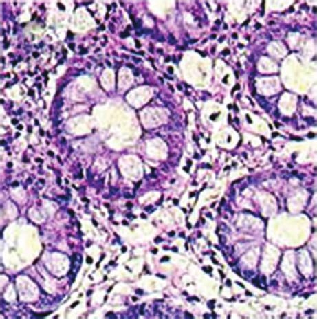
6 ISRN Gastroenterology
Results-endoscopic evaluation
Results-clinical presentation
18
12
16
10 14
Number of cases
12
Number of cases
8 10
6 8
6
4 4
2
2
0
0 1 2 3 4
1 2 3 5 6
4 5 6 7 8 7 8
9 Own classificati 9
Own classificati 10 11 12 13 14 on
on
Before treatment Before treatment
After treatment After treatment
Statistica ranked sign test: (Z = 4.973459, P = 0.000001) Statistica ranked sign test: (Z = 4.929503, P = 0.000001)
hypothesis of equality of distributions should be dismissed hypothesis of equality of distributions should be dismissed
(a) (b)
Results-microscopic presentation
25
20
Number of cases
15
10
5
0
0
1
2
Own classificati 3
on
Before treatment
After treatment
Statistica ranked sign test: (Z = 4.364358, P = 0.000013)
hypothesis of equality of distributions should be dismissed
(c)
Figure 5: Clinical, endoscopic, and microscopic evaluation of children treated with Saccharomyces boulardii (group B).
food, and generally low level of supplementation. Besides, should be treated as a herald of the endoscopic improve-
mucosa defects found in colitis intensify magnesium defi- ment.
ciency through increased apoptosis. Systematic general paediatric and gastroenterological
A favourable effect of magnesium treatment should be control, along with monitoring of number of foamy macro-
probably associated with activating effect of the element phages in microscopic examination, plays a very important
on enzymes necessary for synthesis and utilisation of high- role in diagnosis and therapy of that type of colitis [2]. The
energy compounds inside enterocytes. Further observation suggested own scale for evaluation of microscopic changes
seems important, expecting development of endoscopic based on quantity of foamy macrophages in the lamina
improvement. If it occurs, the microscopic improvement propia shows a complete correlation with the clinical and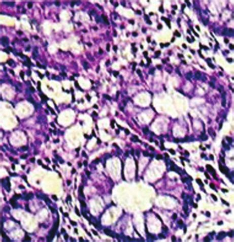
ISRN Gastroenterology 7
Results-endoscopic evaluation
Results-clinical presentation
7 12
6 10
Number of cases
Number of cases
5 8
4
6
3
4
2
1 2
0 0
1 2 3 1 2
4 5 6 3
7 8 9 10 4 5
11 12 13 6 7
Own classifica 14 Own classifica 8 9
tion tion
Before treatment
Before treatment
After treatment
After treatment
Statistica ranked sign test: (Z = 2.752989, P = 0.005905) Statistica ranked sign test: (Z = 1.109400, P = 0.267258)
hypothesis of equality of distributions should be dismissed hypothesis of equality of distributions should be dismissed
(a) (b)
Results-microscopic presentation
35
30
25
Number of cases
20
15
10
5
0
0
1
2
Own classifica 3
tion
Before treatment
After treatment
Statistica ranked sign test: (Z = 1.06066, P = 0.288844)
hypothesis of equality of distributions should be dismissed
(c)
Figure 6: Clinical, endoscopic, and microscopic evaluation of untreated children (group C).
endoscopic evaluation, when analysing the therapeutic effect open new diagnostic and therapeutic horizons. Difficulties
of Saccharomyces boulardii and 5-amino-2-dihydroxybenzoic associated with unanimous classification of a significant
acid and a correlation with the endoscopic presentation in part of colitis to a strictly defined group are a serious
case of no therapeutic intervention. medical problem [5, 16]. Classification of chronic colitis
Prospective clinical observation, periodical endoscopic inadequate to actual condition may negatively influence
examinations of patients with that disease, will allow veri- therapy and prognosis in this large, socially important group
fication of currently available knowledge, and possibly will of civilisation diseases.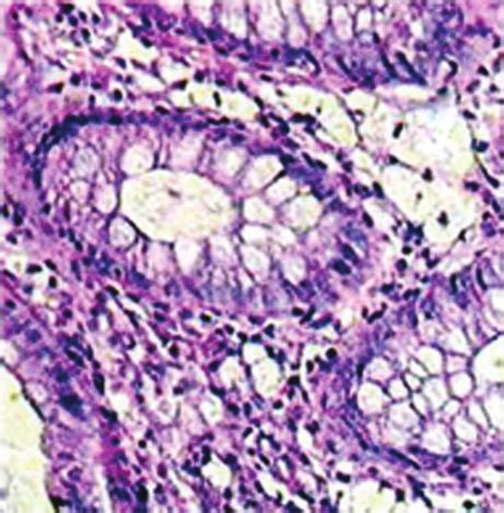
8 ISRN Gastroenterology
Results-endoscopic evaluation
Results-clinical presentation
8 16
7 14
6 12
Number of cases
Number of cases
5 10
4 8
3 6
2 4
1 2
0 0
1 2 3 1
4 5 6 7 8 2 3
9 10 11 4 5
12 13 14 6 7
Own classifica Own classifica 8 9
tion tion
Before treatment Before treatment
After treatment After treatment
Statistica sign test: (Z = 3.198011, P = 0.001384) Statistica sign test: (Z = 2.25, P = 0.024449)
hypothesis of equality of distributions should be dismissed hypothesis of equality of distribution cannot be dismissed
(a) (b)
Results-microscopic presentation
30
25
Number of cases
20
15
10
5
0
0
1
2
Own classifica 3
tion
Before treatment
After treatment
Statistica sign test: (Z = 3.927922, P = 0.000086)
hypothesis of equality of distributions should be dismissed
(c)
Figure 7: Clinical, endoscopic, and microscopic evaluation of children treated with magnesium (group D).
4. Conclusions (3) Efficacy of Saccharomyces boulardii in therapy of mi-
croscopic colitis with presence of foamy macrophages
(1) Presence of foamy macrophages within the lamina was demonstrated.
propria of the large intestine is a deciding factor in
diagnosis of that form of microscopic colitis. (4) No favourable therapeutic effect was achieved follow-
ing use of 5-amino-2-dihydroxybenzoic acid (ASA-5)
(2) Identification of foamy macrophages is possible with in case of the disease.
routine hematoxylin and eosin staining and micro-
scopic evaluation of histological specimens at ×200 (5) The disease shows a tendency for spontaneous remis-
or ×320 magnification. sion, but only in terms of clinical presentation.
ISRN Gastroenterology                                                                                                                         9

    (6) Microscopic and clinical improvement found in case                  [15] I. R. Sanderson, S. Boyle, C. B. Williams, and J. A. Walker-
        of magnesium therapy may be a herald of expected                         Smith, “Histological abnormalities in biopsies from macro-
        endoscopic improvement.                                                  scopically normal colonoscopies,” Archives of Disease in Child-
                                                                                 hood, vol. 61, no. 3, pp. 274–277, 1986.
    (7) Further paediatric and gastroenterological followup                 [16] K. Karolewska-Bochenek, I. Lazowska-Przeorek, P. Albrecht
        of children with that type of microscopic colitis is                     et al., “Epidemiology of inflammatory bowel disease among
        necessary.                                                               children in Poland,” Digestion, vol. 79, no. 2, pp. 121–129,
                                                                                 2009.

References
 [1] L. Librecht, R. Croes, and N. Ectors, “Microscopic colitis with
     giant cells,” Histopatology, vol. 48, pp. 116–132, 2006.
 [2] R. K. Yantiss and R. D. Odze, “Diagnostic difficulties in
     inflammatory bowel disease pathology,” Histopathology, vol.
     48, no. 2, pp. 116–132, 2006.
 [3] B. F. Warren, C. M. Edwards, and S. P. L. Travis, “Microscopic
     colitis, classification and terminology,” Histopathology, vol. 40,
     no. 4, pp. 374–376, 2002.
 [4] J. Józefczuk, B. Woźniewicz, and W. Romańczuk, “Patologia i
     klinika nowej formy zapalenia jelita grubego u dzieci,” Pedi-
     atria Współczesna. Gastroenterologia, Hepatologia i Żywienie
     Dziecka, vol. 5, no. 3, pp. 151–156, 2003.
 [5] J. Józefczuk, B. Woźniewicz, and W. Romańczuk, “Clinico-
     pathology of Foamy Cell Colitis (FCC), the new form non-
     inflamatory bowel disease,” Annals of Diagnostic Paediatric
     Pathology, vol. 5, pp. 71–74, 2001.
 [6] J. Józefczuk and B. M. Wozniewicz, “Clear cell colitis: a
     form of microscopic colitis in children,” World Journal of
     Gastroenterology, vol. 14, no. 2, pp. 231–235, 2008.
 [7] J. Ryżko, J. Socha, and M. Woynarowski, “Validation of disease
     activity indexes Indexes for inflammatory disease,” Surg. Child.
     Int., vol. 4, pp. 17–21, 1966.
 [8] J. P. Buts and P. Bernasconi, “Saccharomyces boulardii:
     basic science and clinical applications in gastroenterology,”
     Gastroenterology Clinics of North America, vol. 34, no. 3, pp.
     515–532, 2005.
 [9] O. Erdeve, U. Tiras, and Y. Dallar, “The probiotic effect of
     Saccharomyce boulardii in a pediatric age group,” Journal of
     Tropical Pediatrics, vol. 50, no. 4, pp. 234–236, 2004.
[10] M. Guslandi, P. Giollo, and P. A. Testoni, “A pilot trial
     of Saccharomyces boulardii in ulcerative colitis,” European
     Journal of Gastroenterology and Hepatology, vol. 15, no. 6, pp.
     697–698, 2003.
[11] M. Guslandi, G. Mezzi, M. Sorghi, and P. A. Testoni, “Sac-
     charomyces boulardii in maintenance treatment of Crohn’s
     disease,” Digestive Diseases and Sciences, vol. 45, no. 7, pp.
     1462–1464, 2000.
[12] J. Ryżko, “Zastosowanie probiotyków i prebiotyków w lecze-
     niu nieswoistych zapaleń jelit oraz zaburzeń czynnościowych
     jelita grubego,” Pediatria Współczesna Gastroenterologia. Hep-
     atologia i Żywienie Dziecka, vol. 4, no. 1, pp. 55–60, 2002.
[13] J. P. Buts, P. Bernasconi, J. P. Vaerman, and C. Dive,
     “Stimulation of secretory IgA and secretory component of
     immunoglobulins in small intestine of rats treated with
     Saccharomyces boulardii,” Digestive Diseases and Sciences, vol.
     35, no. 2, pp. 251–256, 1990.
[14] K. Plein and J. Hotz, “Therapeutic effects of Saccharomyces
     boulardii on mild residual symptoms in a stable phase of
     Crohn’s disease with special respect to chronic diarrhea—a
     pilot study,” Zeitschrift fur Gastroenterologie, vol. 31, no. 2, pp.
     129–134, 1993.
MEDIATORS of
INFLAMMATION
The Scientific Gastroenterology Journal of
World Journal
Hindawi Publishing Corporation
Research and Practice
Hindawi Publishing Corporation
Hindawi Publishing Corporation
Diabetes Research
Hindawi Publishing Corporation
Disease Markers
Hindawi Publishing Corporation
http://www.hindawi.com Volume 2014
http://www.hindawi.com Volume 2014 http://www.hindawi.com Volume 2014 http://www.hindawi.com Volume 2014 http://www.hindawi.com Volume 2014
Journal of International Journal of
Immunology Research
Hindawi Publishing Corporation
Endocrinology
Hindawi Publishing Corporation
http://www.hindawi.com Volume 2014 http://www.hindawi.com Volume 2014
Submit your manuscripts at
http://www.hindawi.com
BioMed
PPAR Research
Hindawi Publishing Corporation
Research International
Hindawi Publishing Corporation
http://www.hindawi.com Volume 2014 http://www.hindawi.com Volume 2014
Journal of
Obesity
Evidence-Based
Journal of Stem Cells Complementary and Journal of
Ophthalmology
Hindawi Publishing Corporation
International
Hindawi Publishing Corporation
Alternative Medicine
Hindawi Publishing Corporation Hindawi Publishing Corporation
Oncology
Hindawi Publishing Corporation
http://www.hindawi.com Volume 2014 http://www.hindawi.com Volume 2014 http://www.hindawi.com Volume 2014 http://www.hindawi.com Volume 2014 http://www.hindawi.com Volume 2014
Parkinson’s
Disease
Computational and
Mathematical Methods
in Medicine
Behavioural
Neurology
AIDS
Research and Treatment
Oxidative Medicine and
Cellular Longevity
Hindawi Publishing Corporation Hindawi Publishing Corporation Hindawi Publishing Corporation Hindawi Publishing Corporation Hindawi Publishing Corporation
http://www.hindawi.com Volume 2014 http://www.hindawi.com Volume 2014 http://www.hindawi.com Volume 2014 http://www.hindawi.com Volume 2014 http://www.hindawi.com Volume 2014You can also read