Case Report Intestinal Myiasis Caused by Sarcophaga spp. in Cusco, Peru: A Case Report and Review of the Literature
←
→
Page content transcription
If your browser does not render page correctly, please read the page content below
Hindawi
Case Reports in Infectious Diseases
Volume 2018, Article ID 3685439, 4 pages
https://doi.org/10.1155/2018/3685439

Case Report
Intestinal Myiasis Caused by Sarcophaga spp. in Cusco, Peru:
A Case Report and Review of the Literature

          Priscilla Ly,1 Adiel Aizenberg ,1 Taylor Martin ,1 Martha Lopez,2
          Miguel Arturo Saldaña,3 Grant Leslie Hughes,4 and Miguel Mauricio Cabada                                               2,5

          1
            School of Medicine, University of Texas Medical Branch, 301 University Boulevard, Galveston, TX 77555, USA
          2
            Cusco Branch, Tropical Medicine Institute, Universidad Peruana Cayetano Heredia, Calle Jose Carlos Mariategui J-6, Wanchaq,
            Cusco, Peru
          3
            Department of Microbiology and Immunology, University of Texas Medical Branch, 301 University Boulevard, Galveston,
            TX 77555, USA
          4
            Department of Pathology, University of Texas Medical Branch, 301 University Boulevard, Galveston, TX 77555, USA
          5
            Division of Infectious Diseases, Department of Internal Medicine, University of Texas Medical Branch,
            301 University Boulevard RT 0435, Galveston, TX 77555, USA

          Correspondence should be addressed to Miguel Mauricio Cabada; micabada@utmb.edu

          Received 12 March 2018; Accepted 6 May 2018; Published 27 May 2018

          Academic Editor: Larry M. Bush

          Copyright © 2018 Priscilla Ly et al. This is an open access article distributed under the Creative Commons Attribution License,
          which permits unrestricted use, distribution, and reproduction in any medium, provided the original work is properly cited.
          Myiasis is the infestation by dipterous fly larvae in humans and animals. The larvae can infect living or necrotic tissue involving the
          skin, nasopharynx, genitourinary, and gastrointestinal tracts. The accidental ingestion of eggs causes infection of the intestinal tract.
          We report a case of intestinal myiasis caused by Sarcophaga spp. larvae in a two-year-old child from Limatambo province in the
          Cusco region of Peru. Live larvae were identified incidentally in this child’s stool sample during the study screening for Strongyloides
          stercoralis. The child did not have any constitutional or abdominal symptoms. The morphological examination of the specimen under
          magnification revealed Sarcophaga spp. larvae. We performed a literature review of publications reporting intestinal myiasis caused
          by Sarcophaga spp. and discussed key aspects of this infestation.

1. Introduction                                                            are considered facultative or accidental myiasis [5]. Fly
                                                                           larvae that can cause a parasitic infestation but can also
Myiasis is the infestation with dipterous fly larvae of                     complete their cycle in the environment are considered
humans and other mammals, typically livestock [1].                         facultative. Although these commonly cause wound or
Myiasis is a natural infection of livestock causing a sig-                 necrotic tissue myiasis, eggs or larvae in the environment
nificant burden on this industry. Occasionally, humans                      can be accidentally ingested causing intestinal myiasis [6].
exposed to endemic areas suffer from zoonotic fly larvae                     The diagnosis is confirmed when the offending larvae are
infestations [2]. The fly larvae feed on living or necrotic                 passed in one or more consecutive stool samples [7]. Intestinal
host tissue, bodily fluids, or ingested food [3]. Human                     myiasis is generally transient and asymptomatic, though
myiasis affects the skin most often and also can affect other                symptoms may include nausea, vomiting, and abdominal
organs, such as the nasopharynx, digestive tract, ear canal,               pain [8, 9]. Intestinal myiasis caused by the larvae of the
orbits, and genitourinary tract [4]. Several classifications of             flesh fly Sarcophaga spp. is reported in several countries,
myiasis have been proposed mostly depending on the                         but to date no case has been reported in Peru. We report
anatomical location of the infestation, the stage of the larval            a case of intestinal myiasis caused by Sarcophaga spp. in the
development, or the existence of an obliged parasitic stage                highlands of Peru and present a review of the literature
[1]. Most of the fly species that cause intestinal infestation              about intestinal myiasis.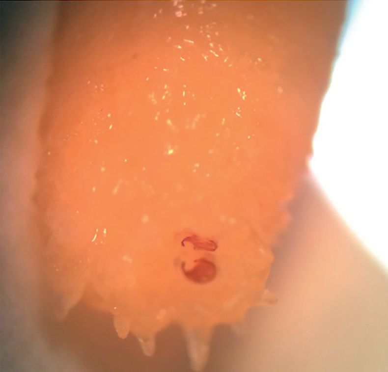
2 Case Reports in Infectious Diseases
(a) (b) (c)
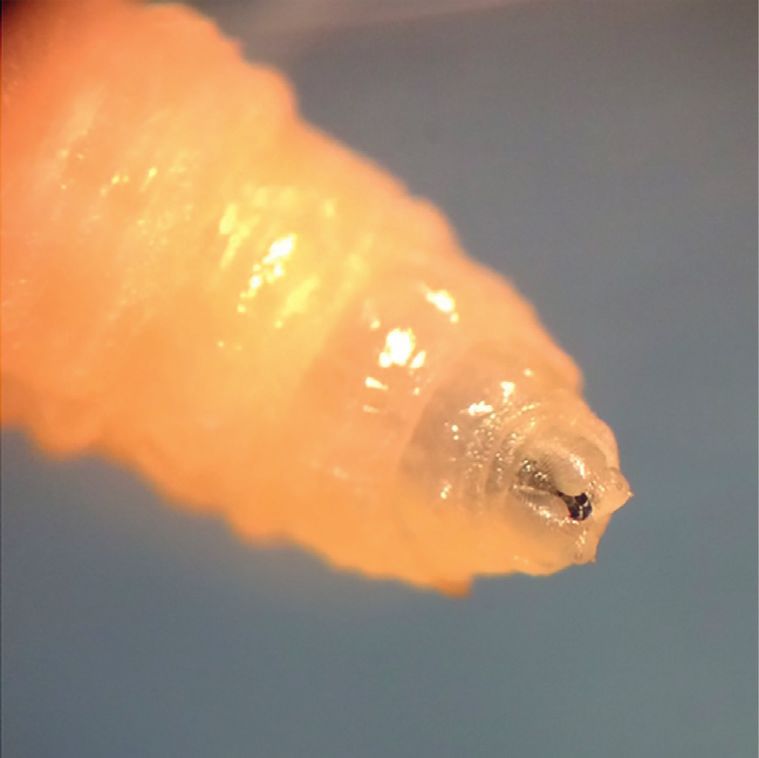
Figure 1: (a) Anterior end of the Sarcophaga spp: larva with two oral hooks. (b) Posterior spiracles located deep inside a fossa with
surrounding tubercles. (c) Three parallel slits surrounded by an incomplete peritreme.
2. Case Presentation 3. Discussion
A two-year-old male from Sauceda, a rural community of Several species of dipterous larvae including Sarcophaga spp.
Limatambo district (elevation 2,550 meters) in the Cusco are capable of producing intestinal myiasis [8]. In most cases,
region of Peru, presented with four white, mobile larvae in his the infection is caused by accidental ingestion of eggs laid on
stool measuring approximately 12 mm. This child was a par- exposed food [12]. As contaminated food passes through the
ticipant in a community study evaluating the prevalence of alimentary tract, eggs will hatch, and released larvae inhabit
Strongyloides stercoralis infection. Participants in this study the lower gastrointestinal tract before being passed in the stool
were instructed to collect freshly produced stool samples di- [12]. Sarcophaga larvae, the genus of the flesh fly family, are
rectly in clean plastic containers and to close them with equipped to feed from tissue and can cause damage to the
a hermetic lid. The stool samples were immediately tested by intestinal mucosa accounting for the symptoms reported in
the agar plate culture method. This method uses a Petri dish some patients [13]. The infestation is self-limited in most cases
with a nutritive agar in which a portion of the stool sample is because larvae are excreted in the stool, where they are often
placed. Then a lid is taped hermetically to prevent Strong- found alive. This infestation does not allow for reproduction
yloides larvae from escaping, and the dish is incubated to of larvae inside the host; however, with repeated ingestion of
observe the track left by Strongyloides larvae in the agar. The fly eggs, protracted cases have been reported with patients passing
larvae were discovered incidentally in the agar culture plate of larvae for months or years [14]. Intestinal myiasis should be
this child. The patient was previously healthy, and the mother differentiated from pseudomyiasis, in which the patients ingest
denied any symptoms including fever, nausea, vomiting, di- larvae rather than eggs and then pass intact larvae in the stool
arrhea, and abdominal pain. He did not have any apparent [15]. The larvae in pseudomyiasis are passed dead and never
skin wounds on his body. The mother reported that the child’s truly colonize the intestinal tract [15]. Larva can also be
diet consisted of poultry, fruit, and vegetables. He lived with deposited in fecal samples left uncovered and typically present
several extended family members in an adobe house with dirt at earlier stages of development upon examination.
floors and opened windows. Several animals including dogs, Our patient presented with multiple, living, L3 larvae in
cats, ducks, and chickens roamed freely in and out of the his stools and thus likely contracted intestinal myiasis from
household. The house was largely infested with flies. accidental ingestion of eggs. He lived in an adobe house from
Morphological examination of the organism revealed a rural community in close contact with several animals.
features consistent with L3 larvae of Sarcophaga spp. The Poor hygienic practices, lack of refrigeration of food, the
larva had smooth body segments with a broad posterior end proximity to a variety of farm animals, and open dwelling
and a tapering anterior end with two oral hooks and mouth allowed for potential sources of infection in our patient [16].
brushes (Figure 1(a)). The posterior spiracles were located In rural areas, domestic animals like dogs and cats are
deep inside a fossa, surrounded by more than 10 tubercles sometimes infested with fly larvae and can be a source for
(Figure 1(b)). The hidden spiracles had the characteristic infection in children [17]. There have been reports of in-
findings of the genus Sarcophaga. The posterior spiracles testinal myiasis caused by ingestion of over ripened fruits
consisted of three parallel slits surrounded by an incomplete such as pears or bananas [18, 19]. Drinking contaminated
peritreme, with the inner slit directed away from the median water has also been reported as a source of infection [20].
line ventrally (Figure 1(c)). These findings are supported by Several cases of intestinal myiasis caused by Sar-
the diagnostic criteria of Sarcophaga spp. reported in the cophaga spp. have been reported in the literature (Table 1).
literature [10, 11]. The four species identified include Sarcophaga crassipalpis,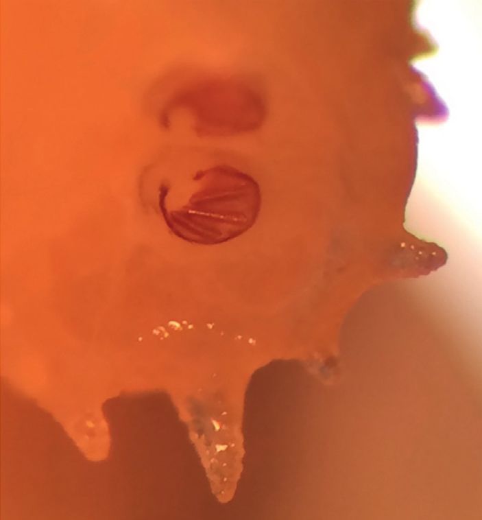
Case Reports in Infectious Diseases                                                                                                        3

                      Table 1: Human cases of intestinal myiasis caused by Sarcophaga spp. reported in the literature.
Author/year                    Species           Age         Sex      Country      Exposure                     Symptoms
Shiota et al.                Sarcophaga                                          Contaminated        Abdominal pain, fever, vomiting, and
                                                   4        Male       Japan
1990 [13]                    crassipalpis                                            food                         diarrhea
                             Sarcophaga                                          Contaminated
                                                   2       Female      Japan                                    Abdominal pain
Nagakura et al.              crassipalpis                                            food
1991 [18]                    Sarcophaga                                          Contaminated
                                                  38        Male       Japan                                     Asymptomatic
                              peregrina                                              food
Tachibana et al.             Sarcophaga                                          Contaminated
                                              Unknown Unknown          Japan                                     Asymptomatic
1987 [21]                     peregrina                                              food
Hasegawa et al.              Sarcophaga
                                               8 months    Female      Japan        Unknown          Change in behavior and bloody stools
1992 [22]                     peregrina
                             Sarcophaga                                                                 Abdominal pain, diarrhea, and
                                                  25        Male       India        Unknown
                           haemorrhoidalis                                                                      flatulence
                             Sarcophaga
                                                  35        Male       India        Unknown             Abdominal pain and flatulence
                           haemorrhoidalis
Udgaonkar et al.             Sarcophaga
                                                  28        Male       India        Unknown             Abdominal pain and flatulence
2012 [14]                  haemorrhoidalis
                             Sarcophaga                                          Living in a rural      Abdominal pain, flatulence, and
                                                  30        Male       India
                           haemorrhoidalis                                             area                 generalized weakness
                             Sarcophaga                                          Living in a rural
                                                  38        Male       India                            Abdominal pain and flatulence
                           haemorrhoidalis                                             area
                             Sarcophaga                                United                        Flatulence, generalized weakness, loss
                                                  37       Female                   Unknown
                               bullata                                 States                             of appetite, and weight loss
                             Sarcophaga                                United                        Generalized weakness, loss of appetite,
                                                  66       Female                   Unknown
                               bullata                                 States                                   and weight loss
Watson 1942 [23]
                             Sarcophaga                                United                           Abdominal pain, flatulence, and
                                                  43        Male                    Unknown
                               bullata                                 States                                generalized weakness
                             Sarcophaga                                United                             Abdominal pain, flatulence,
                                                  51        Male                    Unknown
                               bullata                                 States                          constipation, and loss of appetite
Kenney et al.                                                          United
                           Sarcophaga spp.        60        Male                    Unknown                        Unknown
1976 [24]                                                              States
                                                                                                     Abdominal pain, loss of appetite,
Ahmad et al.                                                                     Living in a rural
                           Sarcophaga spp.        10        Male       Egypt                       nausea, vomiting, diarrhea, and bloody
2011 [25]                                                                              area
                                                                                                                   stools
Das et al. 2010 [8]        Sarcophaga spp.        25        Male       India        Unknown                   Asymptomatic
Watanabe et al.                                                                   Contaminated
                           Sarcophaga spp.        61        Male       Japan                                     Diarrhea
2016 [4]                                                                               food

Sarcophaga peregrina, Sarcophaga haemorrhoidalis, and                   eliminate intestinal larvae almost immediately [14]. Treatment
Sarcophaga bullata. The age of patients reported varies                 with antihelminthic medications such as albendazole has not
widely from 8 months to 66 years, being the average 33                  shown to improve symptoms in patients [8, 9, 14]. Education
years at presentation. The flesh fly belonging to the Sar-                on good food-handling practices and avoiding the con-
cophagidae family has worldwide distribution [8]. Cases                 sumption of food products exposed to flies is important for
have been reported from Japan, India, Egypt, and the                    prevention of this disease [14]. No reports of oral ivermectin
United States. The signs and symptoms of presentation are               use have been published in intestinal myiasis, but this might
nonspecific and vary between reports, with some patients                 be a therapeutic option in severe cases.
being asymptomatic like in this child’s case [13]. Sarcophaga               In conclusion, intestinal myiasis caused by Sarcophaga spp.
species are generally present in rural and urban environments           is a rare occurrence in humans that is often self-limited. Pa-
and are commonly found in houses and indoor dwellings [25].             tients may have a range of presentations going from asymp-
Our review revealed that living in rural areas and the ingestion        tomatic to nonspecific abdominal symptoms. Intestinal myiasis
of contaminated food products were factors that authors as-             can pose a diagnostic challenge for physicians unfamiliar with
sociated with infestation by Sarcophaga spp.                            the condition. Education on good food handling is advised to
    Intestinal myiasis can be largely benign or cause severe            prevent reinfection.
clinical symptoms, depending on the larval species, number,
and location within the digestive tract [14, 26]. In some cases,        Data Availability
like ours, larvae can be passed out in feces without causing
many symptoms [14]. Colonic washes with polyethylene glycol             Data will be freely available through the corresponding
have been reported to relieve gastrointestinal symptoms and             author upon request.
4                                                                                                      Case Reports in Infectious Diseases

Conflicts of Interest                                                      [19] D. E. North, K. L. Matteson, S. D. Helgerson et al., “Intestinal
                                                                                myiasis in a baby attending a public health clinic,” Nurse
The authors declare that they have no conflicts of interest.                     Practitioner, vol. 12, no. 5, pp. 60–62, 1987.
                                                                           [20] A. Clavel, M. Toledo, P. Goni, and C. Aspiroz, “Intestinal
                                                                                myiasis due to Eristalis tenax: report of a new case in Spain,”
References                                                                      New Microbiologia, vol. 34, pp. 335-336, 2011.
                                                                           [21] H. Tachibana, M. Sasao, T. Tanaka et al., “A case of intestinal
 [1] F. Francesconi and O. Lupi, “Myiasis,” Clinical Microbiology               myiasis in Japan,” Tokai Journal of Experimental and Clinical
     Reviews, vol. 25, pp. 79–105, 2012.                                        Medicine, vol. 12, no. 5–6, pp. 349–352, 1988.
 [2] D. John and W. Petri, Markell and Voge’s Medical Parasitology,        [22] S. Hasegawa, H. Miwata, S. Masuda, H. Naruse, and T. Ozaki,
     Saunders Elsevier, Missouri, USA, 9th edition, 2006.                       “An infantile case of intestinal myiasis,” Pediatrics In-
 [3] R. Sehgal, H. P. S. Bhatti, D. K. Bhasin et al., “Intestinal               ternational, vol. 34, no. 1, pp. 87–89, 1992.
     myiasis due to Musca domestica: a report of two cases,” Japan         [23] J. R. Watson, “Sarcophaga bullata parker as a cause of in-
     Journal of Infectious Disease, vol. 55, pp. 191–193, 2002.                 testinal myiasis,” Florida Entomologist, vol. 25, no. 1, pp. 5-6,
 [4] N. Watanabe, T. Kato, Y. Ichiyanagi et al., “A case report of              1942.
     intestinal myiasis in a Japanese man,” Open Journal of Pa-            [24] M. Kenney, L. K. Eveland, V. Yermakov, and D. Y. Kassouny,
     thology, vol. 6, no. 4, pp. 171–176, 2016.                                 “Two cases of enteric myiasis in man: pseudomyiasis and true
 [5] M. T. Shazia, S. Anjum, and M. J. Yousuf, “Systematics and                 intestinal myiasis,” American Journal of Clinical Pathology,
     population of sarcophagid flies in Faisalabad (Pakistan),”                  vol. 66, no. 5, pp. 786–791, 1976.
     International Journal of Agriculture and Biology, vol. 8,             [25] A. K. Ahmad, E. H. Abdel-Hafeez, M. Makhloof, and
     pp. 809–811, 2006.                                                         E. M. Abdel-Raheem, “Gastrointestinal myiasis by larvae of
 [6] V. Kandi, S. K. Lal, Akhila et al., “Persistent pediatric gastro-          Sarcophaga spp. and Oestrus spp. in Egypt: report of cases, and
     intestinal myiasis: a case report of fly larval infestation with            endoscopical and morphological studies,” Korean Journal of
     Musca domestica with review of literature,” Journal of Global              Parasitology, vol. 49, no. 1, pp. 51–57, 2011.
     Infectious Diseases, vol. 5, no. 3, pp. 114–117, 2013.                [26] A. Aguilera, A. Cid, B. J. Regueiro, J. M. Prieto, and M. Noya,
 [7] R. S. Soliman, G. Phillips, and G. Spence, “‘I know an old lady            “Intestinal myiasis caused by Eristalis tenax,” Journal of
     who swallowed a fly’: a case of (hospital-acquired) human                   Clinical Microbiology, vol. 37, p. 3082, 1999.
     intestinal myiasis,” Journal of Hospital Infection, vol. 53, no. 2,
     pp. 157-158, 2003.
 [8] A. Das, A. Pandey, M. Madan, A. Asthana, and A. Gautam,
     “Accidental intestinal myiasis caused by genus Sarcophaga,”
     Indian Journal of Medical Microbiology, vol. 28, no. 2, p. 176,
     2010.
 [9] H. Karabiber, D. G. Oguzkurt, D. G. Dogan, M. Aktas, and
     M. A. Selimoglu, “An unusual case of rectal bleeding: in-
     testinal myiasis,” Journal of Pediatric Gastroenterology and
     Nutrition, vol. 51, no. 4, pp. 530-531, 2010.
[10] J. Sanjean, Taxonomic Studies of Sarcophaga Larvae of New
     York, with Notes on the Adults, Cornell University Agricul-
     tural Experiment Station, Ithaca, NY, USA, 1957.
[11] J. M. Seago, Fly Larvae: Pictorial Key to Some Common Species,
     https://www.cdc.gov/nceh/ehs/docs/pictorial_keys/flies.pdf.
[12] G. Desoubeaux, J. Gaillard, D. Borée-Moreau et al., “Gas-
     trointestinal symptoms resembling ulcerative proctitis caused
     by larvae of the drone fly Eristalis tenax,” Pathogens and
     Global Health, vol. 108, no. 3, pp. 158–163, 2014.
[13] T. Shiota, Y. Yoshida, S. Hirai, and S. Torii, “Intestinal myiasis
     caused by Parasarcophaga crassipalpis (Dipera: Sarcoph-
     agidae),” Pediatrics, vol. 85, pp. 215–217, 1990.
[14] U. S. Udgaonkar, R. Dharamsi, S. A. Kulkarni et al., “Intestinal
     myiasis,” Indian Journal of Medical Microbiology, vol. 30,
     no. 3, pp. 332–337, 2012.
[15] J. J. Laarman and P. H. Van Thiel, “A peculiar case of intestinal
     (pseudo)myiasis and a case of wound myiasis in the Netherlands,”
     Tropical and Geographical Medicine, vol. 19, pp. 187–191, 1967.
[16] E. K. Markel, M. Voge, and D. T. John, Medical Parasitology,
     W.B. Saunders, Philadelphia, PA, USA, 7th edition, 1992.
[17] B. Dik, U. Uslu, and N. Işık, “Myiasis in animals and human
     beings in Turkey,” Journal of the Faculty of Veterinary
     Medicine, Kafkas University, vol. 18, pp. 37–42, 2012.
[18] K. Nagakura, Y. Kawauichi-Kato, H. Tachibana et al., “Three
     cases of intestinal myiasis in Japan,” Journal of Infectious
     Diseases, vol. 163, no. 5, pp. 1170-1171, 1991.
MEDIATORS         of

                                                                                   INFLAMMATION

The Scientific                                 Gastroenterology                                                              Journal of
World Journal
Hindawi Publishing Corporation
                                               Research and Practice
                                               Hindawi
                                                                                   Hindawi
                                                                                                                             Diabetes Research
                                                                                                                             Hindawi
                                                                                                                                                              Disease Markers
                                                                                                                                                              Hindawi
                                                                                   www.hindawi.com        Volume 2018
http://www.hindawi.com
www.hindawi.com                  Volume 2018
                                        2013   www.hindawi.com   Volume 2018                                                 www.hindawi.com    Volume 2018   www.hindawi.com            Volume 2018

Journal of                                                                                                                                                    International Journal of
Immunology Research
Hindawi
                                                                                                                                                              Endocrinology
                                                                                                                                                              Hindawi
www.hindawi.com                  Volume 2018                                                                                                                  www.hindawi.com            Volume 2018

                                                                 Submit your manuscripts at
                                                                    www.hindawi.com

                                                                                                                                                              BioMed
PPAR Research
Hindawi
                                                                                                                                                              Research International
                                                                                                                                                              Hindawi
www.hindawi.com                  Volume 2018                                                                                                                  www.hindawi.com            Volume 2018

                                                                                                                             Journal of
                                                                                                                             Obesity

                                                                               Evidence-Based
Journal of                                     Stem Cells                      Complementary and                                                              Journal of
Ophthalmology
Hindawi
                                               International
                                               Hindawi
                                                                               Alternative Medicine
                                                                               Hindawi                                       Hindawi
                                                                                                                                                              Oncology
                                                                                                                                                              Hindawi
www.hindawi.com                  Volume 2018   www.hindawi.com   Volume 2018   www.hindawi.com                 Volume 2018   www.hindawi.com    Volume 2018   www.hindawi.com            Volume 2013

                                                                               Parkinson’s
                                                                               Disease

Computational and
Mathematical Methods
in Medicine
                                               Behavioural
                                               Neurology
                                                                                                                             AIDS
                                                                                                                             Research and Treatment
                                                                                                                                                              Oxidative Medicine and
                                                                                                                                                              Cellular Longevity
Hindawi                                        Hindawi                         Hindawi                                       Hindawi                          Hindawi
www.hindawi.com                  Volume 2018   www.hindawi.com   Volume 2018   www.hindawi.com                 Volume 2018   www.hindawi.com    Volume 2018   www.hindawi.com            Volume 2018

You can also read