Ectopic palatine tonsil meningioma in type 2 neurofibromatosis: a few times documented phenomenon
←
→
Page content transcription
If your browser does not render page correctly, please read the page content below
tumors of the lip and oral cavity
ISSN 2595-2544
Ectopic palatine tonsil
CASE REPORT
meningioma in type 2
neurofibromatosis: a few times
documented phenomenon
Juan Miguel Alemán-Iñiguez1* , Christian Valencia Padilla2
Abstract
Type 2 neurofibromatosis is a hereditary neurocutaneous entity, characterized by
associations with cranial and spinal meningiomas; extra neural cases are infrequent.
There are oral meningiomas reports, however their presence at the palatine tonsils
have not been described. This is a patient, with type 2 neurofibromatosis inherited
from her father. The guest, presented progressive odyno-dysphagia whose etiology was
WHO I meningioma in palatine tonsils, while the cranial image showed same lineage
meningiomas that were surgically treated. This rare phenomenon in the context of an
uncommon disease and the events that would explain the arrival of menigiomatous
tissue to the palate are discussed.
Keywords: meningioma; neurofibromatosis 2; palatine tonsil.
How to cite: Alemán-Iñiguez JM, Valencia Padilla C. Ectopic palatine tonsil meningioma
in type 2 neurofibromatosis: a few times documented phenomenon. Arch Head Neck
Surg. 2021;50:e202150124. https://doi.org/10.4322/ahns.2021.0005
Universidad San Francisco de Quito,
1
Neurocirugía, Quito, Pichincha, Ecuador
2
Hospital de Especialidades Carlos Introduction
Andrade Marín, Neurocirugía,
Quito, Pichincha, Ecuador
The Neurofibromatosis type 2 (NF-2) (ORPHA: 637) is less frequent than
neurofibromatosis type 1 (NF-1), it has a prevalence of 1-9 / 100,000
inhabitants1; the meningiomas presence constitutes part of the NF-2 minor
Financial support: None. diagnostic criteria, they are the second most common extra-axial tumors after
Conflicts of interest: No conflicts
of interest declared concerning schwannomas2; the natural history characteristics of meningiomas in NF2
the publication of this article. are: slow growth, recurrence and appearance of same nature new tumors,
Submitted: July 08, 2021.
Accepted: July 31, 2021. including in the resected tumor bed. Unlike NF-1, in NF-2lesions have a low
The study was carried out at Hospital rate of malignancy3; the growth pattern and tumor grade are affected by
de Especialidades Carlos Andrade radiotherapy, which may have little benefit4. Between 26 and 36 genetic
Marín, Quito, Pichincha, Ecuador.
mutations have been described in NF2 meningiomas, the main ones: mutation
of the NF-2 gene at the 22q locus and in the cell membrane tumor suppressor
protein, MERLIN5; extraneural and multiple meningiomas are little mentioned,
Copyright Alemán-Iñiguez et al. This
is an Open Access article distributed aggressive and affect hereditary phenotypes and truncated mutations; their
under the terms of the Creative
preferred treatment is extirpation, like those located in neural tissues6. In this
Commons Attribution License,
which permits unrestricted use, case, the patient presented major and minor criteria for NF-2, the meningioma
distribution, and reproduction in any
medium, provided the original work
on the palate was compatible with the same histopathological lineage as on
is properly cited. the intracranial meningiomas. Our purpose is to discuss about extra neural,
Alemán-Iñiguez et al. Arch Head Neck Surg. 2021;50:e202150124. DOI: 10.4322/ahns.2021.0005 1/9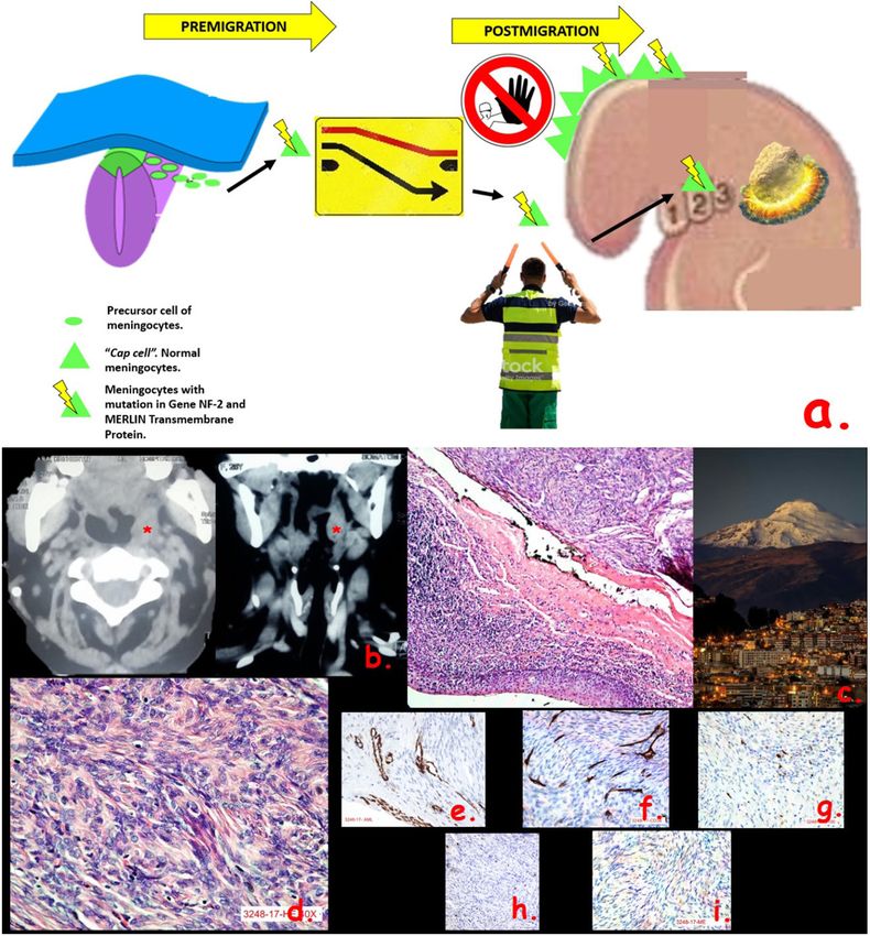
Ectopic palatine tonsil meningioma in type 2 neurofibromatosis: a few times documented EMBRYOLOGY AND CONGENITAL DISEASES | LIP AND ORAL
phenomenon CAVITY TUMORS | MISCELLANEOUS [[Q1: Q1]]
non-metastatic and not associated with invasion from cranial meningiomas
and our objective is to explain the events that would allow menigiomatous
tissue to reach the palate.
Case report
The patient currently in her third decade of life, was diagnosed during her
childhood with hereditary NF-2, had history of cutaneous neurofibromas
surgically treated, there was no exposure to radiotherapy; reported that for
3 years, with no apparent cause, has had a sensation of pharyngeal foreign
body, is accompanied by progressive dysphagia for solids without difficulty
for liquids (HP: 0200136), in addition headache with evening predominance
(HP: 0002315) and walk difficulty (HP: 0002355). Hearing loss and tinnitus
(HP: 0000360), nocturnal snoring, and increased dysphagia are the major
complaints.
The physical examination highlights were: the presence of café-au-lait macules
on the legs, hips and back (HP: 0000957), medialized left tonsillar pillar, uvula
displaced, the tonsil with an exophytic, violaceous upper pole lesion with a
diameter of two centimeters; neurological examination revealed bilateral
sensory hearing loss predominantly on the right ear, peripheral vertiginous
syndrome predominantly on the right (HP: 0002321), non-ophthalmoparesis,
spastic tetraparesis predominantly in the lower limbs. (HP: 0001285). Karnofsky
performance scale (KPS) was 90%. Imaging studies reported intracranial
and oropharyngeal lesions (Figure 1a-f, Figure 2a), contrasted body scan
tomography did not show other lesions.
The upper pole lesion of the palatal tonsil was excised by the
otorhinolaryngology team, in other surgical time than the intracranial
procedure, it was observed to be pale, pedunculated 1.5 x 1.5 cm, with a
histopathological result of WHO I fibrous meningioma (Figure 2c-i)
In the neurosurgical procedure, parasagittal meningiomas were excised
(Figure 1g-m), the result of which was WHO I fibrous type. Eight months later,
the patient presented altered consciousness and tetraplegia (HP: 0030182). A
total excision of brainstem glioma was performed. it was a WHO II astrocytoma,
improving its condition, currently with KPS 80% and in observation of: acoustic
schwannomas, remaining tentorium, convexity and falx meningiomas that
do not present symptoms; she abstains from radiation therapy because of
the risk of detrimentally influencing the natural history of the lesions.
Discussion
The extra neural meningiomas in NF-2 are mostly given by invasion of
intracranial primaries meningiomas from: orbit, pterygopalatine-subtemporal
fossa, nasal-oral cavity and neck, being considered as aggressive forms of
the disease, primary lesions without relation to another intracranial one are
the 2% of meningiomas7; Hoye classified in type 3 extraneural meningiomas
those with no relation to neurophoramens or metastases, he called these
ectopic meningiomas8. The case presented has these characteristics, so is
considered as an oropharynx ectopic meningioma.
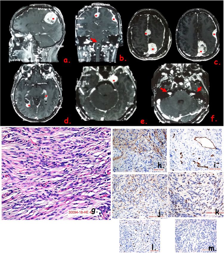
Alemán-Iñiguez et al. Arch Head Neck Surg. 2021;50:e202150124. 2/9Ectopic palatine tonsil meningioma in type 2 neurofibromatosis: a few times documented EMBRYOLOGY AND CONGENITAL DISEASES | LIP AND ORAL phenomenon CAVITY TUMORS | MISCELLANEOUS [[Q1: Q1]] Figure 1. (a) Cerebral Falx meningioma in MRI sagittal section; (b) Cerebral falx, and convexity meningiomas and brainstem glioma (red arrow) in MRI coronal section; (c) Cerebral falx and convexity meningiomas in MRI axial slices; (d) Tentorium meningiomas in MRI axial section; (e) Sphenoidal minor wing meningioma in MRI axial section; (f) Bilateral vestibular schwannomas in MRI axial section; (g) Hematoxylin-eosin X40 stain: swirling cap cells, no mitosis; (h) Epithelial membrane antigen IQ; (i) CD34 IQ; (j) CD68 IQ; (k) Progesterone IQ; (l) KI-67 IQ; (m) S100 IQ. (Source: imaging and pathology center of the Carlos Andrade Marín Hospital, Quito-Ecuador). Menigiomas with contrast enhancement (Red*). Magnetic Resonance image (MRI). Immunohistochemistry (IQ). Alemán-Iñiguez et al. Arch Head Neck Surg. 2021;50:e202150124. 3/9
Ectopic palatine tonsil meningioma in type 2 neurofibromatosis: a few times documented       EMBRYOLOGY AND CONGENITAL DISEASES | LIP AND ORAL

phenomenon                                                                                             CAVITY TUMORS | MISCELLANEOUS [[Q1: Q1]]

Figure 2. (a) Diagram of the pathophysiology of the migration in ectopic meningiomas to the second branchial bag. (Created by:
Author JMAI); (b) Palatine tonsilar meningioma in axial and coronal neck tomography; (c) Hematoxylin-eosin X10 stain image:
palatal tonsil lined by squamous and respiratory epithelium (pseudostratified) in the stroma showing mesenchymal neoplasia
constituted by swirling of spindle cells with large nuclei and small nucleoli of arachnoid tissue, two different tissues (clearly
delimited respiratory and nervous) next to it, an image that exemplifies the contrast of these two tissues; the landscapes contrast
between the Nevado Cayambe and the City of Quito. (Source: Carlos Andrade Marín hospital pathology laboratory and photo
of the Ecuadorian Ministry of Tourism); (d) hematoxylin-eosin X40; (e) Smooth muscle antigen IQ; (f) CD-34 IQ; (g) S-100 IQ; (h)
Ki-67 IQ; (i) Epithelial membrane antigen IQ. (Source: imaging and pathology center of the Carlos Andrade Marín Hospital, Quito-
Ecuador). Ectopic palatine tonsil Menigioma (Red*). Immunohistochemistry (IQ).

Alemán-Iñiguez et al. Arch Head Neck Surg. 2021;50:e202150124.                                                                            4/9
Ectopic palatine tonsil meningioma in type 2 neurofibromatosis: a few times documented        EMBRYOLOGY AND CONGENITAL DISEASES | LIP AND ORAL

phenomenon                                                                                              CAVITY TUMORS | MISCELLANEOUS [[Q1: Q1]]

                                                The pathophysiology is not clarified, several theories have been proposed
                                                in the embryological development of the neural crest arachnoid precursors
                                                cells, their remnants are evidence of the failure in cranial migration, the
                                                mechanisms that originate ectopic meningiomas are: (1) failure in cell signaling
                                                to their destinations in arachnoid tissues, “staying in the middle of the road”
                                                (2) erroneous cell signaling implanting itself in tissues other than neural, (3)
                                                interruption of its migration paths by the presence of other abnormal cells
                                                that will form intracranial lesions, the latter two allow meningocytes to collide
                                                with mesenchymal and endodermal tissues and (4) the dedifferentiation of
                                                normal peripheral neural tissues to meningocytes, the latter less approved9,10;
                                                the crash of neuroectodermal cells to the epithelium (endodermal origin) of
                                                the second pharyngeal bag which is the primordium of the palatine tonsils
                                                could occur between the third and fifth month of intrauterine period, a
                                                time when infiltration of lymphatic tissue in this structure also occurs11; as
                                                a hypothesis applied in our case (Figure 2a).
                                                In the NF-2 embryology, the following play a leading role: the mutation of the
                                                NF-2 gene and the tumor suppressor MERLIN, overexpressed in experimental
                                                models, in extra neural structures such as: the branchial arches, the dorsal
                                                aorta and para-aortic splenopleura, in addition, a high expression in cells that
                                                migrated from the dorsal neural crest, in contrast to almost zero expression
                                                in those same cells in the premigration stage, this corroborates the role of the
                                                mutation of the NF-2 and MERLIN gene in migration, adhesion cell, motility
                                                and proliferation during development9, which supports the explanation of
                                                migration failure and ectopic tumor settlement in our patient.
                                                The associations of neurofibromatosis and anomalies of the second bronchial
                                                arch is an unclear phenomenon, there are reports of persistence of clefts,
                                                cysts and hamartomas in suprahyoid and pharyngeal structures in NF12,
                                                however, ectopic meningiomas are not considered as part of these anomalies.
                                                Symptoms in NF-2 due to intracranial lesions present between the age of 20
                                                and 30 years (minor diagnostic criteria, especially in vestibular schwannomas)3,
                                                this same evolution was found in reports of primary extracranial neck
                                                meningiomas where parapharyngeal locations are mentioned; the main
                                                symptomatology was the mass sensation with dysphagia, which concludes
                                                that presentation is common in people under 30 years old8, including
                                                sporadic extra neural meningiomas, NF-1 and NF-2 parapharyngeal ectopic
                                                primary meningiomas13,14, although it is not exclusive to NF, for instance, a
                                                sporadic pharyngeal ectopic case is mentioned without an association of
                                                any intracranial lesion or any syndrome15; the palatine tonsils are one of the
                                                affected second pharyngeal bag structures, other damaged parts with the
                                                same origin mentioned in NF-2 are: the carotid-thyroid space, the laryngo-
                                                pharynx and the sub and retrolingual space, all of them with evidence of
                                                intracranial lesions16, it is important to study the neck in NF-2 patients with
                                                dysphagia under fourth decade of life, as in our case, because the possibility
                                                of development meningiomatous lesions in the second branchial arch.
                                                The most frequent histotype of meningiomas associated with NF-2 is
                                                meningothelial, while the mutation of the NF-2 gene is the most present
                                                in these, even in sporadic forms17; In the most extensive series of cases
                                                of primary ectopic neck meningiomas, they report the transitional type as

Alemán-Iñiguez et al. Arch Head Neck Surg. 2021;50:e202150124.                                                                             5/9
Ectopic palatine tonsil meningioma in type 2 neurofibromatosis: a few times documented EMBRYOLOGY AND CONGENITAL DISEASES | LIP AND ORAL
phenomenon CAVITY TUMORS | MISCELLANEOUS [[Q1: Q1]]
the most present, they do not mention whether these coincided with the
intracranial or intraspinal lesions lineage in the NF-2 participants8, our case
was fibrous type (the second most frequent type of WHO I meningiomas),
cranial and palatal types were the same nature, with specific differences in
immunohistochemistry (Table 1).
Table 1. Immunohistochemistry comparison between of the NF-2 patient meningiomas.
Intracranial meningioma. Palatal tonsil meningioma.
Vimentin and Epithelial membrane
+++ diffuse. ++ diffuse.
antigen (EMA).
Progesterone. ++ diffuse. + diffuse.
Smooth muscle antigen (SML). +/-. + Focal in accompanying vessels.
CD-34. + Focal in accompanying vessels. + Focal in accompanying vessels.
S-100. + focal. + Weakly focal.
CD-68. +. +/-.
Ki-67. Negative. Negative.
The number of cranial and extracranial lesions reported in similar cases
was not variable in the analysis, but in molecular studies they conclude that
several lesions, including multiple types, MISME Syndrome (Meningiomas,
schwannomas, ependymomas and gliomas) is proportional to the degree of
aggressiveness and recurrence18, but not to malignancy, also more present
in direct hereditary forms19, while series of case studies of extracranial
meningiomas report coincidences of bilateral lesions on the face and neck
in NF-28, our patient inherited her pathology from her father, for therefore,
this trait could be demonstrated with its phenotype of having several lesions:
acoustic schwannomas, supratentorial meningiomas and in the palatal tonsil
and brainstem glioma.
The impact of the presence of ectopic meningiomas on the NF-2 overall
survival and prognosis has not been shown to be related, however there
are molecular bases of mutated aggressive forms that could be associated
with this phenomenon, even neural lesions play a more important role in the
evolution of astrocytomas and ependymomas of the brainstem and spinal
cord6, in this case the greatest impact on the patient morbidity was the
brainstem glioma.
The cornerstone of treatment is the radical excision of the lesions; adjuvant
radiotherapy and chemotherapy are controversial, in general the annual
growth rate and possibility of recurrence is slower than sporadic lesions2,
small lesions are preferably followed with observation since radiotherapy
has the risk of modifying the benign nature and natural history, increasing
its growth rate, even reporting malignancy20-22; In our case, we proceeded
to total excision of the lesion in the palatine tonsils, performing total
tonsillectomy and excision of parasagittal meningioma at the cranial location,
Alemán-Iñiguez et al. Arch Head Neck Surg. 2021;50:e202150124. 6/9Ectopic palatine tonsil meningioma in type 2 neurofibromatosis: a few times documented EMBRYOLOGY AND CONGENITAL DISEASES | LIP AND ORAL
phenomenon CAVITY TUMORS | MISCELLANEOUS [[Q1: Q1]]
as we mentioned before, the patient was complicated with growth of a
brainstem glioma which was performed total exeresis, currently the patient
is in control of other injuries; In view of the increased growth of existing
lesions, surgical intervention will be proposed over other medical procedures
such as radiotherapy.
Conclusions
• Our patient belongs to The NF-2 with strong inheritance features has a
great possibility of expression of meningiomas in ectopic locations;
• The palatine tonsil is part of the neck and the oral cavities meningiomas
that are more frequent after the nervous system;
• Multiple intracranial meningiomas like our case report can be associated
with ectopic locations;
• The physicians who follow NF-2 patients should think about extra neural
locations in presence of atypical symptoms and in ages under 30 years;
• Our case, just like the most NF-2 patients, the most important etiology
are the embryological theories to explain the palate tonsil meningiomas,
because are congenital tumors originating from development and can
be under-diagnosed;
• The total excision is the preferred treatment and the small lesions are
better to observe than to use radiotherapy, this strategy that was used
in our case;
• The recurrence is expected and malignancy is rare, important to maintain
long-term follow-up in our case.
Acknowledgements
To our patient, the main motivation for our research.
References
1. Slattery WH. Neurofibromatosis type 2. Otolaryngol Clin North Am. 2015;48(3):443-
60. http://dx.doi.org/10.1016/j.otc.2015.02.005. PMid:26043141.
2. Goutagny S, Kalamarides M. Medical treatment in neurofibromatosis type 2:
review of the literature and presentation of clinical reports. Neurochirurgie.
2018;64(5):370-4. http://dx.doi.org/10.1016/j.neuchi.2016.09.004.
PMid:28162254.
3. Dirks M, Butman J, Kim H, Wu T, Morgan K, Tran A, Lonser R, Asthagiri A. Long-
term natural history of neurofibromatosis type 2: associated intracranial tumors.
J Neurosurg. 2012;117(1):109-17. http://dx.doi.org/10.3171/2012.3.JNS111649.
PMid:22503123.
4. Evans DG. Neurofibromatosis type 2 (NF2): a clinical and molecular review.
Orphanet J Rare Dis. 2009;4(1):16. http://dx.doi.org/10.1186/1750-1172-4-16.
PMid:19545378.
Alemán-Iñiguez et al. Arch Head Neck Surg. 2021;50:e202150124. 7/9Ectopic palatine tonsil meningioma in type 2 neurofibromatosis: a few times documented EMBRYOLOGY AND CONGENITAL DISEASES | LIP AND ORAL
phenomenon CAVITY TUMORS | MISCELLANEOUS [[Q1: Q1]]
5. Ruttledge M, Andermann A, Phelan C, Claudio J, Han F, Chretien N, Rangaratnam
S, MacCollin M, Short P, Parry D, Michels V, Riccardi VM, Weksberg R, Kitamura
K, Bradburn JM, Hall BD, Propping P, Rouleau GA. Type of mutation in the
neurofibromatosis type 2 gene (NF2) frequently determines severity of disease.
Am J Hum Genet. 1996;59(2):331-42. PMid:8755919.
6. Evans DG, Bowers N, Huson SM, Wallace A. Mutation type and position varies
between mosaic and inherited NF2 and correlates with disease severity. Clin
Genet. 2013;83(6):594-5. http://dx.doi.org/10.1111/cge.12007. PMid:22989157.
7. Friedrich R, Hagel C. Expansive extracranial growth of intracranial meningioma
in neurofibromatosis type. Anticancer Res. 2016;36(6):3161-7. PMid:27272842.
8. Friedman CD, Costantino PD, Teitelbaum B, Berktold RE, Sisson GA Sr. Primary
extracranial meningiomas of the head and neck. Laryngoscope. 1990;100(1):41-8.
http://dx.doi.org/10.1288/00005537-199001000-00010. PMid:2104554.
9. Akhmametyeva E, Mihaylova M, Luo H, Kharzai S, Welling D, Chang L. Regulation
of the neurofibromatosis 2 gene promoter expression during embryonic
development. Dev Dyn. 2006;235(10):2771-85. http://dx.doi.org/10.1002/
dvdy.20883. PMid:16894610.
10. Vital RB, Hamamoto Filho PT, Lapate RL, Martins VZ, de Oliveira Lima F, Romero
FR, Zanini MA. Calvarial ectopic meningothelial meningioma. Int J Surg Case Rep.
2015;10:69-72. http://dx.doi.org/10.1016/j.ijscr.2015.03.033. PMid:25805612.
11. Molina Montes L, Montes de Oca Fernández F, Gamboa Mutuberría B.
Embriología y anatomía de la cavidad oral y faringe. In: Sociedad Española de
Otorrinolaringología y Cirugía de Cabeza y Cuello, editor. Cavidad oral y faringe.
1. ed. Alcalá de Henares: Hospital Universitario Príncipe de Asturias. Libro virtual
de formación. Capítulo 67; p. 2.
12. Zaifullah S, Yunus MR, See GB. Diagnosis and treatment of branchial cleft
anomalies in UKMMC: a 10-year retrospective study. Eur Arch Otorhinolaryngol.
2013;270(4):1501-6. http://dx.doi.org/10.1007/s00405-012-2200-7.
PMid:23053382.
13. Shetty C, Avinash KR, Auluck A, Mupparapu M. Extracranial meningioma
of the parapharyngeal space: report of a case and review of the literature.
Dentomaxillofac Radiol. 2007;36(2):117-20. http://dx.doi.org/10.1259/
dmfr/56368887. PMid:17403892.
14. Albsoul N, Rawashdeh B, Albsoul A, Abdullah M, Golestani S, Rawshdeh
A, Mohammad M, Alzoubi M. A rare case of extracranial meningioma in
parapharyngeal space presented as a neck mass. Int J Surg Case Rep. 2015;11:40-3.
http://dx.doi.org/10.1016/j.ijscr.2015.04.012. PMid:25912007.
15. Kumar S, Hasija S, Goyal R, Kataria SP, Sen R, Wadhera R. Ectopic parapharyngeal
meningioma: diagnosis of a rare entity on FNAC. J Surg Case Rep.
2012;2012(12):rjs024. http://dx.doi.org/10.1093/jscr/rjs024. PMid:24968421.
16. Amer HW, Hafed L, Ibrahim S, Shaker S. Case report: rare site for
intraoral meningioma. F1000 Res. 2020;9:95. http://dx.doi.org/10.12688/
f1000research.21999.2. PMid:32850120.
Alemán-Iñiguez et al. Arch Head Neck Surg. 2021;50:e202150124. 8/9Ectopic palatine tonsil meningioma in type 2 neurofibromatosis: a few times documented EMBRYOLOGY AND CONGENITAL DISEASES | LIP AND ORAL
phenomenon CAVITY TUMORS | MISCELLANEOUS [[Q1: Q1]]
*Correspondence 17. Evans JJ, Jeun SS, Lee JH, Harwalkar JA, Shoshan Y, Cowell JK, Golubic M. Molecular
Juan Miguel Alemán-Iñiguez alterations in the neurofibromatosis type 2 gene and its protein rarely occurring
Universidad San Francisco de Quito
(USFQ), Escuela de Postgrados, in meningothelial meningiomas. J Neurosurg. 2001;94(1):111-7. http://dx.doi.
Neurocirugía org/10.3171/jns.2001.94.1.0111. PMid:11147878.
Diego de Robles y Vía Interoceánica,
Medical Specialties Building, HDLV 18. Durand A, Champier J, Jouvet A, Labrousse F, Honnorat J, Guyotat J, Fèvre-Montange
111 – B. M. Expression of c-Myc, neurofibromatosis Type 2, somatostatin receptor 2 and
Postal Code 170901, Quito, Ecuador
erb-B2 in human meningiomas: relation to grades or histotypes. Clin Neuropathol.
Tel: +593 995534351
E-mail: juanmig_18@hotmail.com 2008;27(5):334-45. http://dx.doi.org/10.5414/NPP27334. PMid:18808065.
19. Coy S, Rashid R, Stemmer-Rachamimov A, Santagata S. An update on the CNS
Authors information
manifestations of neurofibromatosis type 2. Acta Neuropathol. 2020;139(4):643-
JMAI - Neurocirugía, Universidad San
Francisco de Quito. CVP - Neurocirigía, 65. http://dx.doi.org/10.1007/s00401-019-02029-5. PMid:31161239.
Hospital de Especialidades Carlos
Andrade Marín. 20. Grill J, Dhermain F, Habrand JL. Risks of radiation therapy in patients with
neurofibromatosis. Int J Radiat Oncol Biol Phys. 2009;75(2):632. http://dx.doi.
org/10.1016/j.ijrobp.2009.03.076. PMid:19735889.
21. Miao R, Wang H, Jacobson A, Lietz A, Choy E, Raskin K, Schwab J, Deshpande V,
Nielsen GP, DeLaney TF, Cote GM, Hornicek FJ, Chen YE. Radiation-induced and
neurofibromatosis-associated malignant peripheral nerve sheath tumors (MPNST)
have worse outcomes than sporadic MPNST. Radiother Oncol. 2019;137:61-70.
http://dx.doi.org/10.1016/j.radonc.2019.03.015. PMid:31078939.
22. Evans DG, Birch JM, Ramsden RT, Sharif S, Baser ME. Malignant transformation and
new primary tumours after therapeutic radiation for benign disease: substantial
risks in certain tumour prone syndromes. J Med Genet. 2005;43(4):289-94. http://
dx.doi.org/10.1136/jmg.2005.036319. PMid:16155191.
Alemán-Iñiguez et al. Arch Head Neck Surg. 2021;50:e202150124. 9/9You can also read