The cerebellum in children with spastic cerebral palsy: Volumetrics MRI study
←
→
Page content transcription
If your browser does not render page correctly, please read the page content below
Prog Health Sci 2011, Vol 1 , No2 Cerebellum cerebral palsy volumetrics study

The cerebellum in children with spastic cerebral palsy: Volumetrics MRI
study

Gościk E.1*, Kułak W.2, Gościk J.3, Gościk J.4, Okurowska-Zawada B.2, Tarasow E.5

1
  Department of Children’s Radiology, Medical University of Bialystok, Poland
2
  Department of Pediatric Rehabilitation, Medical University of Bialystok, Poland
3
  Faculty of Computer Science, Bialystok University of Technology, Poland
4
  Faculty of Mechanical Engineering, Bialystok University of Technology, Poland
5
  Department of Radiology, Medical University of Bialystok, Poland

ABSTRACT
__________________________________________________________________________________________

Purpose: To determine the volume of the              difference between the total cerebellar volume and
cerebellum in children with spastic cerebral palsy   gender in patients with CP was found. No
(CP) in relation to risk factors and motor           significant relationship between cerebellar volume
development.                                         and birth weight, Apgar score, gestational age, and
Material and methods: The present study included     Gross Motor Function Classification System
30 children with spastic CP, aged 2-17 years. The    (GMFCS) level were noted. Positive correlations
volume of the cerebellum was examined on sagittal    between birth weight, Apgar score, gestational age,
magnetic resonance images (MRI) of the CP            and GMFCS level, between Apgar score and
patients and on 33 healthy subjects. To estimate the gestational age, or between gestational age and
total cerebellum volume of each subject we used      GMFCS level were found.
Analyze 10 Biomedical Imaging Software.              Conclusion: Our results show that children with
Results: Children with spastic CP (129726,2 ±        spastic CP had smaller volumes of the cerebellum
26040,72 mm3) had a significantly smaller mean of    as compared to controls.
the cerebellum volume compared to controls           Key words: MRI, semi-automatic cerebellar
(143122,5 ± 12351,10 mm3). No significant            volume estimation, spastic cerebral palsy
__________________________________________________________________________________________

*Corresponding author:
Department of Children’s Radiology, Medical University of Bialystok
17 Waszyngtona str.
15-274 Bialystok, Poland
Tel. +48 85 7450627
E-mail : elzbieta.goscik@umwb.edu.pl (Elżbieta Gościk)

Received: 10.10.2011
Accepted: 5.12.2011
Progress in Health Sciences
Vol. 1(2) C 2011 · pp 67-75.
© Medical University of Bialystok, Poland
                                                                                                     67
INTRODUCTION prospective MRI scans that were performed for
clinical diagnostic purposes at Children’s
According to different statistics, cerebral University Clinical Hospital of Białystok. The
palsy (CP) occurs with frequency from 1.5 to 3.0 study was approved by the ethics committee of the
per 1000 live births. Clinically, the disease is Medical University of Bialystok. The subjects
dominated by signs of injury: motor cortex (limb consisted of two groups - a patient group and
paresis), basal ganglia (involuntary movements), controls. The patient group consisted of 30 subjects
cerebellum (congruity disorder of movement and with CP (aged 2-17 years, 21 males and nine
balance). Approximately, 30-40% of children with females). All CP individuals included into the study
CP finds varying degrees of mental retardation in were had CP (diplegia spastica, tetraplegia,
about 35% of cases of epilepsy. Impaired vision hemiplegia dextra or sinistra). In each case of CP,
(strabismus, eye disease) is seen in 50% of children the diagnosis was confirmed by authors. Children
and approximately 25% of hearing dysfunction [1- with postnatal meningitis, encephalitis, trauma, and
3]. In recent years, there has been an increase in the metabolic or degenerative disorders were excluded
number of new cases of CP by improving the care from the study. The control group included 33
of a mother and a child. CP, due to the incidence healthy children (aged 4-19 years with mean value
and multiplicity of symptoms, is important medical of age 11.12 and 4.77 SD; 16 males, 17 females).
and social problem [4-6]. Contemporary diagnostics The subjects in the control group were healthy
of children with CP, apart from the neurological individuals without any known history of any
examination, psychological and speech therapy as disease as well as were free of any psychiatric or
well as the neurophysiologic evaluation EEG also neurological abnormalities. MRI were normal. All
uses neuroimaging such as magnetic resonance the control subjects were right-handed.
imaging (MRI) [7-9]. Reports of recent years
Motor Function
indicate the potential to differentiate changes in the
Each child was classified according to the
various forms of CP by the technique of MRI [9-
Gross Motor Function Classification System
12]. Changes in white matter can be demonstrated
(GMFCS): level 1, walks without restrictions; level
in children with spastic diplegia, and tetraplegia in
2, walks without assistive devices, limitations in
the sequences of T1 and T2 and FLAIR [10-12].
walking outdoor; level 3, walks with assistive
The cerebellum plays the most important role in
devices; level 4, self-mobility with limitations,
motor coordination gathers information from many
children are transported or use powered mobility;
centers of the brain, analyzing them quickly and
and level 5, self-mobility is severely limited.
appropriately modulates that movements were
smooth and accurate [13-14]. The cerebellum Definitions
constantly monitors the course of the movement Cerebral palsy was classified as spastic
and coordinates the auto-patch as well as changes tetraplegia (spasticity of all four limbs and of about
the voltage of other skeletal muscles to restore equal involvement), and spastic diplegia –
balance. The cerebellum also regulates: tone spasticity of lower limbs more affected than the
(tension) muscle, balance, learning motor behavior upper. Hemiplegic CP refers to one arm and leg on
(i.e. cycling), smoothness and precision of either the right or left side of the body being
voluntary movements (by motor interacts with the affected [1]. Prematurity was defined by the World
surroundings of the cerebral cortex), to name of Health Organization as an infant with a gestation of
few. Despite the great importance of the less than 37 weeks from the first day of the last
cerebellum, there is still insufficient knowledge to menstrual period. Asphyxia is defined as an Apgar
justify the reports of its dysfunction in patients with score ≤ 4. Diagnosis of mental abnormality was
CP. The cerebellum is the subject of various studies based on clinical assessment, supplemented by
and research in this field [6,7,13,14]. Based on the standard tests if available at the time of diagnosis,
existing literature. We hypothesized that CP and the need for special education. Epilepsy was
children would demonstrate smaller cerebellum defined as a separate occurrence of two or more
volume and that these changes would be associated apparently unprovoked seizures [15].
with more severe motor delay and mental
development. In the present study, we present the Magnetic Resonance Imaging and Acquisition
results of volumetric studies of children with CP The MRI’s brain datasets have been
with the relation to age gender, Apgar score, obtained at Department of Imaging Diagnostic in
gestational age, and motor development of patients. Children’s University Clinical Hospital of
Bialystok on the same 0.35 T Siemens Magnetom C
MATERIAL AND METHODS scanner using the standard 4-channel head coil. A
set of sagittal T1-weighted images were acquired
Subjects with the following scan parameters: a time
Our quantitative MRI measurements were repetition (TR) of 20 ms, a time echo (TE) of 8.9
made on existing MRI data sets from previous, ms and a flip angle of 45. Each volume consists of
68Prog Health Sci 2011, Vol 1 , No2 Cerebellum cerebral palsy volumetrics study
between 124 and 128 sagittal slices, and each slice Statistics
has dimensions 256 x 256 pixels. Voxel's To assess the effects of age and gender on
dimensions were 0.9 x 0.9 x 5 mm. Routinely, brain the cerebellum volume (CV), the absolute CVs
data sets were saved and stored in a standard were firstly analyzed to examine the linear
DICOM format controlled by Kodak DirectView relationship between CV and age using regression
OSM (Eastman Kodak Company, USA) storing analysis. Then one- and multifactor analysis of
system. For post acquisition processing of MR variance with gender and CONDITION – CP as
images, the DICOM files were transferred into a factors and cerebellum volume as a dependent
Window controlled desktop PC where they were variable was carried out. Finally non-parametric
processed using specialized image data processing analysis of variance (ANOVA) was performed to
software. verify the results of multifactor analysis of variance
(MANOVA) due to statistically significant result of
Image Analysis variance homogeneity test. All analyses were
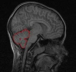
To estimate the total cerebellum volume of considered as statistically significant when a p-
each subject there were two important stages to value was less than 0.05. The data analyses were
perform, MRI data pre-processing and the performed with statistical data analysis software
cerebellum segmentation (Figure 1). For both of STATISTICA Ver. 9.0 (StatSoft, Inc., Tulsa, OK,
them dedicated tools implemented in the Analyze USA).
10 Biomedical Imaging Software (AnalyzeDirect,
Overland Park, KS, USA) were used. In the first RESULTS
stage, two databases have been created by using
DICOM Tool of the Analyze 10. Collected the The study group was composed of 30
original MRI DICOM volumetric data were then children with CP (21 boys, nine girls). There were
converted to internal volumetric AVW format. To more boys than girls in the CP group, but did not
prepare for further brain segmentation each set has differ significantly (p=0.077). The ages of the
been also linearly interpolated to isotropic matrix. children when first seen ranged from 2 to 17 years,
In the second stage, the cerebellar volume of the with a mean age of 6.33 ± 4.06 years. The control
individual was manually estimated in a slice-by- group included 33 healthy children (aged 4-19
slice fashion according to the following procedure. years with mean value of age 11.12 ± 4.77; 16
After loading a set of T1-weighted sagittal images males, 17 females). There was the significant age
into the Analyze 10 workspace the midline slice difference between groups (pbetween gender and the cerebellum volume in CP
patients and in controls were found (Table 3).
Analysis of variance
One-way analysis of variance was
conducted on two data sets defined by levels of
variable CONDITION. Variable gender was taken
as a factor. Two-way analysis of variance was
conducted on a data set containing all cases, with
levels of factors defined by variables gender and
CONDITION. The results of one-way and two-way
analysis of variance are summarized in Table 4
including values of F statistic (including degrees of
freedom) and p-values. Results were considered
significant when (pProg Health Sci 2011, Vol 1 , No2 Cerebellum cerebral palsy volumetrics study
DISCUSSION involve motor skill acquisition [18,19] and varied
cognitive and sensory discrimination tasks [20].
This study has demonstrated that CP group Furthermore, in the present study, we did not
had a significantly smaller mean of the cerebellum include CP children with ataxia. We evaluated only
volume than did the control group. Positive children with spastic CP. Many children with CP
relationship between cerebellar volume and age in have poor walking abilities and manipulation skills.
children with CP and controls was found. No Balance and motor coordination in CP patients is
significant difference between total cerebellar often impaired [21]. One contributing factor to their
volume and gender in patients with CP was problems with gait and reaching movement is poor
detected. In the control group, boys had a lager total balance control because the maintenance of stability
cerebellar volume than girls. Our findings are in is critical to all movements. There is a general
agreement with previous studies [11,16,17]. agreement that the cerebellum is involved in motor
Bodensteiner and Johnsen [11] reviewed 94 brains coordination and learning, although the exact role
MRIs of children with CP whose birth weight was of the cerebellum is still a matter of discussion [22].
less than 1000 g and whose gestational age was less More recently, an additional role of the cerebellum
than 28 weeks. The common cerebral abnormalities in pure cognitive function has been proposed
included decreased white-matter volume without [20,23]. In the last decade, an increasing number of
gliosis (n=36), periventricular leukomalacia (n=16), studies in cerebellar patients and in healthy subjects
and a thin corpus callosum (n=18). Cerebellar using functional brain imaging techniques claimed
abnormalities were found in 32. The cerebellar evidence for an involvement of the cerebellum in
findings included destruction of major portions of mental skills [23,24]. In the current study, we did
the cerebellum (usually the inferior vermis and not examine the mental development of children
hemispheres) and focal or unilateral loss of with CP. To estimate the total cerebellum volume
cerebellar tissue. The pattern of cerebellar injury of each subject, there were two important stages to
suggests a vascular insult, and the deficient white perform, MRI data pre-processing and the
matter without gliosis suggests immaturity of cerebellum segmentation. For both of them,
oligodendrogliocytes with the limited response to dedicated tools implemented in the Analyze 10
injury. In our study, common MRI finding was also Biomedical Imaging Software were used. In
periventricular leukomalacia - 19 cases. And all principle, there are a number of sources of potential
patients had normal cerebellar imaging. Similar error in the data acquisition and analysis system
findings described Srinivasan et al. [15] employed here [25,26]. These include image
determining the absolute cerebellar volumes in term artifacts (intensity drift, motion and pulsatile phase
and preterm infants in correlation with risk factors. artifacts, susceptibility effects), errors in segmen-
The median cerebellar volume of preterm was tation, and the use of multiple scanners with
significantly smaller as comparing to term-born variation in imaging sequence parameters. Our use
infants. Moreover, in the multiple regression of a semi-automated segmentation system gives the
analysis of perinatal variables showed that only segmenter the flexibility to adjust for the majority
infants with supratentorial lesions were of minor image anomalies such as intensity artifacts
significantly associated with the reduction in due to susceptibility changes and phase artifacts.
cerebellar volumes. In other study, [17] children Images which possess an unacceptable level of an
with spastic CP had a significantly smaller mean of artifact are not analyzed and the imaging is
the cerebellar hemispheres and the brain stem than reprogrammed These and previous studies
did controls. The cerebellar volumes were [16,27,28] have demonstrated no significant
positively correlated with age of children with CP differences in volumes determined using the
and the control group. No significant correlations scanners and parameters similar to ones used in this
between gender and the volumes of cerebellar study. Caviness et al. [16] performed volumetric
hemispheres and the brain stem in controls and in MRI-based morphometry on the brains of 30
the CP group were found. No significant normal children with a mean age of nine years
correlations between asphyxia and the volumes of (range 7-11 years). They found that females, unlike
cerebellar hemispheres and the brain stem in the CP males, reach adult cerebellar volume earlier in
group were noted. Positive correlation between the childhood, at this time both genders have equivalent
cerebellum volume and mental development in relative cerebellar volumes. The brain at this age is
children with CP was found. Negative relationship 95% the volume of the adult brain. The brain of the
between the cerebellar hemispheres volume and female child is 93% the volume of the male child.
GMFCS in patients with CP was found. Results The development of the human brain is a dynamic
from animal studies suggest a causal relationship process that continues throughout gestation.
exists between motor skill acquisition and practice Cortioconeurogenesis refers to the process of neural
and structural changes in the cerebellum [18,19]. development and the connectivity between the
The traditional view of cerebellar function has various layers of the brain. Subcortical neurons are
expanded from one of the motor coordination to detectable as early as 10-week gestation. However,
71neuronal proliferation continues through mid- CP. In addition, cerebellar atrophy was shown to be
gestation, and the maturation process continues to a bad prognostic factor considering the cognitive
the end of gestation [29]. There is a linear increase outcome in children after brain trauma. Our results
in brain volume with increasing gestational age in are in agreement with previous clinical and
cerebral and cerebellar tissues. During the last five neuroradiologic reports on CP [7,8,9,11,12]. The
weeks of gestation, there is a significant increase in MRI findings may help us to understand not only
dendritic connections and sulci formation. In the the type of lesion but also the timing of insult. It is
present study, one-third of patients was born particularly important to perform MRI in all
preterm, on the other hand most 60% was born in children with motor impairment (e.g., difficulty in
term. The preoligodendroglia are the precursors to sitting or standing) to detect brain abnormalities
the developing white matter. They are uniquely such as periventricular leukomalacia, cerebral
vulnerable to injury in infants born prematurely. atrophy, or porencepahly. The association between
The magnitude of potential injury is significant cerebellum volume in the early childhood and
when one considers that this is the dominant cell neurodevelopmental outcome highlights its impor-
lineage in the developing white matter between 28 tance as a useful clinical indicator in the follow-up
weeks and 41 weeks gestation [29,30]. of high-risk preterm and term infants. Early
Furthermore, the critical phases of axonal synapto- detection of brain abnormalities in children with CP
genesis, maturation and elongation, still need to may help in prognosis and in the initiation of
occur during the latter half of gestation. These data appropriate therapy (rehabilitation of motor
are in part responsible for the increased risk for impairment or language therapy). This study has
white matter injury in the preterm infant [11,31]. some limitations such as lack of the mental
Over the last decade, increasing evidence of developpment, visual and language studies of
cognitive functions of the cerebellum during patients with CP.
development and learning processes could be
ascertained [32,33]. Posterior fossa malformations
such as cerebellar hypoplasia are known to be
related to developmental problems in a marked to CONCLUSION
moderate extent. More detailed analyses reveal
special deficits in attention, processing speed, The present study demonstrated that children with
visuospatial functions, and language. Acquired spastic CP had smaller volumes of the cerebellum
cerebellar or vermis atrophy was found in groups of as compared to controls.
children with developmental problems such as
prenatal alcohol exposure, extreme prematurity or
Table 1. Clinical data of children with spastic cerebral palsy (CP).
Birth weight (kg)
Gestational age
history (weeks)
/Pre-perinatal
Age (years)
Epilepsy
GMFCS
CP type
Patient
Apgar
MRI
Sex
1 8 M 1050 1 27 Tetra Yes PVL 4
2 13 F 3700 6 39, CS HemD Yes MCAI 1
3 8 F 2700 1 35, S Tetra Yes PVL 3
4 5 M 3900 10 40, CS FDS HemD No PVL 1
5 3 F 2480 6 37, TP Dip No N 1
6 5 M 2800 8 34 Dip No PVL, CCT 1
7 10 F 2680 9 38, S Tetra Yes CA 4
8 7 M 2950 9 38, CS FD Tetra Yes CA 4
9 18 F 2500 1 38 Tetra No PVL 2
10 7 M 3800 9 37 Dip No PVL 1
11 9 M 3050 4 40, CS Dip No PVL 2
12 7 M 900 2 25 CS, B Tetra No PVL 4
13 3 M 2800 3 32 HemS No PVL 1
14 1 M 3040 10 37 Dip No N 1
72Prog Health Sci 2011, Vol 1 , No2 Cerebellum cerebral palsy volumetrics study
Gestational age /Pre-
Birth weight (kg)
perinatal history
Age (years)
Epilepsy
GMFCS
CP type
(weeks)
Patient
Apgar
MRI
Sex
15 11 M 2600 4 34 Dip Yes PVL 2
16 4 M 3650 10 39 Dip No PVL 1
17 13 M 2100 7 37 Tetra No CA 4
18 3 M 2800 6 34 Dip No PVL 2
19 2 M 3500 10 38 HemD No PVL 1
20 8 F 3300 5 38 HemD No PVL 1
21 4 M 4350 10 42 HemD No PVL LS 1
22 1 M 3350 9 40 HemD No PVL LS 1
23 2 F 1100 4 28 Dip No PVL 3
24 11 F 3600 3 38, CS HemS No PLL 1
25 2 M 3000 8 38 Dip No PVL 2
26 3 M 2800 7 37, CS Dip No PVL 2
M-male, F- female, B - bleeding, S - sepsis, N - normal, CP- cerebral palsy, CS-cesaren sectio, CA-cerebral
atrophy, Dip-diplegia, TP - twin pregnancy, LS - left side, CCT - corpus callosum thin, PLL -porencephaly left
lobe, FDS-fetal distress, HemD-hemiplegia dextra, HemS-hemiplegia sinistra, Tetra- tetraplegia, Hem-
hemiplegia, PVL- Periventricular leukomalacia, MCAI- Middle cerebral artery infarct.
Table 2. Cerebellum volume in children with spastic cerebral palsy (CP) and controls related to gender.
No
Mean Median Minimum Maximum Variance Std. Dev.
subjects
CP
Volume of the cerebellum (mm3)
30 129726,2 132721,5 74029,00 184156,0 678119144 26040,72
Controls
33 143122,5 139655,0 112950,0 171219,0 152549657 12351,10
CP, Gender = F
11 122279,7 122522,0 74029,00 162288,0 842632482 29028,13
CP, Gender = M
21 133626,8 135382,0 94133,00 184156,0 583295854 24151,52
Controls, Gender = F
17 135515,6 136599,0 112950,0 149757,0 79479164 8915,109
Controls, Gender = M
16 151204,8 152690,5 134426,0 171219,0 105403359 10266,61
CP- cerebral palsy, M-male, F-female
Table 3. Correlations between the cerebellar volume and age with respect to gender in patients with spastic
cerebral palsy (CP).
CP Controls
GENDER=F GENDER=M No grouping GENDER=F GENDER=M No grouping
R=0.33 R=0.26 R=0.19 R=0.39 R=0.36 R=0.06
P=0.31 P=0.26 P=0.29 P=0.13 P=0.17 P=0.73
N=11 N=21 N=32 N=17 N=16 N=33
73Table 4. Results of one-way and two-way analysis
One-way ANOVA Two-way ANOVA
Factor = CONDITION Factors
CP Controls GENDER CONDITION Interaction
F(1,30)=1.39 F(1,31)=22.05 F(1,61)=7.48 F(1,61)=9.71 F(1,61)=0.19
P>0.24 PProg Health Sci 2011, Vol 1 , No2 Cerebellum cerebral palsy volumetrics study
12. Cioni G, Di Paco MC, Bertuccelli B, movements. Neuroimage. 2005 Sep; 27(3):
Paolicelli PB, Canapicchi R. MRI findings 505-19.
and sensorimotor development in infants with 25. Gurleyik K, Haacke EM. Quantification of
bilateral spastic cerebral palsy. Brain Dev. errors in volume measurements of the caudate
1997 Jun;19(4):245-53. nucleus using magnetic resonance imaging. J
13. Kleim JA, Swain RA, Czerlanis CM, Kelly Magn Reson Imaging. 2002 Apr; 15(4): 353-
JL, Pipitone MA, Greenough WT. Learning- 63.
dependent dendritic hypertrophy of cerebellar 26. Cercignani M, Alexander DC. Optimal
stellate cells: plasticity of local circuit acquisition schemes for in vivo quantitative
neurons. Neurobiol Learn Mem. 1997 Jan; magnetization transfer MRI. Magn Reson
67(1): 29-33. Med. 2006 Oct; 56(4): 803-10.
14. Ito M. Mechanisms of motor learning in the 27. Filipek PA, Richelme C, Kennedy DN,
cerebellum. Brain Res. 2000; 886: 237-245. Caviness VS Jr. The young adult human brain:
15. Commission on Classification and an MRI-based morphometric analysis. Cereb
Terminology of the International League Cortex. 1994 Jul-Aug; 4(4): 344-60.
Against Epilepsy. Proposal for revised 28. Caviness VS Jr, Kennedy DN, Richelme C,
classification of epilepsies and epileptic Rademacher J, Filipek PA. The human brain
syndromes. Epilepsia 1989; 30: 389-99. age 7-11 years: a volumetric analysis based on
16. Srinivasan L, Allsop J, Counsell SJ, magnetic resonance images. Cereb Cortex.
Boardman JP, Edwards AD, Rutherford M. 1996 Sep-Oct; 6(5): 726-36.
Smaller cerebellar volumes in very preterm 29. Kinney HC. The near-term (late preterm)
infants at term-equivalent age are associated human brain and risk for periventricular
with the presence of supratentorial lesions. leukomalacia: a review. Semin Perinatol. 2006
AJNR Am J Neuroradiol. 2006 Mar; 27(3): Apr; 30(2): 81-8.
573-9. 30. Back SA, Luo NL, Borenstein NS, Levine JM,
17. Kułak W, Sobaniec W. Magnetic resonance Volpe JJ, Kinney HC. Late oligodendrocyte
imaging of the cerebellum and brain stem in progenitors coincide with the developmental
children with cerebral palsy. Adv Med Sci. window of vulnerability for human perinatal
2007; 52 Suppl 1:180-2. white matter injury. J Neurosci. 2001 Feb;
18. Anderson BJ, Eckburg PB, Relucio KI. 21(4): 1302-12.
Alterations in the thickness of motor cortical 31. Rezaie P, Dean A. Periventricular leuko-
subregions after motor-skill learning and malacia, inflammation and white matter
exercise. Learn Mem. 2002 Jan-Feb; 9(1): 1-9. lesions within the developing nervous system.
19. Mauk MD, Garcia KS, Medina JF, Steele PM. Neuropathology. 2002 Sep; 22(3): 106-32.
Does cerebellar LTD mediate motor learning? 32. Kleim JA, Swain RA, Czerlanis CM, Kelly
Toward a resolution without a smoking gun. JL, Pipitone MA, Greenough WT. Learning-
Neuron 1998 Mar; 20(3): 359-62. dependent dendritic hypertrophy of cerebellar
20. Timmann D, Drepper J, Maschke M, Kolb FP, stellate cells: plasticity of local circuit
Boring D, Thilman AF, Diener HC. Motor neurons. Neurobiol Learn Mem. 1997 Jan;
deficits cannot explain impaired cognitive 67(1): 29-33.
associative learning in cerebellar patients. 33. Steinlin M. Cerebellar disorders in childhood:
Neuropsychologia. 2002; 40(7): 788-800. cognitive problems. Cerebellum. 2008; 7(4):
21. Woollacott MH, Shumway-Cook A. Postural 607-10.
dysfunction during standing and walking in
children with cerebral palsy: what are the
underlying problems and what new therapies
might improve balance? Neural Plast. 2005;
12: 211-9.
22. Llinás R, Lang EJ, Welsh JP. The cerebellum,
LTD, and memory: alternative views. Learn
Mem. 1997 Mar-Apr; 3(6): 445-55.
23. Tiemeier H, Lenroot RK, Greenstein DK,
Tran L, Pierson R, Giedd JN. Cerebellum
development during childhood and
adolescence: a longitudinal morphometric
MRI study. Neuroimage. 2010 Jan; 49(1): 63-
70.
24. Lacourse MG, Orr EL, Cramer SC, Cohen MJ.
Brain activation during execution and motor
imagery of novel and skilled sequential hand
75You can also read