Prosthetic Dentistry Possibilities in Patients with Cleft Deformities of The Palate and Alveolar Ridge: A Case Report
←
→
Page content transcription
If your browser does not render page correctly, please read the page content below
Prosthetic Dentistry Possibilities in Patients with Cleft
 Deformities of The Palate and Alveolar Ridge: A Case
                        Report
               Albertas Kriaučiūnas;Prof. Alvydas Gleiznys;Oskaras Godvaišas

Abstract
A 56 year old man presented with one-sided cleft palate, affecting alveolar ridge in the upper left
canine region. Patient was unable to eat and drink without having the food fall out through his mouths
opening into his nose. During first visit, patient underwent x-ray analysis to identify how deeply the
bone damage has affected his maxilla. It revealed deep bone penetration, connecting the base of the
nose with the mouth. We suggested one of the most effective methods for treating these types of
cases – a removable partial denture, which was made to work as an obturator at the same time, closing
the space, which causes difficulties to the patient. This case report represents a rare presentation and
treatment of cleft palate by using removable partial dentures (RPDs) to close the gap, connecting the
patient’s mouth with the nose structures.

Keyword: Cleft palate, dental, prosthodontics, dentures, removable dentures;
Published Date: 5/31/2019                            Page.275-281                       Vol 7 No 5 2019

DOI: https://doi.org/10.31686/ijier.Vol7.Iss5.1510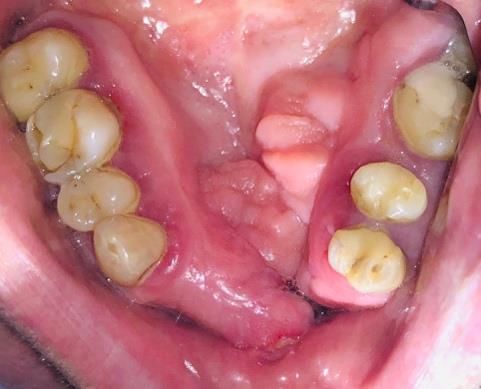
International Journal of Innovation Education and Research         www.ijier.net         Vol:-7 No-5, 2019

    Prosthetic Dentistry Possibilities in Patients with Cleft Deformities of
                    The Palate and Alveolar Ridge: A Case Report

                             Albertas Kriaučiūnas (Corresponding author)
                                E-mail: albertas.kriauciunas@gmail.com
       Department of Dental and Maxillofacial Orthopedics, Lithuanian University of Health Sciences
                                           Kaunas, Lithuania.

                                        Prof. Alvydas Gleiznys
       Department of Dental and Maxillofacial Orthopedics, Lithuanian University of Health Sciences
                                           Kaunas, Lithuania.

                                              Oskaras Godvaišas
                              Student of Lithuanian University of Health Sciences
                                               Kaunas, Lithuania

Abstract
A 56 year old man presented with one-sided cleft palate, affecting alveolar ridge in the upper left canine
region. Patient was unable to eat and drink without having the food fall out through his mouths opening
into his nose. During first visit, patient underwent x-ray analysis to identify how deeply the bone damage
has affected his maxilla. It revealed deep bone penetration, connecting the base of the nose with the
mouth. We suggested one of the most effective methods for treating these types of cases – a removable
partial denture, which was made to work as an obturator at the same time, closing the space, which causes
difficulties to the patient. This case report represents a rare presentation and treatment of cleft palate by
using removable partial dentures (RPDs) to close the gap, connecting the patient’s mouth with the nose
structures.

Keywords: Cleft palate, dental, prosthodontics, dentures, removable dentures;

1. Introduction
The most common congenital defects involving face and jaws are orofacial clefts, affecting approximately
1,2/1000 births [1, 2]. Mostly, clefts come as isolated nonsyndromic deformities (~70%) , but it can also
be seen as frequent symptoms of monogenic syndromes (~6%) [3, 4]. Although often causes cannot be
identified [3], it is noted that certain teratogens (aspirin, cigarette smoke (hypoxia), 275 ilantin, 6 –
mercaptopurine, ethyl alcohol) play major role in facial deformities development [1].
People with facial cleft deformities tend to have broad spectrum of physical dysfunctions, including

International Educative Research Foundation and Publisher © 2019                                  pg. 275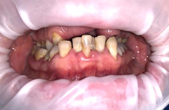
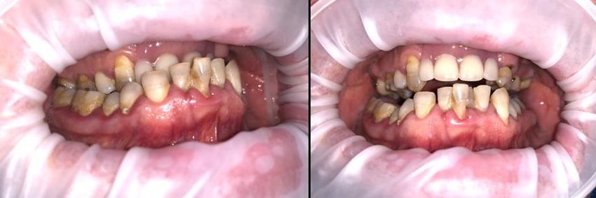
International Journal of Innovation Education and Research          www.ijier.net         Vol:-7 No-5, 2019

speaking, biting and chewing foods, drinking through straw, food falling out of their mouths. However,
speech impairment is distinguished as main factor affecting the quality of life [5].
Today medicine offers numerous surgical and non – surgical treatment options at early stages of life that
have minimal residual manifestations and help to retrieve physiological functions and aesthetics [6, 7]. The
American Cleft Palate – Craniofacial Association highlights the importance of assembling interdisciplinary
team in order to achieve adequate treatment, successful results and longevity [8].
Prosthetic treatment should be considered as part of rehabilitation since various prosthetic obturators can
be provided in order to close existing defect both in newborns and adults [9, 10]. The purpose of this case
report is to present possible prosthetic treatment in patient with cleft palate and alveolar ridge.

2. Case presentation:
The clinical case we report here is of a 56-year-old male patient, with previous medical history of heart
diseases and pulmonary tract problems, which were insignificant, for current pathology. Patient came into
Lithuanian University of Health Sciences Maxillofacial Orthopaedics clinic with problems during
mastication. Patient has asked for help, because every time he masticates, food and fluids art flowing
through his nose. Extraoral examination has shown protruding lower jaw, lower lip was bigger than the
upper one, side profile shown protruding occlusion of the lower jaw teeth. Intraorally, multiple caries
lesions were found, gums were inflamed, they were bleeding on probing, patient’s hygiene was poor.
Patient had no canine in his left side in upper jaw and no incisors. In frontal teeth region, we could observe
a hole connecting the mouth with the nose. Bite was pathological with an Angle’s third class malocclusion
(Picture 1).

Picture 1.Patients bite and upper jaw.

Orthopantomogram has shown both soft tissue and bone defect of the upper jaw alveolar ridge in the front
teeth area connecting mouth and nasal cavities of the left nostril. Nasal septum was intact with slight
deformation. (Picture 2).

International Educative Research Foundation and Publisher © 2019                                   pg. 276
International Journal of Innovation Education and Research www.ijier.net Vol:-7 No-5, 2019
Picture 2. Orthopantomogram.
To extend clinical evaluation, both, upper and lower jaw alginate (“Alligat fast set”, Kulzer)
impressions were taken in order to manufacture diagnostic plaster (I class) model casts (Picture 3). Casts
were thoroughly examined and after consulting with dental technician, possible treatment ways were
discussed. Depending on patient’s disagreement with any treatment that would involve surgery and his
current financial situation, we suggested treatment with removable partial dentures.
Picture 3. Plaster cast.
Treatment was begun by taking alginate (“Alligat fast set”, Kulzer) functional impression of upper jaw
with individual tray that was made on diagnostic plaster model. All functional movements were performed
during the making of impression in pursuance of clear soft tissue contour. Functional impression was used
as a mold to make a working plaster (III class) cast.
On the next appointment teeth color was determined. Then, using individual wax bite registration material
patient’s central occlusion was fixated and transferred onto articulator. Initial wax partial denture was made
by dental technician and tested in vivo by performing horizontal, sagittal and vertical lower jaw movements.
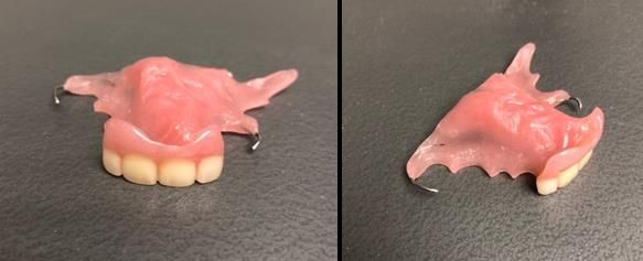
The final denture was made with an obturating part, to close the space which leads to the food being pushed
into the nasal cavity (Picture 4).
International Educative Research Foundation and Publisher © 2019 pg. 277International Journal of Innovation Education and Research www.ijier.net Vol:-7 No-5, 2019
Picture 4. Removable partial dentures with obturating parts.
By making this RPD, patient’s smile aesthetics were highly improved considering that the patient was very
happy with the final result. RPD restored biting function and greatly enhanced phonetic ability (Picture 5).
After 24 hours few base corrections were made. As one week of usage has passed, patient reported no
longer having a problem of food flowing to the nasal cavity. No further corrections were needed.
Picture 5. Removable partial dentures in the mouth of the patient.
3. Discussion:
Alveolar cleft treatment should be performed as early in the patients life as possible – if available, within
the first few days of birth [8]. There are various methods, that are being used to treat these cases, when
people are still young, one of them being computer assisted distraction osteogenesis, which was concluded
as an effective method, to completely close the cleft by Zhang et al. [11]. Although, this method is invasive
and requires intervention.
Alveolar cleft should always be treated with the help of a team of specialists, which should involve not
only prosthodontists, but also audiologists, neurosurgeons, psychologists, plastic surgeons, etc. [8].
When treating kids with cleft palate and holoprosencephaly (they have wide midline clefts) hard-
setting acryl is very useful, because they have protrusive positioning of tongue (which affects the surgical
result) [15]. In our case we used removable partial denture made of hard plastic, which helped closing the
oronasal communication.
Also, orthodontic treatment can be very challenging, take a long time and often requires orthognathic
surgery. Nevertheless, this type of treatment can be successful and can yield positive results, one of which
was described by Hameed et al. when the full course of orthodontic treatment for a cleft lip/palate patient
International Educative Research Foundation and Publisher © 2019 pg. 278International Journal of Innovation Education and Research www.ijier.net Vol:-7 No-5, 2019
took 3.4 years and 44 visits, but it was successful and gave positive results to the patient [12].
Plastic surgeons can be involved by treating cleft palate with fat grafting procedure, to help these
patients without the need of removable partial dentures, although, this is also an invasive surgical procedure,
which can cause complications, and should be considered very carefully according to its specific
indications [13]. To repair maxillary cleft, hybrid obturator can be used to improve quality of life, by
providing adequate functional and aesthetic conditions and reducing airspace through sealing of the
oronasal communication [14].
Some patients can be treated with non-removable obturators, one of which was described in Borzabadi
et al. article, as an obturator which can be used in times when the surgical closure of the fistula is not
feasible and a removable device fails to succeed - such non-removable obturator can also be used for
treatment of malocclusion, it maintains space and also preserves molar anchorage [16]. Fixed prosthesis on
implants can also be used as a treatment method for cleft palate obturations, as Lopes et al. described in
their work, a case of 6 implants were used for fixed prosthetic dentures, and in order to make a guiding
plane for the insertion of a removable palatal obturator, which improved speech and patients ability to
swallow [17].
4. Conclusion.
All cleft palate treatments and obturations should be treated as early in persons life as possible. However,
undiagnosed, and untreated patients need their space between the nose and mouth space closed with
prosthetic obturators, which can also be in a form of removable partial dentures. Clinicians should consider
the bone structure deformations around the cleft, and customize the denture accordingly.
Soft polyvinylsiloxane materials can be used to reline the denture if hard plastics fail to obturate the space,
and food continues to fall through the space connecting mouth with patients nose. While treating cleft
palate in adulthood can be challenging, prompt treatment, and obturators can be the only way to assure a
qualitative life of the patient.
7. References
1. William R. Proffit, Henry W. Fields, Jr. Et al „Contemporary orthodontics, sixth edition“ p. 108
2. Rahimov, Fedik, et al. “Genetics of Nonsyndromic Orofacial Clefts.” The Cleft Palate-Craniofacial
Journal, vol. 49, no. 1, Jan. 2012, pp. 73–91, doi:10.1597/10-178. URL:
https://journals.sagepub.com/doi/abs/10.1597/10-178?journalCode=cpca
3. Tolarova MM, Cervenka J. Classification and birth prevalence of orofacial clefts. American journal
of medical genetics. 1998;75(2):126-37. URL: https://www.ncbi.nlm.nih.gov/pubmed/9450872
4. Rahimov F, Jugessur A, Murray JC. Genetics of nonsyndromic orofacial clefts. Cleft Palate
Craniofac J. 2012;49(1):73–91. doi:10.1597/10-178.
https://www.ncbi.nlm.nih.gov/pmc/articles/PMC3437188/
5. Klassen AF, Riff KWW, Longmire NM, Albert A, Allen GC, Aydin MA, et al. Psychometric
findings and normative values for the CLEFT-Q based on 2434 children and young adult patients
International Educative Research Foundation and Publisher © 2019 pg. 279International Journal of Innovation Education and Research www.ijier.net Vol:-7 No-5, 2019
with cleft lip and/or palate from 12 countries. Canadian Medical Association Journal.
2018;190(15):E455-E62. URL: http://www.cmaj.ca/content/190/15/E455
6. Worley ML, Patel KG, Kilpatrick LA. Cleft Lip and Palate. Clinics in perinatology. 2018;45(4):661-
78. URL: https://www.clinicalkey.com/#!/content/playContent/1-s2.0-
S0095510818313927?returnurl=null&referrer=null
7. Kitchin S, Grames L, Naidoo SD, Skolnick G, Schoenborn A, Snyder-Warwick A, et al. Surgical,
Speech, and Audiologic Outcomes in Patients With Orofacial Cleft and Van der Woude Syndrome.
The Journal of craniofacial surgery. 2019. URL: https://www.ncbi.nlm.nih.gov/pubmed/31058728
8. “Parameters For Evaluation and Treatment of Patients With Cleft Lip/Palate or Other Craniofacial
Differences.” The Cleft Palate-Craniofacial Journal, vol. 55, no. 1, Jan. 2018, pp. 137–156,
doi:10.1177/1055665617739564. URL:
https://journals.sagepub.com/doi/full/10.1177/1055665617739564
9. Goyal S, Rani S, Pawah S, Sharma P. A novel approach for prosthodontic management of patient
with cleft of palate. Journal of the Indian Society of Pedodontics and Preventive Dentistry.
2017;35(3):279-81. URL:https://www.ncbi.nlm.nih.gov/pubmed/28762357
10. Valizadeh B, Barzanji SA, Patel M, Shahdad S. Rehabilitation of an Edentulous Maxilla in a Patient
with Isolated Cleft Palate. Dental update. 2016;43(3):214-6. URL:
https://www.ncbi.nlm.nih.gov/pubmed/27439268
11. Zhang B, Liu SH, Zhao ZJ, Bai XF, Li ZJ, Liu Q. Clinical research of extensive alveolar cleft
treatment with computer assistant distraction osteogenesis. Zhonghua kou qiang yi xue za zhi =
Zhonghua kouqiang yixue zazhi = Chinese journal of stomatology. 2019;54(2):112-7.
12. Hameed O, Amin N, Haria P, Patel B, Hay N. Orthodontic burden of care for patients with a cleft
lip and/or palate. Journal of orthodontics. 2019;46(1):63-7.
13. Jones CM, Mackay DR. Autologous Fat Grafting in Cleft Lip and Palate. The Journal of craniofacial
surgery. 2019;30(3):686-91.
14. Goiato MC, dos Santos DM, Magri FM, Rahal V, Andreotti AM, Moreno A, et al. Rehabilitation of
maxillary cleft with hybrid obturator prosthesis. The Journal of craniofacial surgery.
2013;24(5):e517-21. URL: https://www.ncbi.nlm.nih.gov/pubmed/24036832
15. Acharya BS, Chen EA, Lewis RL, Teichgraeber JF, Lypka MA. A Postsurgical Obturator After Cleft
Lip Repair in Patients With Holoprosencephaly. The Cleft palate-craniofacial journal : official
publication of the American Cleft Palate-Craniofacial Association. 2015;52(4):480-3. URL:
https://www.ncbi.nlm.nih.gov/pubmed/24524206
16. Borzabadi-Farahani A, Groper JN, Tanner AM, Urata MM, Panossian A, Yen SL. The nance
obturator, a new fixed obturator for patients with cleft palate and fistula. Journal of prosthodontics:
official journal of the American College of Prosthodontists. 2012;21(5):400-3. URL:
https://www.ncbi.nlm.nih.gov/pubmed/22738139
17. Lopes JF, Pinto JH, de Almeida AL, Lopes MM, da Silva Dalben G. Cleft palate obturation with
Branemark protocol implant-supported fixed denture and removable obturator. The Cleft palate-
craniofacial journal : official publication of the American Cleft Palate-Craniofacial Association.
International Educative Research Foundation and Publisher © 2019 pg. 280International Journal of Innovation Education and Research www.ijier.net Vol:-7 No-5, 2019
2010;47(2):211-5. URL: https://www.ncbi.nlm.nih.gov/pubmed/20210640
Copyright Disclaimer
Copyright for this article is retained by the author(s), with first publication rights granted to the journal.
This is an open-access article distributed under the terms and conditions of the Creative Commons
Attribution License (http://creativecommons.org/licenses/by/4.0/).
International Educative Research Foundation and Publisher © 2019 pg. 281You can also read