Repeated cerebellar infarction in the affected nondominant vertebral artery distribution with reversible vertebral artery occlusion elicited by ...
←
→
Page content transcription
If your browser does not render page correctly, please read the page content below
J Neurosurg Case Lessons 1(8):CASE2061, 2021
DOI: 10.3171/CASE2061

Repeated cerebellar infarction in the affected nondominant vertebral artery distribution with
reversible vertebral artery occlusion elicited by head tilt: illustrative case
Takanori Nozawa, MD, PhD,1,3 Kouichirou Okamoto, MD, PhD,2 Shinji Nakazato, MD, PhD,1 Kunio Motohashi, MD,1,3
Tomoaki Suzuki, MD, PhD,3 Kotaro Morita, MD, PhD,1 Hideki Tashi, MD, PhD,4 Kei Watanabe, MD, PhD,4 Hitoshi Hasegawa, MD, PhD,3
Masato Watanabe, MD, PhD,1 Hiroyuki Kawashima, MD, PhD,4 and Yukihiko Fujii, MD, PhD3
1Department of Neurosurgery, Kuwana Hospital, Niigata City, Japan; Departments of 2Translational Research and 3Neurosurgery, Brain Research Institute,

Niigata University, Niigata City, Japan; and 4Division of Orthopedic Surgery, Department of Regenerative and Transplant Medicine, Graduate School of Medical and
Dental Sciences, Niigata University, Niigata City, Japan

   BACKGROUND Bow hunter’s syndrome or stroke (BHS) is characterized by rotational vertebrobasilar insufficiency elicited by rotation of the neck.
   It is caused by dynamic and reversible occlusion of the vertebral artery (VA). Reversible symptoms of rotational vertebrobasilar insufficiency are
   described as bow hunter’s syndrome, although brain infarction is rarely reported as bow hunter’s stroke.
   OBSERVATIONS A 70-year-old man experienced repeated cerebellar infarctions three times in the posterior inferior cerebellar artery (PICA)
   distribution of the nondominant right VA connecting the basilar artery. The onset of symptoms indicating cerebellar infarcts and the patient’s head
   position changes were unrelated. Dynamic digital angiography (DA) revealed that the nondominant right VA was occluded by an osteophyte from the
   C4 vertebral body, and the right PICA branches were shown to be passing through the distal right VA from the left VA. These findings were observed
   when the patient’s head was tilted to the right. An arterio-arterial embolic mechanism was suggested as the cause of repeated cerebellar infarctions.
   LESSONS Transient nondominant VA occlusion has been rarely reported as a cause of BHS when the head is tilted. To confirm the diagnosis of
   BHS, additional head tilt is recommended when performing dynamic DA in patients with a cervical osteophyte.

   https://thejns.org/doi/abs/10.3171/CASE2061
   KEYWORDS repeated cerebellar infarction; bow hunter’s syndrome; stroke; cervical osteophyte; transient vertebral artery occlusion; head tilt

    Bow hunter’s syndrome or stroke (BHS) is characterized by ro-                          the last 2 years after undergoing a cervical left laminoplasty procedure
tational vertebrobasilar insufficiency. This condition is elicited by neck                 that had been performed 5 years earlier. The infarctions occurred in the
rotation in most cases and by neck extension or flexion in some cases.                     posterior inferior cerebellar artery (PICA) distribution of the affected
BHS is caused by dynamic and reversible occlusion of the vertebral                         nondominant VA, with no infarction occurring in any other region.
artery (VA) and a lack of collateral blood supply to the brainstem.1–4                     Transient occlusion of the right VA by an osteophyte at the level of C4 was
Although the term “bow hunter’s stroke” was coined by Sorensen for a                       demonstrated during dynamic digital angiography (DA) when the pa-
patient with suspected brainstem infarction,5 reversible symptoms of                       tient’s head was tilted to the right. No symptoms or additional infarction
rotational vertebrobasilar insufficiency are usually described as bow                      occurred after surgery with posterior fixation of the cervical spine
hunter’s syndrome.4                                                                        (C3–C6).
    BHS associated with recurrent cerebellar infarction and transient
occlusion of the nondominant VA induced by head tilt has not been                          Illustrative Case
reported in the literature. In this study, we report a case involving an                       A 70-year-old man underwent left laminoplasty at C3–C6 in a
older man who experienced recurrent cerebellar infarctions three times in                  different hospital 5 years ago for ossification of the posterior

ABBREVIATIONS 3D-CTA = three-dimensional computed tomography angiography; BHS = bow hunter’s syndrome or stroke; DA = digital angiography; DSA = digital
subtraction angiography; DWI = diffusion-weighted imaging; MRA = magnetic resonance angiography; MRI = magnetic resonance imaging; PICA = posterior inferior cerebellar
artery; VA = vertebral artery.
INCLUDE WHEN CITING Published February 22, 2021; DOI: 10.3171/CASE2061.
SUBMITTED December 10, 2020. ACCEPTED December 17, 2020.
© 2021 The authors, CC BY-NC-ND 4.0 (http://creativecommons.org/licenses/by-nc-nd/4.0/).

                                                                                                          J Neurosurg Case Lessons | Vol 1 | Issue 8 | February 22, 2021 | 1

                                                                                                                       Unauthenticated | Downloaded 07/18/21 02:09 AM UTC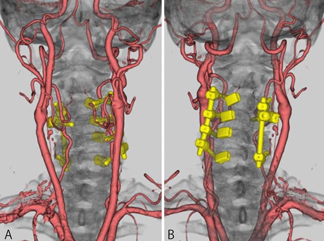
longitudinal ligament. He had a prominent medical history of cerebellar               admission, including dizziness, nausea, and vomiting. MRA revealed
infarction in the lateral region of the right PICA distribution that was              an identical right VA occlusion with no new infarction on DWI. During
detected 2 years ago. For this he had started taking oral clopidogrel (75             subsequent dynamic digital subtraction angiography (DSA) and DA
mg/day). He was admitted to our hospital with complaints of the sudden                procedures with the patient’s head in the neutral position, the non-
onset of dizziness, nausea, and vomiting. Diffusion-weighted imaging                  dominant right VA and the vermian and hemispheric branches of the
(DWI) revealed an additional cerebellar infarction in the medial region               right PICA were injected without stenosis (Fig. 4A–D). No severe
of the right PICA distribution, and magnetic resonance angiography                    stenosis or occlusion was revealed in the right VA when the neck was
(MRA) revealed that the right VA was occluded (Fig. 1A, B, D, and E).                 rotated to the right (Fig. 4E) or left (Fig. 4F) or when the neck was flexed
Three days later, repeated MRA showed recanalization of the non-                      or extended. However, the right VA was occluded by an osteophyte at
dominant right VA connecting the basilar artery (Fig. 1C and F).                      the C4 level when his head was tilted to the right, which elicited no
Echocardiography and 24-hour Holter monitor electrocardiography                       complaints of dizziness, nausea, or vomiting (Fig. 4G). A similar oc-
results were negative, and the etiology of his recurrent cerebellar                   clusion was not induced by head tilt to the left (Fig. 4H). No stenosis or
infarction could not be elucidated. He was discharged from the hospital,              occlusion was demonstrated in the dominant left VA on dynamic DA.
and the following medications were prescribed without anticoagulants:                 The distal right VA was demonstrated in a retrograde fashion by in-
aspirin (100 mg/day) and clopidogrel (75 mg/day).                                     jection of the left VA when the patient’s head was tilted to the right
    Two months later, the man returned to our hospital with symptoms                  (Fig. 4I), and both the vermian and hemispheric branches of the right
identical to those reported during the previous admission. DWI and                    PICA were subsequently opacified (Fig. 4J and K). The man was
MRA showed a new small cerebellar infarction in the right side of the                 asymptomatic while his head was tilted to the right for more than 1
inferior cerebellar vermis, with no infarction in any other region, as well           minute.
as another right VA occlusion (Fig. 2A, B, D, and E). The right VA                        The patient denied that changes in the head or neck position caused
appeared patent on MRA on the 12th day after the second admission to                  the symptoms or the stroke. An arterio-arterial embolism from the
our hospital (Fig. 2C and F). Three-dimensional computed tomography                   transiently occluded right VA to the right PICA distribution was sug-
angiography (3D-CTA) showed that the right VA was laterally com-                      gested as a cause of the repeated cerebellar infarction in this patient.
pressed by an osteophyte from the C4 vertebral body with no stenosis                  He underwent posterior cervical fixation at C3–C6 at Niigata University
(Fig. 3B, arrow). Nineteen days later, during the man’s second ad-                    Medical and Dental Hospital. After the posterior cervical fixation, both
mission, he complained of symptoms similar to those in his first                      VAs were shown to operate continuously without stenosis from the

                          FIG. 1. MRI and MRA on the patient’s first admission to our hospital (A, B, D, and E) and follow-up MRA at his
                          first admission (C and F). A: DWI showing a high signal intensity lesion in the medial right PICA distribution.
                          Brain (B) and cervical (E) MRA on admission. The right VA is not demonstrated. D: FLAIR image showing a
                          previous right cerebellar infarction as a small hypointensity lesion in the lateral right PICA distribution. Brain
                          (C) and cervical (F) MRA 3 days later. The nondominant right VA is observed from the origin to the junction of
                          the left VA and basilar artery. FLAIR = fluid-attenuated inversion recovery.

2 | J Neurosurg Case Lessons | Vol 1 | Issue 8 | February 22, 2021

                                                                                                                Unauthenticated | Downloaded 07/18/21 02:09 AM UTC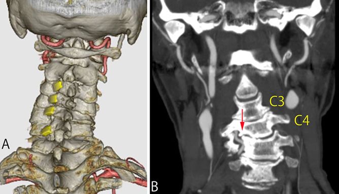
FIG. 2. MRI and MRA on the patient’s second admission to our hospital (A, B, D, and E) and follow-up MRA at
his second admission (C and F). A: DWI showing a small, high signal intensity lesion in the right side of the
inferior cerebellar vermis. Brain (B) and cervical (E) MRA on admission. The right VA is not observed as it
was on his previous admission (see Fig. 1B and E). D: FLAIR image showing past cerebellar infarcts in the
right PICA distribution as hypointense lesions. Brain (C) and cervical (F) MRA 12 days later. The right VA
appears again as previously demonstrated (see Fig. 1C and F).
origin to the basilar artery on 3D-CTA (Fig. 5). He had no symptoms, nondominant right VA and was demonstrated three times on MRI and
and no additional cerebellar infarction developed for 1 year 6 months MRA in the past 2 years after a C3–C6 left laminoplasty that had been
after the surgery. performed 5 years earlier. 3D-CTA showed an osteophyte of C4
compressing the right VA laterally without stenosis. On dynamic DA,
Discussion occlusion of the right VA was demonstrated when the patient tilted his
In this patient, repeated cerebellar infarction was restricted to head to the right, although no severe stenosis or occlusion was ob-
the PICA distribution with reversible occlusion of the ipsilateral served in other head and neck positions. No stenosis or occlusion was
shown in the dominant left VA. In this patient, without demonstration of
the right VA occlusion elicited by head tilt to the right, diagnosis of BHS
could not be made, even after dynamic DA was performed. In elderly
male patients (most ≥40 years), reversible symptomatic VA stenosis/
occlusion caused by an osteophyte of the degenerative cervical
vertebral body is one of the common characteristics in BHS when
changing head and neck positions.1–6 In our patient, left laminoplasty of
C3–C6 might have predisposed him to contralateral cervical osteo-
phyte formation or growth at the C4 vertebral body.
BHS is the descriptive term for rotational vertebrobasilar insuffi-
ciency. This condition is elicited by neck rotation in most cases and by
neck extension or, rarely, by neck flexion in some patients. BHS is
caused by dynamic and reversible occlusion of the VA and a lack of
collateral blood supply to the brainstem, either because of a hypoplastic
or an absent contralateral VA or because of a deficient circle of Willis.4
Both VAs have adequate flow, and the patient remains asymptomatic
FIG. 3. 3D-CTA. A: Posterior view with volume-rendering method when one VA is positionally occluded.7 In 1978, Sorensen was the first
showing C3–C6 left laminoplasty performed at a previous clinic. to use the term “bow hunter’s stroke” in a case of suspected brainstem
B: Coronal reformatted image. An osteophyte of C4 (red arrow)
infarction due to a VA injury,5 although brain infarction is rare in BHS.7–10
compresses the right VA laterally without stenosis.
The suspected cause of cerebral or cerebellar infarction in BHS is
J Neurosurg Case Lessons | Vol 1 | Issue 8 | February 22, 2021 | 3
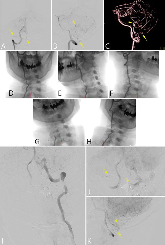
Unauthenticated | Downloaded 07/18/21 02:09 AM UTCFIG. 4. Dynamic bilateral vertebral arteriography. A–C: DSA of the right VA at the neutral position of the head
and neck. The hemispheric (arrows) and vermian (arrowheads) branches of the right PICA are demonstrated
by injection of the right VA. A: Anterior-posterior view. B: Lateral view. C: Lateral view of 3D-rotation
angiography with the volume-rendering method. D–H: Dynamic DA of the right VA. No stenosis is seen in the
right VA at the neutral position (D), rotation of the neck to the right (E) and to the left (F), and head tilt to the left
(H). Occlusion of the right VA is demonstrated at C4 level during head tilt to the right (G). I–K: Dynamic DSA
of the left VA during head tilt to the right. I and J: Anterior-posterior view. K: Lateral view. The distal portion of
the right VA is opacified in a retrograde fashion from the left VA (I), and both PICA hemispheric (arrows) and
vermian (arrowheads) branches are subsequently shown (J and K).
4 | J Neurosurg Case Lessons | Vol 1 | Issue 8 | February 22, 2021
Unauthenticated | Downloaded 07/18/21 02:09 AM UTCaspirin and clopidogrel was used in our patient. The combination of
these antiplatelet agents synergistically inhibits platelet aggrega-
tion.17 In comparison with aspirin monotherapy, such dual-antiplatelet
therapy reduces the rate of stroke occurrence from 11.7% to 8.2% in
patients who have transient ischemic attack or minor stroke.17
However, repeated cerebellar infarction occurred in our patient
while on the therapy. Some possible reasons for these repeated
occurrences include the limited contribution of these antiplatelet
agents because the affected nondominant VA showed no stenosis in
the neutral head position and/or antiplatelet resistance to one or both
antiplatelet agents.18 Nearly 50% of patients who receive conser-
vative treatment are vulnerable to infarcts or have residual neuro-
logical deficits.16
Surgical intervention is proposed if medical treatment fails.14 All
previously reported patients with BHS caused by nondominant VA
compression required surgical management.14 Therefore, surgical
treatment was considered in the current patient. An anterior approach is
FIG. 5. 3D-CTA with volume-rendering method after C3–C6 posterior often used to remove osteophytes at C4–C6.1,2,4,9,10,14,19–21 PICA-to-
cervical fixation. A: Anterior view. B: Posterior view. Both VAs are PICA bypass surgery has also been performed in a patient.22 Recently,
demonstrated without stenosis. successful endovascular treatment with angioplasty and stenting for
contralateral VA stenosis was reported as an attempt to augment
collateral flow.23 In a patient with transient occlusions of the VA by an
osteophyte from a cervical vertebral body while changing neck po-
sitions and associated with repeated embolic infarction in the posterior
hemodynamic impairment1–4,8 or arterio-arterial embolism from the circulation, a coil embolization procedure was performed at the distal
affected VA.7–10 In our patient, the hemodynamic mechanism for re- portion of the affected VA to prevent further embolic infarction in the
peated cerebellar infarction in the right PICA distribution was less likely posterior circulation.24 The current patient underwent C3–C6 left lam-
given that he was asymptomatic when the right VA was occluded at the inoplasty for ossification of the posterior longitudinal ligament 5 years ago,
C4 level. Moreover, the right intracranial VA and branches of the right and posterior fusion of C3–C6 was subsequently selected. After
PICA were demonstrated during dynamic DA of the left VA when the surgery, he experienced no additional symptoms or further cerebellar
patient’s head was tilted to the right. However, repeated cerebellar infarctions.
infarcts developed in the right PICA distribution in the 2 years after the
C3–C6 left laminoplasty procedure that had been performed 5 years Observations
earlier; nonetheless, he denied that these events occurred during neck Repeated cerebellar infarction developed in the right PICA dis-
positional changes. Arterio-arterial embolism from the transiently tribution in our patient. No VA stenosis was observed on 3D-CTA and
occluded right VA was a likely mechanism for cerebellar infarction in DSA in the neutral head position, although DA revealed that the
this patient, although the precise mechanism remains unexplained for nondominant right VA was occluded by an osteophyte from the C4
repeated infarction restricted to the ipsilateral PICA distribution. vertebral body and that the right PICA branches passed through the
BHS is caused by an atypical pattern of transient occlusion of a distal right VA from the left VA without producing any symptoms when
nondominant VA terminating in the PICA.6,11–13 In addition, BHS that the patient’s head was tilted to the right. Transient nondominant VA
results from nondominant VA compression, similar to that in our occlusion has been rarely reported as a cause of BHS when the head is
patient, does not terminate in the PICA and has recently been re- tilted.
ported in 20 cases.14 Among these cases, BHS caused by throm-
boembolism without VA dissection was extremely rare in patients with Lessons
degenerative cervical spondylosis.9,10,14 Such a condition was re- In patients who show an association between repeated cerebellar
ported in two patients with wake-up stroke in the PICA distribution: infarction of unknown cause and degenerative cervical spondylosis,
ipsilateral in one patient and contralateral to the affected nondom- the occurrence of BHS should be considered even in the following
inant right VA in the other.9,10 The affected nondominant right VA was instances: VAs are not stenotic in the neutral head position, a non-
stenotic at C4–C5 or C5–C6 in the neutral position but completely dominant VA connects to the opposite VA and the basilar artery, and
occluded with head rotation to the right.9,10 However, in our patient, no characteristic symptoms are induced by head and neck positional
repeated cerebellar infarction was not a wake-up stroke, and the changes. To gain further information, additional head tilt is re-
nondominant right VA was not stenotic in the neutral head position commended during a dynamic DA study for patients with suspected
(Fig. 4D), although complete occlusion of the right VA was induced by BHS.
head tilt to the right (Fig. 4G). BHS induced by head tilt is an extremely
rare condition.6,15 Acknowledgments
Treatment for BHS includes conservative treatment and surgery. We thank Dr. Takashi Kumagai (Department of Neurosurgery, Ya-
Conservative treatment includes the avoidance of head rotation, magata Prefectural Central Hospital) and Dr. Kazuhiro Ando (Department
cervical collars, and/or antiplatelet or anticoagulation therapy.4,6,16 of Neurosurgery, Brain Research Institute, Niigata University) for their
Conservative management with antiplatelet agents may be con- initial help with this case report. We also thank Editage for assistance with
sidered the first line of treatment for BHS.6 Dual-antiplatelet therapy with English language editing.
J Neurosurg Case Lessons | Vol 1 | Issue 8 | February 22, 2021 | 5
Unauthenticated | Downloaded 07/18/21 02:09 AM UTCReferences 17. Wang Y, Wang Y, Zhao X, et al. Clopidogrel with aspirin in acute
1. Bakay L, Leslie EV. Surgical treatment of vertebral artery in- minor stroke or transient ischemic attack. N Engl J Med. 2013;
sufficiency caused by cervical spondylosis. J Neurosurg. 1965; 369(1):11–19.
23(6):596–602. 18. Topçuoglu MA, Arsava EM, Ay H. Antiplatelet resistance in stroke.
2. Nagashima C. Surgical treatment of vertebral artery insufficiency Expert Rev Neurother. 2011;11(2):251–263.
caused by cervical spondylosis. J Neurosurg. 1970;32(5):512–521. 19. Lu DC, Zador Z, Mummaneni PV, et al. Rotational vertebral artery
3. Husni EA, Bell HS, Storer J. Mechanical occlusion of the vertebral occlusion—series of 9 cases. Neurosurgery. 2010;67(4):
artery. A new clinical concept. JAMA. 1966;196(6):475–478. 1066–1072.
4. Jost GF, Dalley AT. Bow hunter’s syndrome revisited: 2 new cases 20. Vilela MD, Goodkin R, Lundin DA, et al. Rotational vertebrobasilar
and literature review of 124 cases. Neurosurg Focus. 2015;38(4):E7. ischemia: hemodynamic assessment and surgical treatment.
5. Sorensen BF. Bow hunter’s stroke. Neurosurgery. 1978;2(3): Neurosurgery. 2005;56(1):36–45.
259–261. 21. Zaidi HA, Albuquerque FC, Chowdhry SA, et al. Diagnosis and
6. Choi KD, Choi JH, Kim JS, et al. Rotational vertebral artery management of bow hunter’s syndrome: 15-year experience at
occlusion: mechanisms and long-term outcome. Stroke. 2013; Barrow Neurological Institute. World Neurosurg. 2014;82(5):
44(7):1817–1824. 733–738.
7. Grossmann RI, Davis KR. Positional occlusion of the vertebral artery: 22. Kan P, Yashar P, Langer DJ, et al. Posterior inferior cerebellar
a rare cause of embolic stroke. Neuroradiology. 1982;23(4):227–230. artery to posterior inferior cerebellar artery in situ bypass for the
8. Andereggen L, Arnold M, Andres RH, et al. Bow hunter’s stroke treatment of bow hunter’s-type dynamic ischemia in hol-
due to prominent degenerative spinal disorder. Clin Neuroradiol. overtebral dissection. World Neurosurg. 2012;78(5):
2012;22(4):355–358. 553.e15–553.e17.
9. Okawa M, Amamoto T, Abe H, et al. Wake-up stroke in a young 23. Sugiu K, Agari T, Tokunaga K, et al. Endovascular treatment for
woman with rotational vertebral artery occlusion due to far-lateral bow hunter’s syndrome: case report. Minim Invasive Neurosurg.
cervical disc herniation. J Neurosurg Spine. 2015;23(2): 2009;52(4):193–195.
166–169. 24. Mori K, Ishikawa K, Fukui I, et al. A subtype of bow hunter’s
10. Nishikawa H, Miya F, Kitano Y, et al. Positional occlusion of syndrome requiring specific method for detection: a case of re-
vertebral artery due to cervical spondylosis as rare cause of wake- current posterior circulation embolism due to “hidden bow
up stroke: report of two cases. World Neurosurg. 2017;98: hunter’s syndrome.” JNET J Neuronendovasc Ther. 2018;12(6):
877.e13–877.e21. 295–302.
11. Matsuyama T, Morimoto T, Sakaki T. Bow hunter’s stroke caused
by a nondominant vertebral artery occlusion: case report. Disclosures
Neurosurgery. 1997;41(6):1393–1395. The authors report no conflict of interest concerning the materials or
12. Yeh JF, Lin YJ, Po HL, et al. A case of bow hunter’s stroke caused methods used in this study or the findings specified in this paper.
by non-dominant vertebral artery. Acta Neurol Taiwan. 2005;14(2):
69–73. Author Contributions
13. Noh Y, Kwon OK, Kim HJ, et al. Rotational vertebral artery Conception and design: Nozawa, Okamoto, Nakazato, Morita, Fujii.
syndrome due to compression of nondominant vertebral artery Acquisition of data: Motohashi, Tashi, Fujii. Analysis and interpretation
terminating in posterior inferior cerebellar artery. J Neurol. 2011;
of data: Okamoto, K Watanabe, Fujii. Drafting the article: Okamoto,
258(10):1775–1780.
Hasegawa, Fujii. Critically revising the article: Okamoto, Fujii.
14. Iida Y, Murata H, Johkura K, et al. Bow hunter’s syndrome by
nondominant vertebral artery compression: a case report, literature Reviewed submitted version of manuscript: Okamoto, Suzuki, Tashi,
review, and significance of downbeat nystagmus as the diag- Hasegawa, M Watanabe, Kawashima, Fujii. Administrative/
nostic clue. World Neurosurg. 2018;111:367–372. technical/material support: K Watanabe, Kawashima. Study
15. Choi JH, Kim MJ, Lee TH, et al. Dominant vertebral artery oc- supervision: Okamoto, Hasegawa, Kawashima, Fujii.
clusion during ipsilateral head tilt. Neurology. 2011;76(19):1679.
16. Kuether TA, Nesbit GM, Clark WM, et al. Rotational vertebral Correspondence
artery occlusion: a mechanism of vertebrobasilar insufficiency. Takanori Nozawa: Yamagata Prefectural Central Hospital, Yamagata
Neurosurgery. 1997;41(2):427–433. City, Japan. tnozawa@bri.niigata-u.ac.jp.
6 | J Neurosurg Case Lessons | Vol 1 | Issue 8 | February 22, 2021
Unauthenticated | Downloaded 07/18/21 02:09 AM UTCYou can also read