Case 8057 Occlusion or Near-Occlusion of the ICA?
←
→
Page content transcription
If your browser does not render page correctly, please read the page content below
Case 8057
 Occlusion or Near-Occlusion of the ICA?

  Santiago I1, Canelas A2, Pinto E2
  1Hospital Infante D. Pedro - Aveiro, Portugal
  2Clinica Universitária de Imagiologia dos HUC - Coimbra, Portugal

  Section: Cardiovascular
  Published: 2010, Jan. 11
  Patient: 84 year(s), female

Clinical History
  An 84-year-old woman presented to the ER with dysarthria, left hemianopsia and hemiplegia. Head
  CT excluded a haemorrhagic stroke.
  Although at B-Mode US the right ICA appeared to be completely filled with echogenic thombus,
  Color and Power Doppler evaluation showed a string of flow within it, diagnostic of near-occlusion.

Imaging Findings
  An 84-year-old woman was brought to the ER due to sudden loss of consciousness while standing,
  followed by a fall with head trauma. Family members who witnessed the episode stated that she
  regained consciousness soon after the fall but was drowsy ever since.
  Physical examination revealed a frontal head wound, dysarthria, left hemianopsia and hemiplegia.
  Pulse was rhythmic and arterial tension was normal.
  Head CT, performed without contrast administration, revealed no signs of recent intracranial
  haemorrhage.
  The patient was admitted to the Neurology Department with the diagnosis of ischemic cerebral
  infarction, and was treated accordingly.
  Doppler Ultrasound was performed the following day. On the B mode examination, echogenic
  material seemed to fill the whole lumen of the right internal carotid artery, starting at its emergence
  and extending along the full length of observable vessel. This finding suggested the presence of
  total occlusion due to an intraluminal thrombus (Figs. 1 and 2). Colour and Power Doppler US
  evaluation, though, revealed a narrow and irregular flowpath within the thrombus. This finding is
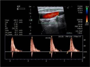
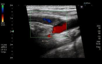
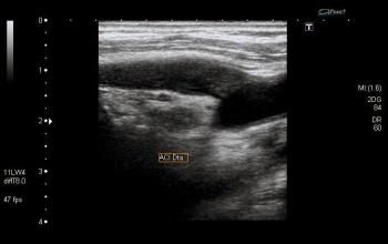
known as "trickle flow" or "string sign" and is diagnostic of near-occlusion (Figs. 3 and 4).
  Pulse Wave (PW) Doppler analysis showed high-flow-resistance spectral morphology and low peak
  systolic velocity in the right common carotid artery. Spectral morphology and flow velocities were
  normal in the left common carotid artery (Figs. 5 and 6).
  Despite the near-occlusion of the right internal carotid artery, right subclavian artery maintained
  normal flow velocity and direction (Fig. 7).

Discussion
  Internal carotid artery (ICA) occlusion is most commonly caused by atherosclerosis. Dissection and
  fibromuscular dysplasia are much less frequent causes.
  On US, ICA occlusion presents as echogenic material completely filling the arterial lumen
  accompanied by absence of arterial pulsations or flow at Colour, Power and PW Doppler
  interrogation. Other findings include a characteristic to-and-fro" flow pattern at the point of
  occlusion, known as thud flow", seen at Colour and PW Doppler imaging and damped resistive
  flow in the common carotid artery (CCA) at PW Doppler imaging, known as externalization of the
  CCA.
  Near occlusion typically presents with a string sign" or trickle flow" at both Colour and Power
  Doppler imaging. The advantages of Power over Colour Doppler in this particular clinical setting
  are not unanimous.
  The distinction between occlusion and near occlusion is of extreme importance because patients
  with near occlusion may be treated with endarterectomy, while total occlusion precludes it.
  To avoid false-positive diagnosis of total occlusion, optimisation of Doppler parameters if of utmost
  importance. It involves:
  . Using a low frequency (< 7 MHz) transducer;
  . Obtaining the best possible views of the ICA, in both longitudinal and transverse planes, with
  adequate box size and steering;
  . Lowering the pulse repetition frequency to values as low as possible, preferably below 15 cm/sec;
  . Lowering the low frequency filter to avoid missing low frequency signals;
  . Increasing Colour and PW gain settings to the point of visible background noise;
  . Increasing Colour threshold to over 80%;
  . Increasing the sample volume gate to 2.5 mm or more.
  Doubtful cases may require alternative imaging modalities, such as catheter or CT angiography.

Final Diagnosis
  Near-occlusion of the right internal carotid artery

Figures

  Figure 1 B Mode US
B Mode examination of the carotid arteries revealed echogenic material filling the lumen of
 the right internal carotid artery, not associated with caliber reduction.

Figure 2 B Mode US

 Intraluminal filling of the right internal carotid artery with echogenic material, here seen in a
 transverse plane.

Figure 3 Colour Doppler US

 Colour Doppler evaluation revealed the string sign" or trickle flow", characteristic of near
 occlusion.
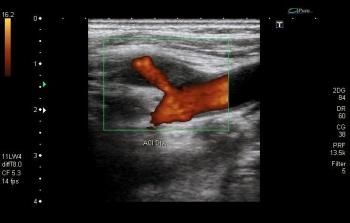
Figure 4 Power Doppler US

 Trickle flow" as seen on power Doppler US.

Figure 5 Spectral Doppler US

 Right common carotid artery showed a high-flow-resistance spectral morphology, with zero
 flow in the end-diastole. Note peak systolic velocity of only 48 cm/s.

Figure 6 Spectral Doppler US
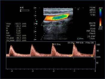
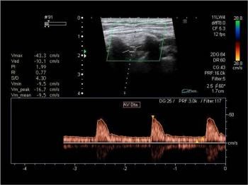
Flow velocities were considerably higher (103 cm/s) in the contralateral common carotid
   artery, with preserved low-flow-resistance spectral morphology.

  Figure 7 Spectral Doppler US

   Despite the pre-occlusive stenosis of the right internal carotid artery, flow was not reversed
   and velocities were normal in the homolateral vertebral artery.

MeSH
  Carotid Stenosis [C14.907.137.230]
  Narrowing or stricture of the internal, common, or external carotid artery, most often as a result of
  atherosclerotic plaque formation. Ulcerations may form in atherosclerotic plaques and induce
  thrombus formation. Platelet or cholesterol emboli may arise from stenotic carotid lesions and
  induce a transient ischemic attack (ISCHEMIC ATTACK, TRANSIENT) or
  CEREBROVASCULAR ACCIDENT. Emboli which travel to the eye may manifest as
  AMAUROSIS FUGAX (temporary blindness). (From Adams et al., Principles of Neurology, 6th
  ed, pp822-3)

References
  [1] Tahmasebpour HR (2005) Sonographic Examination of the Carotid Arteries. BSc, RDMS et al;
  RadioGraphics 25:15611575

  [2] Zwiebel, Pellerito (2005) Introduction to Vascular Ultrasonography. Elsevier Saunders; 5th
  Edition

Citation
  Santiago I1, Canelas A2, Pinto E2
  1Hospital Infante D. Pedro - Aveiro, Portugal
  2Clinica Universitária de Imagiologia dos HUC - Coimbra, Portugal (2010, Jan. 11)
Occlusion or Near-Occlusion of the ICA? {Online}
URL: http://www.eurorad.org/case.php?id=8057
You can also read