Revision of asterolepidoid antiarch remains from the Ogre Formation (Upper Devonian) of Latvia - Estonian Academy Publishers
←
→
Page content transcription
If your browser does not render page correctly, please read the page content below
Estonian Journal of Earth Sciences, 2021, 70, 1, 3–17 https://doi.org/10.3176/earth.2021.01
Revision of asterolepidoid antiarch remains from the Ogre Formation
(Upper Devonian) of Latvia
Ervīns Lukševičs
Department of Geology, University of Latvia, Raiņa Boulevard 19, Riga LV1586, Latvia; ervins.luksevics@lu.lv
Received 3 June 2020, accepted 8 September 2020, available online 15 December 2020
Abstract. The Frasnian (Upper Devonian) antiarch Walterilepis speciosa was first described in 1933 (as Taeniolepis) on the basis of
a single specimen. The newly collected material has allowed the head to be described in a more detail, especially the nuchal and
paranuchal plates. Other newly described elements include bones of the pectoral appendages and trunk armour, demonstrating a
rather high and short trunk armour. The shape and proportions of the head and trunk armour suggest the attribution of Walterilepis
to the family Pterichthyodidae; it is most probably closely related to Lepadolepis from the Late Frasnian of Germany. Whilst W.
speciosa is endemic to the Latvian part of the Baltic Devonian Basin, the connection to the Rheinisches Schiefergebirge is probably
closer than previously presumed. Walterilepis fits into the biostratigraphical column at the same level as Bothriolepis maxima and B.
evaldi, indicating the high diversity of antiarchs during Pamūšis time.
Key words: Frasnian, biostratigraphy, morphology, placoderm, Baltic Devonian basin.
INTRODUCTION noted that the generic name of Gross (1932, 1933a) is
preoccupied by the sarcopterygian name Taeniolepis
The wellknown Baltic German palaeontologist Walter Trautschold, 1874, and he erected the replacement name
Gross, who was born in the close vicinity of Riga, made Walterilepis (Moloshnikov 2001). Following Denison
a great contribution to research on Devonian vertebrates (1978), Moloshnikov erroneously gave 1932 as the year
of Estonia and Latvia. He collected fossil fishes in Latvia of description, but this is a nomen nudum; the description
in the 1930s with his brother, local collector Roland of this taxon was not published until 1933. Recently a new
Gross, and Dr Nikolajs Delle from the University of material of Walterilepis has been collected from Eastern
Latvia (Gross 1933a). It is not clearly stated who collected Latvia enabling the revision of this taxon. The main aim
a single specimen (Museum für Naturkunde Berlin No. of this article is to revise all asterolepidoid material from
MB f 136), consisting of articulated nuchal and postpineal the Ogre Formation, to provide a more accurate de
plates, at the left bank of the Daugava River close to the scription of the species Walterilepis speciosa and to
former Bramberģe manor house (Brambergshof in establish its systematic position within antiarchs.
German: Gross 1933a; fig. 1; locality C, Fig. 1A). The
specimen without description was first named by Gross
Taeniolepis speciosa in his catalogue of antiarchs (1932) GEOLOGICAL SETTING
and the description was published a year later (Gross
1933a). He determined that it was a new genus and species The Upper Devonian is the most complete and widely
of antiarchan fish, differing from both Asterolepis and exposed part of the Devonian section in Latvia where
Bothriolepis in characteristic ornamentation consisting of both the Frasnian and Famennian stages are present.
smooth, radially arranged ridges. Later Gross (1942) The lithostratigraphy of this sequence in Latvia is
provided details of the stratigraphic level for the rather well established (Sorokin 1981a): the Frasnian
specimen; it came from the e/f beds (e/f Stufe in German: section consists of the Amata(?), Pļaviņas, Salaspils,
Gross 1942, p. 400), corresponding to the lower part of Dubnik (a lateral equivalent of the Salaspils Formation),
the Ogre Formation, upper part of the Frasnian, in the Daugava, Katleši, Ogre, Stipinai and Amula formations
modern stratigraphic chart of Latvia. Moloshnikov (2001) (Fig. 2).
© 2021 Author. This is an Open Access article distributed under the terms and conditions of the Creative Commons Attribution 4.0
International Licence (http://creativecommons.org/licenses/by/4.0).
3
Estonian Journal of Earth Sciences, 2021, 70, 1, 3–17
#
%
#
!" $
%&
Fig. 1. A, generalized geological map of Latvia with fossil fish sites (circles designated by letters) where Walterilepis speciosa was
collected; B, a sketch map showing the position of outcrops of the Ogre Formation along the Gurova River. A, modified from Stinkule
& Stinkulis (2013, fig. 3). Legend for part A: 1, Middle Devonian Narva, Aruküla and Burtnieki formations; 2, Middle–Upper
Devonian Gauja, Sietiņi, Lode and Amata formations; 3–6, Upper Devonian; 3, interval of the Pļaviņas–Daugava formations; 4,
Ogre Formation; 5, Stipinai and Amula formations; 6, deposits of the Famennian Stage; 7, Carboniferous; 8, Permian; 9, Triassic;
10, Middle–Upper Jurassic. Fossil localities: A, Langsēde site along the Imula River; B, Kalnamuiža Mill site along the Amula River;
C, Bramberģe site along the Daugava River (type locality); D, localities along the Gurova River. Numbers in part B designate the
outcrops of the Ogre Formation along the Gurova River.
sedimentation was predominant in the interval of the
Pļaviņas–Daugava regional stages and in the Stipinai
Regional Stage. Several comparatively short episodes of
clastic sedimentation occurred during the Frasnian, and
the deposits of the Ogre Formation represent the results
! of one of such events. The Ogre Formation is composed
of the sandstone, siltstone, dolomitic marl, sandy dolomite
" #$
% # and rarely dolomite sandstone, clay and gypsum,
% % "
overlying the eroded surface of the Katleši Formation, but
-
% in the northwestern and northeastern parts of Latvia in
& # & # places it disconformably covers the Daugava Formation
'
&
(Lukševičs et al. 2011).
The Ogre Formation consists of a 15–18 m (western
Latvia) up to 50mthick (eastern Latvia) mainly silici
( # ' ( # ' ) *
clastic sequence, composed of three members: the
%
Lielvārde Member at the base, the Rembate Member in
+ * + * ," the middle and the uppermost Suntaži Member. The
Lielvārde Member consists of sandstone, clay, dolomitic
marl, siltstone, dolomite and gypsumrich dolomite. Fish
Fig. 2. Stratigraphic chart of the uppermost Givetian–Frasnian
remains often form clusters, the socalled ‘fish breccia’
deposits of Latvia. Modified from Stinkulis (2004), Lukševičs
et al. (2012) and Lukševičs & Stinkulis (2018). Black point (Sorokin 1978). The middle part of the Ogre Formation,
designates the stratigraphic level of this study. the Rembate Member, is composed of crossstratified
feldsparquartz sandstone with a high mica content and
carbonate cement, rhythmically alternating with argillite
Carbonate sedimentation dominated during the about (clay), siltstone and dolomitic marl in eastern Latvia. The
7.5Myrlong time span of the Frasnian, while the territory sandstone is found at the base of the rhythms. In the study
was covered by a shallow epicontinental, sometimes area, in the Gurova Ravine, the Rembate Member is
restricted basin with changing salinity. The carbonate represented mainly by sandstone containing vertebrate
4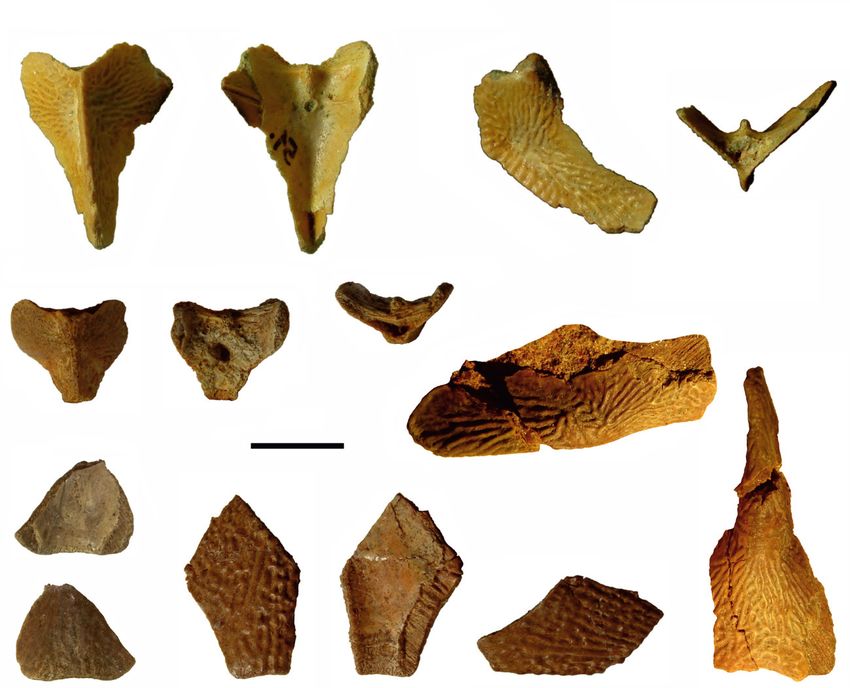
E. Lukševičs: Revision of Walterilepis speciosa
fossils. The Suntaži Member comprises clay and dolomitic the material collected in 1981 at the Langsēde locality
marl with siltstone and sandstone interbeds; most pro (Lukševičs et al. 2011). Considering that three bothriolepid
bably this member is missing in the study area. species, namely Bothriolepis maxima Gross, 1933, B. evaldi
The vertebrate assemblage of the Ogre Formation Lyarskaya, 1986 and Grossilepis spinosa (Gross, 1942),
corresponds to the Psammosteus falcatus (now the zonal have previously been described from the Ogre Formation
taxon is known as Traquairosteus? falcatus: Glinskiy (Lukševičs 2001), the total number of antiarch taxa from
2018) and Bothriolepis maxima biozones, which correlate this formation is unusually high and needs to be revised.
with the late rhenana Standard Conodont Biozone (Esin That was the situation until the author together with
et al. 2000); conchostracans and lingulid brachiopods, the team of students and the graduates from the University
although rare, have also been found in this formation of Latvia collected fish remains from a new locality of the
(Sorokin 1981b). The vertebrate assemblage from the Devonian fish remains and identified some new speci
Ogre Formation in Latvia yields the heterostracomorphs mens of Walterilepis speciosa. The new material comes
Traquairosteus? falcatus (Gross) and Psammosteus sp., from several small outcrops along the Gurova creek in the
acanthodians Devononchus laevis Gross and Acanthodii picturesque Gurova Ravine in eastern Latvia (Stinkulis et
gen. et sp. indet., porolepiform Holoptychius cf. nobilissimus al. 2020). This material includes plates that Gross (1933a)
Agassiz, dipnoans ‘Dipterus’ cf. marginalis Agassiz did not have available to study, such as an anterior median
and Dipteriformes gen. et sp. indet., osteolepiform dorsal plate, a posterior median dorsal plate, an anterior
Platycephalichthys bischoffi Vorobyeva and tetrapod ventrolateral plate, a central dorsal plate of the pectoral
Obruchevichthys gracilis Vorobyeva (Lukševičs et al. fin armour, a lateral plate and a paranuchal plate. Thus, a
2011). Besides these taxa, Psammosteus tenuis Obruchev, more accurate description of the species Walterilepis
Obruchevia heckeri (Obruchev) and Webererpeton speciosa became possible. The detailed comparison with
sondalensis Clément & Lebedev have been reported from the other asterolepidoid antiarch species allowed the
the timeequivalent deposits of northwestern Russia attribution of this species to the family Pterichthyodidae.
(Esin et al. 2000; Clément & Lebedev 2014).
The Ogre Formation is well exposed in many outcrops
along the Daugava, Ogre, Pededze and Tirza rivers in MATERIAL AND METHODS
central and eastern Latvia (mostly Vidzeme), the Lielupe,
Mūsa and Tērvete rivers in southern Latvia (Zemgale) and The Gurova Ravine in eastern Latvia is in the vicinity of
the Abava, Amula, Imula and Venta rivers in western Latvia Aizgalīne village, Medņeva Parish, Viļaka municipality.
(Kurzeme), as well as some other rivers. Fish remains from Nine outcrops of the Upper Devonian, Frasnian Ogre
these various localities have been collected by several Formation lie at a distance of 600 m on both banks of the
authors (Gross 1933a, 1942; Lyarskaya 1986; Lukševičs Gurova River in the interval from about 1.5 km to
2001; Lukševičs et al. 2011). However, no new specimens approximately 2.1 km upstream from its confluence with
of Walterilepis speciosa were reported until 2017, despite the Kira River (Fig. 1). The detailed geological sections
extensive collection efforts. Thus, most researchers have of four outcrops of the Ogre Formation from the Gurova
treated Walterilepis as incertae sedis among Antiarcha (e.g. Ravine were compiled by Ģirts Stinkulis, Simona Mačute,
Denison 1978; Lyarskaya 1981), or Familia incertae sedis Terēze Reķe and the author of this article in 2017 and
among Asterolepiformes (KaratajūtėTalimaa 1963; Gross 2019. Five outcrops of sandstone, of the total of nine
1965), or among Asterolepidoidei (Moloshnikov 2008). exposures, yield vertebrate remains at the Gurova site; the
Yet, two additional specimens of small antiarch fishes have remains of Walterilepis have been found in all of them,
been described from the same stratigraphic level of the namely in outcrops Nos 1, 5, 6, 7 and 8. The vertebrate
Ogre Formation where Walterilepis comes from: one is remains (about 700 specimens identified to the specific or
Antiarchi gen. indet. from the Langsēde locality along the generic level) were collected by the participants in the
Imula River (Gross 1942), and the other is Asterolepis? Summer school in field palaeontology in 2017 and
amulensis Lyarskaya, 1981 from the sandstone deposits mechanically prepared by the author and T. Reķe using an
outcropping along the Amula River 500 m downstream optical microscope and a mounted needle. Most of the fish
from the Kalnamuiža watermill (Lyarskaya 1981). In the material occurs in the light grey, yellowish and light red
description of the latter Lyarskaya (1981) noted that the sandstone, generally as disarticulated and fragmented
attribution of a single anterior ventral lateral (AVL) plate to plates, scales and spines. The bone tissue of Walterilepis
the genus Asterolepis is conditional based only on several is usually well preserved, brown or dark brown and shows
features resembling those of Asterolepis syasiensis, little or no sign of abrasion by the current. Plates were
Byssacanthus and Stegolepis. Two additional specimens measured with a digital Vernier calliper, studied under
provisionally attributed to Asterolepis? amulensis were optical zoom in the binocular microscope Stemi 508 with
found by the author of this paper during the preparation of the builtin photocamera Axiocam 208 Color and
5Estonian Journal of Earth Sciences, 2021, 70, 1, 3–17
photographed with a Sony DSCHX350 digital camera. suborbital lamina; sot, supraoptic thickening; vlc, ventro
Photogrammetry of the nuchal plate LDM Pl 10/523, lateral corner of AVL; vlr, ventrolateral ridge.
paranuchal plate LDM Pl 10/524 and a partial lateral plate
LDM Pl 10/526 was performed by Jurijs Ješkins; several
hundreds of photos have been used to produce the 3D SYSTEMATIC PALAEONTOLOGY
images of these bones. Digital models of the plates were
exported into the modelling programme Blender 2.74 and Suborder ASTEROLEPIDOIDEI Miles, 1968
used to produce the 3D reconstruction of a partial head Family PTERICHTHYODIDAE Stensiö, 1948
shield. The studied material from the Gurova Ravine is
kept in the Latvian National Museum of Natural History Definition (from Young & Gorter 1981). Asterolepidoids
(LDM), Riga; collection No. LDM Pl 10. with a high and short trunk shield, the component plates
Two specimens previously attributed to Asterolepis? being broad in proportion to their length. Distal segment
amulensis are referred below to Walterilepis speciosa. of pectoral fin with two dorsal central and two ventral
These specimens come from the Langsēde locality, which central plates.
has been previously described in detail (Lukševičs et al. Genus Walterilepis Moloshnikov, 2001
2011). These specimens are also kept in the LDM,
collection No. LDM G 99. A partial and damaged anterior Type species. Walterilepis speciosa (Gross, 1933).
median dorsal plate from the same Langsēde locality,
Diagnosis. As for the type species (by monotypy).
described by Gross (Langserde in Gross 1942) and kept
in the Natural History Museum in Stockholm, is also Walterilepis speciosa (Gross, 1933)
referred to Walterilepis. Figures 3–6
Institutional abbreviations. LDM, Latvian National
Museum of Natural History, Riga, Latvia; MB, Natural 1932 Taeniolepis speciosa sp. nov.; Gross, p. 35 (nomen
History Museum (Museum für Naturkunde), Berlin, nudum).
Germany; NRM, Natural History Museum (Naturhistoriska 1933a Taeniolepis speciosa n. g. n. sp.; Gross, p. 43, abb.
riksmuseet), Stockholm, Sweden. 24, taf. IV, fig. 11.
Anatomical abbreviations. The following abbreviations 1942 Antiarchi gen. indet.; Gross, pp. 422–423, abb. 12.
are used in the text: adc, anterodorsal corner of AVL; ADL, 1964 Taeniolepis speciosa Gross; Obruchev, pl. VI,
anterior dorsal lateral plate; alr, anterolateral ridge; AMD, fig. 9.
anterior median dorsal plate; AVL, anterior ventral lateral 1965 Taeniolepis speciosa; Gross, abb. 3G.
plate; Cd1, dorsal central plate 1; Cd2, dorsal central plate 2; 1981 Asterolepis? amulensis Lyarskaja, pp. 137–138,
cf.ADL, area overlapping the anterior dorsal lateral plate; fig. 104; pl. XXXVII, fig. 5.
cf.AMD, area overlapping the anterior median dorsal plate; 2001 Walterilepis speciosa (Gross); Moloshnikov,
cf.MxL, area overlapping the mixilateral plate; cf.PVL, p. 214.
area overlapping the posterior ventral lateral plate; cit1, the 2011 Asterolepis? amulensis Lyarskaja; Lukševičs et
anterior branch of the anterior transverse internal crest; cit2, al., p. 362.
the posterior branch of the anterior transverse internal Material and localities. Holotype: Museum für Naturkunde
crest; cr.d, dorsal crest; cr.dm, dorsomesial crista; cr.pm, Berlin, No. MB f 136 (originally referred to without the
paramarginal crest; cr.pto, postorbital crest; cr.tp, trans number in Gross (1933a)), articulated nuchal and
verse posterior internal crest; d.end1, internal openings of postpineal plates in dorsal view. Material from the Gurova
the endolymphatic ducts; d.end2, external openings of the Ravine, Latgale, eastern Latvia: LDM Pl 10/518–10/523,
endolymphatic ducts; f.ar, articular facet; f.artd, dorsal six nuchal plates; LDM Pl 10/524 and 10/525, paranuchal
crest of the articular facet; f.ax, axillary foramen; f.ax1, plates; LDM Pl 10/526, an incomplete left lateral plate;
axillary foramen on the visceral surface; fmp, protractor LDM Pl 10/527 and 10/533, fragmentary anterior median
area of processus brachialis; f.retr, levator fossa; l, lateral dorsal plates; LDM Pl 10/528 and 10/529, partial posterior
corner of AMD; La, lateral plate; Ml2, lateral marginal median dorsal plates; LDM Pl 10/534, an incomplete,
plate 2; Mm1, mesial marginal plate 1; Mm2, mesial badly preserved trunk armour plate, possibly mixilateral
marginal plate 2; moc, median occipital crista; mpg, plate; LDM Pl 10/530, the proximal part of the right
middle pitline groove; MxL, mixilateral plate; Nu, nuchal central dorsal plate 1 of the pectoral fin, and LDM Pl
plate; pbr, brachial process; p.co, pars condyloidea; pma, 10/531, the left central dorsal plate 1 of the pectoral fin;
posterior marginal area; PMD, posterior median dorsal LDM Pl 10/532, processus brachialis of the fragmentary
plate; Pn, paranuchal plate; Pp, postpineal plate; p.pe, pars anterior ventral lateral plate. From the Kalnamuiža
pedalis of processus brachialis; pr, ventral process on the locality on the Amula River, Kurzeme, western Latvia:
PMD; pt, ventral funnel pit; sna, supranuchal area; sol, LDM G 671, the left anterior ventral lateral plate
6E. Lukševičs: Revision of Walterilepis speciosa
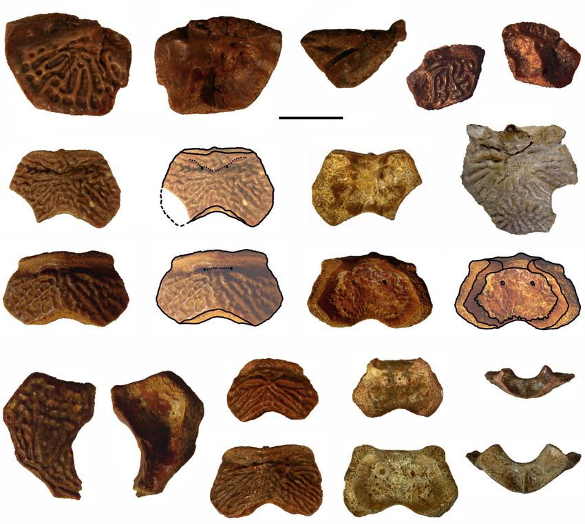
Fig. 3. Walterilepis speciosa (Gross) skeletal elements of the head armour. A, LDM Pl 10/526, partial left lateral plate in dorsal
(A1) and visceral (A2) view; B, LDM Pl 10/518, nuchal plate in dorsal (B1), visceral (B2) and posterior (B3) view; C, LDM
Pl 10/520, nuchal plate in dorsal (C1), visceral (C2) and posterior (C3) view; D, LDM Pl 10/523, nuchal plate in dorsal (D1)
and visceral (D3) view, as well as labelled drawings in dorsal (D2) and visceral (D4) view; E, LDM Pl 10/521, nuchal plate in
dorsal (E1) and visceral (E3) view, as well as labelled drawing in dorsal (E2) view; F, holotype MB f 136, articulated nuchal
and postpineal plates in dorsal view; G, LDM Pl 10/525, left paranuchal plate in dorsal (G1) and visceral (G2) view; H, LDM
Pl 10/524, right paranuchal plate in dorsal (H1), visceral (H2) and posterior (H3) view. A–E, G, H, from the Gurova Ravine;
F, from the type locality on the Daugava River close to Bramberģe. Abbreviations: cf.La, area overlapping lateral plate; cf.Pn,
area overlapping paranuchal plate; cr.o, median occipital crest; cr.pm, paramarginal crest of head shield; cr.pto, postorbital
crest of head shield; cr.tv, transverse nuchal crest; d.end1, ventral foramina of endolymphatic ducts on head shield; d.end2,
dorsal foramina of endolymphatic ducts on head shield; fm, insertion fossa on head shield for levator muscles; ifc1, principal
section of infraorbital sensory line on head shield; mc, lateral corner of Nu plate; mpg, middle pitline groove; nm, smooth
obtected nuchal area of head shield; npp, postpineal notch in nuchal plate; Nu, nuchal plate; pc, posterolateral corner of Nu
plate; Pp, postpineal plate; pr.nm, nuchal process of head shield; pr.po, anterolateral angle of oticooccipital depression of head
shield; sol, suborbital lamina of head shield; sot, supraoptic thickening.
7Estonian Journal of Earth Sciences, 2021, 70, 1, 3–17
Fig. 4. A, B, model of the partial head shield of Walterilepis speciosa (Gross) based on the photogrammetric models of the left La
plate LDM Pl 10/526 and its mirror image as the right La plate, the Nu plate LDM Pl 10/523, the right Pn plate LDM Pl 10/524 and
its mirror image as the left Pn plate. All specimens from the Gurova Ravine. C, D, tentative reconstruction of the head shield of
Walterilepis speciosa (Gross) based on the model and the holotype MB f 136, in dorsal (C) and posterior (D) view. Abbreviations:
La, lateral plate; Nu, nuchal plate; Pmg, postmarginal plate; Pn, paranuchal plate; Pp, postpineal plate; Prm, premedian plate.
(holotype of Asterolepis? amulensis). From the Langsēde wall of the Pn plate is very high. A high trunkshield has
locality on the Imula River, Kurzeme, western Latvia: strongly arched dorsal wall and flat ventral wall. The
LDM G 99/50, a right anterior ventral lateral plate; LDM AMD plate with a high dorsal crest caudally from the
G 99/51, a posterior median dorsal plate; NRMPZ P4586, tergal angle. PMD plate high and short, and conical in
a partial anterior median dorsal plate. shape, with the dorsal crest in the anterior third of the
plate. Lateral lamina of the AVL plate is highest at the
Type locality. Outcrop at the left bank of the Daugava anterodorsal corner; it is higher than the ventral lamina is
River near Bramberģe, Daugmale Parish, central Latvia. wide; lateral lamina about 1.6–1.8 times as long as high,
Note that the level of the Daugava River has risen due to with a short posterodorsal contact face for the MxL plate.
the construction of the dam of the Riga hydroelectric The angle between the ventral and lateral laminae reaches
station, thus the main part of the type locality nowadays 90°–100°. The foramen axillare is very small, smaller than
is below the water level. 1 mm, oval; it opens dorsally on the visceral surface of
Emended diagnosis. A small pterichthyodid with a dorsal the AVL plate. The plates of the head shield and dorsal
length of head and trunkshields reaching at least 60 mm. trunk armour are ornamented with smooth, radially
The Nu plate is quite strongly vaulted, with breadth/length arranged tubercles; the AVL plate is weakly ornamented
index of 1.42–1.70. The endolymphatic ducts open not far with very low and smooth radiating ridges.
from each other. The posterior margin of the Nu plate
bears a pointed posterior projection. The pits for insertion Description
of the head muscles are well pronounced on the high
Head
posterior wall of the Nu plate. The triangular incision for
the postpineal is not deep. The anterior margin of the Pp Walterilepis was a small pterichthyodid. The available
plate is narrower than the posterior margin. The posterior material indicates a similar maximum size to Gerdalepis
8E. Lukševičs: Revision of Walterilepis speciosa
Fig. 5. Walterilepis speciosa (Gross) skeletal elements of the dorsal part of the trunk armour: A–C, partial AMD plates: A, LDM Pl
10/527, in dorsal (A1) and visceral (A2) view; B, LDM Pl 10/533, in dorsal (B1), visceral (B2) and left lateral (B3) view; C, NRM
PZ P4586, in dorsal (C1) and right lateral (C2) view; D, E, partial PMD plates: D, LDM Pl 10/528, in dorsal (D1), visceral (D2) and
posterior (D3) view; E, LDM G 99/51, in dorsal (E1), visceral (E2), right lateral (E3) and posterior (E4) view. A, B, D, from the
Gurova Ravine; C, E, from the Langsēde locality at the Imula River. Abbreviations: alr, anterolateral ridge on AMD; cf.ADL, area
overlapping ADL; cf.ADL1, imprint of area overlapping ADL; cf.AMD, area overlapping AMD; cf.MxL, area overlapping MxL;
cr.d, dorsal median crest of trunk shield; cr.tp, internal posterior transversal crest; dlg2, posterior oblique dorsal sensory line groove;
dmr, dorsal median ridge of trunk shield; f.retr, levator fossa of AMD; l, lateral corner of AMD; lc, lateral corner of PMD; plc,
posterior lateral corner of PMD; pma, posterior marginal area of PMD; pop, posterior pit of PMD; pr, posterior ventral process of
PMD; pt, posterior ventral pit of dorsal wall of trunk shield; sna, supranuchal area. Arrows point anteriorly.
rhenana, Pterichthyodes milleri or Sherbonaspis was rather vaulted, relatively long and narrow; most
andreannae (Gross 1941; Hemmings 1978; Panteleyev probably it was longer than broad. The orbital fenestra of
1993). Byssacanthus dilatatus, Stegolepis jugata, Gerdalepis the model is relatively large and broad; the anterior
dohmi and Sherbonaspis hillsi were somewhat larger portion of the head shield is about half as long as the
species (KaratajūtėTalimaa 1960; Denison 1978; Young posterior portion.
& Gorter 1981). Of the dermal bones of the head shield
Postpineal plate (Pp). The only known Pp plate (MB f
only the postpineal, nuchal, paranuchal and partial lateral
136) is wider than long with a B/L index of 1.78 (Fig. 3F).
plates are known (Fig. 3).
The lateral margin contacting with the La plate is long, thus
The tentative 3D reconstruction of the head produced
the plate is relatively more elongated than the Pp plate in
using Blender 2.74 (Fig. 4) suggests that the head shield
9Estonian Journal of Earth Sciences, 2021, 70, 1, 3–17
Fig. 6. Walterilepis speciosa (Gross) skeletal elements of the ventral part of the trunk armour (A–C) and pectoral appendage (D, E):
A, left AVL LDM G 67/1, in lateral (A1), visceral (A2) and ventral (A3) view; B, right AVL LDM G 99/50, in lateral (B1), visceral
(B2), ventral (B3) and anterior (B4) view; C, partial processus brachialis of the AVL LDM Pl 10/532 in posterior view; D, LDM Pl
10/531, left central dorsal plate 1 in dorsal view; E, LDM Pl 10/530, partial right central dorsal plate 1 in dorsal view. A, B, from the
Langsēde locality at the Imula River; C–E, from the Gurova Ravine. Abbreviations: adc, anterodorsal corner of AVL plate; ar3d,
external articular area of Cd1; cf.ADL, area overlapping ADL; cf.MxL, area overlapping MxL; cf.PVL, area overlapping PVL; cit1,
postbranchial crest (anterior division of crista transversalis interna anterior); cit2, posterior division of crista transversalis interna
anterior; cr.dm, dorsomesial crest of pectoral fin; f.ar, articular facet; f.artd, dorsal part of pectoral articular fossa; f.ax, axillary
foramen; f.ax1, opening of axillary foramen on visceral surface; fmp, protractor area of brachial process; fp, funnel pit; p.co, pars
condyloidea; pdc, posterodorsal corner of AVL plate; p.pe, pars pedalis of brachial process; pbr, processus brachialis; ri, ridge on
AVL plate; vlc, ventrolateral corner; vlr, ventrolateral ridge.
Byssacanthus, Pterichthyodes and Stegolepis (Karatajūtė maximum breadth of the plate occurs also in the Pp plate
Talimaa 1960; Malinovskaya 1973; Hemmings 1978). The of Pterichthyodes (Hemmings 1978, textfig. 1B, C); how
anterior margin is slightly convex, and it is shorter than the ever, its lateral margin is always very short and never forms
maximum width of the plate along the posterior lateral an acute angle with the posterior margin. The posterior
corners. Hence the anterior lateral margins are oblique, margin in Walterilepis forms a shallow Vshape, strongly
forming an acute angle with the right and left portions of differing in shape from all other pterichthyodids. Smooth
the posterior margin. The anterior margin shorter than the rounded ridges radiate from the centre of the plate.
10E. Lukševičs: Revision of Walterilepis speciosa
Nuchal plate (Nu). The nuchal plate (Fig. 3B–F) is Paranuchal plate (Pn). The Pn plate (Fig. 3G, H) is sub
morphologically variable but usually its shape resembles square in shape, with the maximum width in its posterior
that of the Nu plate in some species of Asterolepis part slightly exceeding the length. The B/L index is 1.12–1.34
(KaratajūtėTalimaa 1963; Lyarskaya 1981) and (measured in two specimens). The posterior margin of the
Sherbonaspis (Young & Gorter 1981). The Nu plate is bone is slightly convex, with a wide and welldeveloped
well preserved in LDM Pl 10/518 and 10/523 (Fig. 3B, D). smooth obtected nuchal area. The middle pitline groove
The plate is rather strongly vaulted; it is short and wide (mpg) is short and weakly seen. The lateral segment of the
with a B/L index of 1.42–1.70 (1.54 on an average of six bone is 0.35–0.49 times as wide as the medial segment.
measured specimens) at the lateral corners (1.63 in the The posterior wall is very high, much higher than in
holotype). It is proportionately shorter and broader than Asterolepis. The internal surface shows a very high
in Gerdalepis, Stegolepis jugata and Sherbonaspis (Gross paramarginal crest (cr.pm) dividing the surface into two
1941; Malinovskaya 1973; Young & Gorter 1981; regions approximately equal in width. The posterolateral
Panteleyev 1993), but similar to that of Byssacanthus corner of the oticooccipital depression is rounded. The
(KaratajūtėTalimaa 1960) and relatively longer and external ornamentation consists of vermiculating ridges
narrower than that in Pterichthyodes (Hemmings 1978). and pits.
The lateral corners are situated anteriorly from the middle
Lateral plate (La). The single lateral plate (LDM Pl
of the length of the lateral edges so that the anterior
10/526) is incomplete, missing the anterior part, but the
division of the lateral margin is 1.4–2.3 times shorter than
orbital, lateral, posterolateral and posteromesial margins
the posterior lateral division. This feature is similar to
are well preserved. As reconstructed (Fig. 4) the plate is
Sherbonaspis hillsi (Young & Gorter 1981). The occipital
about twice as long as broad. The preserved length is
(posterior) margin usually is slightly concave or almost
21.2 mm. The plate is thickest (6.0 mm) at the middle of
straight (holotype). It is slightly longer than the anterior
the orbital margin. The preserved part of the paramarginal
margin and 1.28–1.41 times shorter than the maximum
crest (cr.pm) is so situated as to suggest a quite lateral
breadth through the lateral corners. A relatively short,
position for the infraorbital sensory canal, with a cor
smooth obtected nuchal area continues along this margin.
respondingly narrow lateral division of the bone, as in all
The bone has a small sharp posterior middle projection.
asterolepidoids (Stensiö 1938, p. 82). In its elongate shape
The rounded triangular notch for the Pp plate is shallow,
the specimen resembles the La plate of Asterolepis or
not reaching posteriorly the line crossing the lateral
Pterichthyodes and differs from Stegolepis jugata
corners of the Nu plate. The external openings of the
(Malinovskaya 1973, fig. 1, p. 74) with a much broader
endolymphatic ducts (d.end2) are very small, sometimes
plate. The prominent postorbital crest (cr.pto) continues
not well seen (e.g. in LDM Pl 10/523, Fig. 3D), and
beneath the orbital opening as a high suborbital lamina
relatively closely spaced (about 3.5–5.6 mm apart). Gross
(sol), but there is no indication of a lateral division of the
(1933a) noted the absence of sensory line canal grooves
preorbital recess, a characteristic feature of Bothriolepis
in the holotype, where neither the middle pitline groove
(Stensiö 1948, p. 49). The postorbital crest (cr.pto) is much
nor the Vshaped central sensory line canal is visible (Fig.
closer to the orbital margin than in Sherbonaspis (Young
3F). This can partly be explained by the poor preservation
& Gorter 1981, fig. 13) or Asterolepis (Lyarskaya 1981,
of the holotype, because the middle pitline groove (mpg)
fig. 71). Judging from the structure of the posteromesial
is well discernible in the newly collected material.
margin, the postorbital crest extended posteriorly onto the
Usually it is represented by short branches between the
endolymphatic duct openings, however, in LDM Pl Pp and Nu plates along the suture between these plates,
and not onto the Nu plate as in bothriolepids and
10/521 it continues also laterally from the openings
Pterichthyodes (see Hemmings 1978, p. 13), nor onto the
(Fig. 3E) but not reaching the posterolateral margin. On
Pp plate as in Asterolepis (Lyarskaya 1981, fig. 68) and
the posterior wall of the plate, the paired parasagittal
presumably Sherbonaspis (Young & Gorter 1981). The Pp
fossae lateral to the median occipital crista (moc) are
relatively large and deep (Fig. 3B3, C3). These were plate overlaps the La plate along a substantially longer
section of its margin than in Pterichthyodes (Hemmings
interpreted by Stensiö (1931) to be attachment areas for
1978, textfigs 1, 2), but in this respect is similar to
the cranial levator muscles. The internal surface of the
Asterolepis (e.g. Lyarskaya 1981, figs 62, 68). The
plate exhibits a rough large supraotic thickening (sot). The
paramarginal crest is moderately high and lower than the
internal foramina of the endolymphatic ducts (d.end1) are
postorbital crest, thus differing from its form in Asterolepis
large (Fig. 3D3, D4) and between 2.9 and 5.0 mm apart.
(e.g. Lyarskaya 1981, fig. 71). The external ornamentation
Sometimes the distance is slightly shorter than between
consists of vermiculating ridges running in rows parallel
the external foramina. The tubercles on the outer surface
to the lateral edge in the lateral part of the plate and a fine
form ridges in rows radiating from the centre point of the
meshed network of pits in the medial part of the plate.
plate.
11Estonian Journal of Earth Sciences, 2021, 70, 1, 3–17
Trunk shield contact face overlapping the AMD (cf.AMD) along the
anterior edge of the PMD plate is partially preserved only
Anterior median dorsal plate (AMD). Only three speci in LDM G 99/51. The transverse posterior internal crest
mens are known: LDM Pl 10/527 (Fig. 5A) from a smaller (cr.tp) is low, with a slight anterior curvature reflecting the
individual, LDM Pl 10/533 (Fig. 5B) and NRMPZ P4586 concave posterior margin of the plate. This crest bears a
(Fig. 5C) from slightly larger individuals. All are very prominent, sharp conical tubercle (pr) in the middle, and
incomplete, so the proportions of the plate cannot be a deep ventral funnel pit (pt) is well developed anteriorly
estimated. However, the plate seems relatively shorter and from the crest. The posterior marginal area (pma) is
broader than that of Pterichthyodes and Sherbonaspis narrow in the middle and expanded at the lateral margins.
(Hemmings 1978, textfig. 9; Young & Gorter 1981,
fig. 15A). The AMD plate is strongly vaulted, with the Anterior ventral lateral plate (AVL). Two relatively well
dorsal median ridge seen in LDM Pl 10/527 as a slight preserved AVL plates are known from Kurzeme. The left
elevation along the posterior broken margin. There is a AVL plate LDM G 67/1 was previously described as the
high and wellpronounced keel in NRMPZ P4586 holotype of Asterolepis? amulensis Lyarskaja (Fig. 6A).
(Fig. 5C). The anterior margin is concave, as is the LDM G 99/50 (Fig. 6B) is a right AVL plate, and one
anterior lateral margin that formed a contact with the broken processus brachialis of the AVL plate LDM Pl
ADL plate. The visceral surface of LDM Pl 10/533 10/532 comes from the Gurova site (Fig. 6C). Specimen
(Fig. 5B) shows a distinct supranuchal area (sna), and a LDM G 67/1 was erroneously mentioned and figured as a
very low anterolateral ridge (alr) bordering a long narrow right AVL plate by Lyarskaya (1981, p. 137; fig. 104, a
depressed area corresponding to the levator fossa in mirror image; compare with the photo of this specimen in
Bothriolepis (f.retr). This is developed similarly to Lyarskaya’s plate XXXVII, fig. 5). The two complete AVL
Sherbonaspis (Young & Gorter 1981, fig. 15A). The area plates are of similar size (length 35.3 mm in LDM G 67/1
overlapping the anterior dorsal lateral plate (cf.ADL) is and 34.7 mm in LDM G 99/50). The processus brachialis
narrow, terminating posteriorly from the lateral corner (l). LDM Pl 10/532 is from a much larger individual. All
Smooth rounded ornamental ridges radiate from the margins of the lateral lamina in the two complete plates
ossification centre of the plate on the outer surface. Two are well preserved, except for a slightly damaged posterior
short branches of the posterior oblique dorsal sensory line edge in LDM G 99/50. In contrast, the margins of the
groove are weakly developed in NRMPZ P4586, as in ventral lamina are damaged, particularly in LDM G 67/1,
some species of Asterolepis (e.g. Asterolepis sp. 1 and probably because these edges were very thin. The angle
Asterolepis ornata: Lyarskaya 1981). between ventral and lateral laminae is 90° in LDM G
99/50 and 100° in LDM G 67/1. The lateral lamina is
Posterior median dorsal plate (PMD). The three known higher than the ventral lamina is wide, suggesting a rather
PMD plates (Fig. 5D, E) are rather incomplete, but all are high and narrow trunk shield in Walterilepis speciosa,
preserved in three dimensions. LDM G 99/51 is the most resembling that of Byssacanthus (KaratajūtėTalimaa
complete (Fig. 5E), with a B/L index of 0.87. Thus, the 1960), and differing from the proportions in Asterolepis,
plate was high and moderately short, like the PMD plate Stegolepis and Sherbonaspis (e.g. Malinovskaya 1973,
in Gerdalepis rhenana (Gross 1941). The dorsal median p.76; Lyarskaya 1981; Young & Gorter 1981). In the latter
ridge is developed as a moderately high crest (cr.d, Fig. 5E3) forms the ventral lamina is wider than the lateral lamina
along the whole length of the plate, and would have been is high. The ventral lamina of the plate is elongate (B/L
continuous with that on the AMD plate. The posterior index about 0.41 in LDM G 99/50). The ventral lamina is
margin is concave with no posterior angle, resembling that flat, as in Byssacanthus, Stegolepis and Sherbonaspis, and
in Pterichthyodes (Hemmings 1978, textfig. 10). This in contrast to Gerdalepis. The subcephalic division is
margin is swollen due to the crest of the dorsal median relatively short, probably comprising about 22–24% of the
ridge and shows the posterior pit between the dorsal length of the ventral lamina, but the anterior margin in
surface of the plate and the transverse posterior crest LDM G 99/50 is damaged so a precise measurement is
on the internal surface (cr.tp). However, the structure of impossible. The anterolateral corner of the anterior margin
the posterior margin differs from that in Gerdalepis, and the notch for the semilunar plate are not well
Grossaspis and Lepadolepis in the absence of regular, preserved. The left AVL plate probably overlapped the
prismatic spongiosa (cf. Gross 1965). The dorsal surface right one. The lateral margin is slightly convex in both
shows the network of low anastomosing ridges radiating specimens. The area overlapping the median ventral plate
from the posterior central part of the plate. The visceral is missing in both specimens. The area overlapping the
surface of LDM G 99/51 (Fig. 5E2) shows the two posterior ventral lateral plate (cf.PVL) is rather long; the
overlapping areas for the mixilateral plates (cf.MxL) on ventrolateral corner (vlc) is sharp and well pronounced.
the lateral edges, but both are broken and incomplete. The The ventrolateral ridge (vlr) is sharp along the subcephalic
12E. Lukševičs: Revision of Walterilepis speciosa
division and rounded but well developed along the Pectoral appendages. Only two disarticulated dorsal
remaining length of the plate (damaged in the anterior part central plates 1 (Cd1) are known from the pectoral
of LDM G 99/50). The ventral division of the crista appendage. The Cd1 is about 1.8 times as long as broad at
transversalis interna anterior is very high laterally and the broadest point in LDM Pl 10/531 (Fig. 6D) and is
immediately decreases in height mesially. As in therefore slightly longer and narrower than in
Asterolepis and Pterichthyodes (Hemmings 1978, p. 23), Sherbonaspis (Young & Gorter 1981, p. 107). This is
but in contrast to Sherbonaspis (Young & Gorter 1981, within the interval of variability for Pterichthyodes
fig. 18), it is divided into anterior and posterior branches. (Hemmings 1978). The dorsal lamina is widest in the
The anterior branch (cit1) is sharpedged and high laterally proximal part, as in Sherbonaspis (Young & Gorter 1981,
but decreases in height anteromesially. The posterior fig. 18D, E), and contrary to Pterichthyodes (Hemmings
branch (cit2) forms a very low rounded ridge mesially. 1978, textfig. 20). It sutures with the lateral marginal
The lateral lamina of the AVL plate is about 1.6–1.8 plate 2 (Ml2), mesial marginal plate 1 (Mm1), mesial
times as long as high, thus being proportionately higher marginal plate 2 (Mm2) and dorsal central plate 2 (Cd2).
even than in Byssacanthus (KaratajūtėTalimaa 1960, pl. The dorsomesial crista (cr.dm) is quite distinct but is
3, fig. 2). It is highest at the anterodorsal corner (adc), in smooth without any spines along the edge (Fig. 6E). The
contrast to Pterichthyodes (Hemmings 1978, figs. 13, 14), mesial lamina lacks ornament, as in Asterolepis and
Stegolepis (Malinovskaya 1973, p. 77) and Sherbonaspis Sherbonaspis, but on the dorsal lamina it consists of rows
(Young & Gorter 1981, fig. 18, p. 106) where it is highest of elongated tubercles and low ridges radiating from the
at the posterodorsal corner. The dorsal margin is slightly most proximal mesial point.
concave. The area overlapping the anterior dorsal lateral
plate (cf.ADL) is wide anteriorly and tapers posteriorly,
passing into the posterodorsal area overlapping the DISCUSSION
mixilateral plate (cf.MxL). This continues ventrally into
the posterior area overlapping the posterior ventral lateral Morphology and systematics
plate (cf.PVL).
The foramen axillare (f.ax) is very small, only 0.7 mm × The basis on which Walterilepis speciosa is referred here
0.5 mm in LDM G 67/1. It is oval as in Pterichthyodes to the Pterichthyodidae requires some comment. Gross,
(Hemmings 1978, p. 31) rather than subcircular as in with only the Nu and Pp plates available for the study,
Sherbonaspis (cf. Young & Gorter 1981, p. 106). The initially placed ‘Taeniolepis’ (now Walterilepis) among the
axillary foramen on the visceral surface (f.ax1) opens Antiarchi incertae sedis (Gross 1933a). Gross (1965)
dorsally, rather than mesially as in Asterolepis ornata revised this opinion and referred ‘Taeniolepis’ to the
Eichwald (pers. observation). The brachial process (pbr) Asterolepiformes incertae familiae. The suborder
in the three preserved examples shows the articular facet Asterolepidoidei (family Asterolepidae of Denison 1978)
(f.ar) which is rather small and weakly defined caudally is one of three widely accepted higher taxa within the order
from the processus brachialis. It is more clearly Antiarcha. Denison (1978) did not recognize a
delimited dorsally by a low crest, which separates the pterichthyodid grouping and listed six genera in his ‘family
articular facet from the dorsal part of the pectoral Asterolepidae’ (Asterolepis, Byssacanthus, Gerdalepis,
articular fossa (f.artd). This crest is better preserved in Pterichthyodes, Remigolepis and Stegolepis). Denison
LDM G 99/50. The prepectoral corner is damaged in all (1978) listed other forms as Antiarchi incertae sedis (e.g.
specimens. The pars condyloidea (p.co) of the brachial Lepadolepis and Taeniolepis). New genera described since
process is of rounded triangular shape, as is typical in then include Sherbonaspis Young & Gorter (1981),
asterolepidoids (cf. Lyarskaya 1981, fig. 86). The wide Pambulaspis Young (1983) and Wurungulepis Young
and flat triangular protractor area (fmp) is oriented (1990). A broad and long trunk shield distinguishes the
anteriorly; the pars pedalis (p.pe) in front of this area is genera Asterolepis and Remigolepis from other
very narrow. asterolepidoids that have a high and short trunk shield
The AVL plates are weakly ornamented, with very low composed of proportionally broader plates (exemplified by
and smooth ridges radiating mesially and caudally from Pterichthyodes, Byssacanthus or Gerdalepis). On this basis
the articular pectoral fossa on the ventral lamina, and a two families can be distinguished, namely the
finemeshed network in the anterior part of the lamina. Asterolepididae and Pterichthyodidae, the latter previously
The lateral lamina is almost smooth, with faint ridges seen proposed by Stensiö (1948) and later accepted by various
only in the very oblique light. The ornamentation of the authors (KaratajūtėTalimaa 1960; Hemmings 1978; Young
AVL plate in Walterilepis resembles that of the PVL plate & Gorter 1981; Young 1990; Moloshnikov 2008).
and other skeletal elements in Asterolepis syasiensis However, the detailed character analysis based on a
(Lyarskaya 1981, pp. 133–136). comprehensive character matrix of antiarchs (Zhu 1996)
13Estonian Journal of Earth Sciences, 2021, 70, 1, 3–17
supported the opinion that Pterichthyodidae is a para proportions of the lateral lamina of the AVL plate and the
phyletic (Janvier & Pan 1982) or even polyphyletic proportion of the dorsal lamina of the dorsal central plate 1.
grouping (Zhu 1996). The presence of an apical chamber, Byssacanthus differs from Walterilepis in having a dorsal
a single semilunar plate and the characteristic shape of the median spine instead of a dorsal median keel; it differs
armour of Gerdalepis were used to classify this genus in a also in a much larger size, in the relatively shorter Pp plate
separate subfamily by Stensiö (1948). This classification and many other features concerning the shape and
was accepted by Gross (1965), Miles (1968) and proportions of the trunk shield plates.
Hemmings (1978). Friman (1982) recognized two pter Walterilepis resembles Sherbonaspis in the presence
ichthyodid subfamilies (of three asterolepid subfamilies in of a dorsal median crest on the AMD and PMD plates and
Miles 1968): Pterichthyodinae containing Pterichthyodes the shape of the Nu plate. Walterilepis differs clearly from
and Byssacanthus and Gerdalepidinae containing Sherbonaspis in the following: its relatively shorter and
Gerdalepis, Grossaspis and Lepadolepis. Zhu (1996) united broader AMD plate, the proportions of a more elongated
the three latter genera into the family Gerdalepididae. This ventral wall of the trunk shield, the much shorter branches
family is characterized by the similar dorsal spongy layer of the middle pitline groove of the Nu plate, the shape
in the dermal bone of the trunk shield. However, this and proportions of the lateral lamina of the AVL plate and
subdivision is not well supported as the taxa are rather the division of the internal transverse crest into two
poorly known. Besides, Walterilepis shows no spongy layer branches. Besides, Sherbonaspis hillsi is a much larger
in the PMD and AVL plates, and the presence of this feature species than Walterilepis speciosa. However, Sherbonaspis
in the AMD plate is unknown. Long’s (1983) antiarchs andreannae is of a similar maximum size as Walterilepis
classification lists the following genera belonging to the (Panteleyev 1993). Walterilepis differs from Stegolepis in
family Pterichthyodidae: Pterichthyodes, Sherbonaspis, almost all aspects of its morphology except the radially
Stegolepis, Gerdalepis, Lepadolepis, Grossaspis and arranged ornamentation. However, in Stegolepis it
Byssacanthus. Later Wurungulepis was also demonstrated consists of ridges, whereas in Walterilepis it is composed
to belong to the Pterichthyodidae (Young 1990). of the radially arranged tubercles, rather smooth on the
Walterilepis speciosa is not readily compared with lateral wall of the trunk shield. Relevant here is the
other genera within the Asterolepidoidea because of the suggestion by Janvier & Pan (1982) that the genera
limited available material. However, the broad AMD and Stegolepis and closely similar and related Hunanolepis
PMD plates with a distinct dorsal crest, a strongly vaulted from China (Wang 1991; Young 1993) may be more
Nu plate, a very high posterior wall of the Pn plate and a primitive than other known pterichthyodids, including
relatively high AVL plate suggest a high and short trunk Pterichthyodes and Sherbonaspis, even though these have
shield with a welldeveloped dorsal median crest. Thus, been found from younger strata (Givetian/Frasnian).
Walterilepis clearly differs from the Asterolepididae Walterilepis shows many similarities with Gerdalepis
characterized by a low and long trunk shield. All the rhenana, including a similar maximum size (G. dohmi
abovementioned features clearly indicate that Walterilepis (Gross, 1933) is a much larger species; G. jesseni Friman,
should be assigned to the Pterichthyodidae. A high and 1982 is of a similar size as G. rhenana), the presence of
short trunk shield could be the synapomorphy uniting the median dorsal crest forming a keel on the AMD and
Walterilepis with the other Pterichthyodidae. PMD plates, the shape and proportions of the PMD plate,
Pterichthyodes, Byssacanthus, Sherbonaspis and the proportions of the lateral lamina of the AVL plate and
Stegolepis are the best described genera within the the tuberculated ornamentation. Walterilepis differs from
Pterichthyodidae (KaratajūtėTalimaa 1960; Malinovskaya Gerdalepis in the absence of regular, prismatic spongiosa
1973; Hemmings 1978; Young & Gorter 1981; Panteleyev along the posterior margin of the PMD plate, in the flat
1993). Detailed comparison with Walterilepis shows a ventral lamina of the AVL plate (convex in Gerdalepis)
small number of resemblances to Pterichthyodes: the and in the shorter and broader Nu plate. Evidence on the
similar small maximum size of the armour, the shape of crosssection shape of the trunk shield in Walterilepis is
the La plate, the shape of the posterior margin of the PMD limited; it is triangular in Gerdalepis, with an acute dorsal
plate and the oval shape of the axillary foramen. median crest, but this is not the case in Walterilepis.
Walterilepis differs readily from Pterichthyodes and Despite the limited material for the comparison,
Byssacanthus in the presence of a high and well Walterilepis demonstrates strong similarity with
pronounced keel on the AMD plate; it differs also from Lepadolepis from Germany (Gross 1933b) in the trunk
Pterichthyodes in the following: the more elongated Pp shield morphology although the trunk shield is slightly
plate with a very long lateral margin of the plate, the larger in Lepadolepis. Due to the limited material of
longer and narrower Nu plate, the position of the Lepadolepis, it is difficult to make more detailed
postorbital crest on the posterior part of the La plate, the comparisons. The head shield of Lepadolepis is unknown,
relatively shorter and broader AMD plate, the shape and so on available evidence no conclusion can be drawn as
14E. Lukševičs: Revision of Walterilepis speciosa
to whether these two genera are synonymous or only 1978), and the occurrence of the abovementioned
closely related. antiarchs in marine and deltaic or estuarine facies may
indicate that the youngest pterichthyodid representatives
Biostratigraphy, biogeography and palaeoecology of the Asterolepidoidei inhabited the shallow marine
rather than the freshwater environment.
As noted above, at least six taxa of antiarch placoderms
have been previously listed from the Ogre Formation,
namely Bothriolepis maxima, B. evaldi, Asterolepis? CONCLUSIONS
amulensis (referred here to Walterilepis), Grossilepis
spinosa, Walterilepis speciosa and Antiarchi gen. et sp. New data on Walterilepis speciosa indicate its assign
indet. (Gross 1942). Such a total number of antiarch taxa ment to the family Pterychthyoididae and probably a
is unusually high for a relatively thin lithostratigraphic close relationship to Lepadolepis from the Frasnian of
unit which formed within a rather short time span most Germany. Walterilepis speciosa is so far endemic to the
probably corresponding to about one third of the rhenana Baltic Devonian Basin, but the distribution of the latest
conodont Zone (Lukševičs 2001). The above revision of representatives of pterichthyodids suggests closer palaeo
asterolepidoid material from the Ogre Formation places it geographical connections between the Baltic Devonian
all in one species Walterilepis speciosa, representing the Basin and Rheinisches Schiefergebirge during the
youngest record to date of asterolepidoids in the Baltic Frasnian than realized previously.
Devonian Basin. Thus, four antiarch species from this
formation are comparable to other formations in the Baltic
Devonian Basin, e.g., three from the Amata Formation Acknowledgements. Many thanks to all participants in the
(Asterolepis radiata, Bothriolepis prima and B. obrutschewi) Summer school in field palaeontology in 2017, students and
and the Pļaviņas Formation (Asterolepis radiata, graduates from the University of Latvia Valters Alksnītis, Dārta
Ansaberga, Laura Bērtiņa, Mikus Daugavvanags, Sandra
Bothriolepis cellulosa [or vicariant species B. trauds-
Dombrovska, Simona Mačute, Evita Maderniece, Līva Matisone,
choldi in NW Russia] and Grossilepis tuberculata;
Jekaterina Matuko, Kristīne Molnare, Edgars Novickis, Līga
Lukševičs 2001). Paparde, Terēze Reķe, Amanda Stūrmane, as well as Dr Sandijs
Many forms of the Pterichthyodidae are known from Mešķis. Jana Būdniece, S. Mačute, K. Molnare, S. Mešķis and
Eifelian and Givetian strata, including Byssacanthus in the Ģirts Stinkulis participated in the field work in April of 2019.
Baltic States and NW Russia, Pterichthyodes in Scotland, Deputy Director and curator of palaeontological collections of
Gerdalepis and Grossaspis in Germany and Belgium, the Latvian Museum of Natural History Ivars Zupiņš gave access
Wurungulepis and Sherbonaspis hillsi in Australia, to the specimens of Asterolepis? amulensis. Dr Thomas Mörs
Stegolepis and Sherbonaspis andreannae in Kazakhstan, (Natural History Museum, Stockholm) kindly provided access to
Hunanolepis in China (for ages see Gross 1965; the original specimen of Antiarchi gen. et sp. indet. described by
W. Gross and photographs of the specimen. My thanks are also
Malinovskaya 1973; Hemmings 1978; Lyarskaya 1978;
due to Dr Florian Witzmann (Museum für Naturkunde Berlin)
Young & Gorter 1981; Wang 1991; Panteleyev 1993). The for the photograph of the holotype of Walterilepis speciosa.
diversity of younger pterichthyodid antiarchs is much Cordial thanks to Prof. Vita Kalnbērziņa (University of Latvia)
more restricted and yields Lepadolepis from the Upper for linguistic support. The referees Prof. Gavin C. Young (ANU,
Frasnian Kellwasserkalk of the Manticoceras Beds, Bad Canberra, Australia) and Dr Zhu Min (IVPP, Beijing, China) are
Wildungen, Germany (Gross 1933b). The occurrence of thanked for their corrections, constructive remarks and helpful
Lepadolepis most probably coincides with the Upper comments. The publication costs were covered by the grant No.
Kellwasser Horizon and hence the linguiformis conodont lzp2018/20231 ‘Influence of tidal regime and climate on the
Zone (e.g. Feist & Schindler 1994). Thus Lepadolepis is Middle–Late Devonian biota in the epeiric Baltic palaeobasin’ of
only slightly younger than Walterilepis, which is consistent the Latvian Council of Science and by the Estonian Academy of
Sciences.
with the suggestion that these genera are closely related. The
distribution of Walterilepis, Gerdalepis and Lepadolepis
suggests also a closer palaeogeographical connection
REFERENCES
between the Baltic Devonian Basin and Rheinisches
Schiefergebirge during the early Late Devonian.
Clément, G. & Lebedev, O. 2014. Revision of the early tetrapod
As noted above, Walterilepis comes from siliciclastic Obruchevichthys Vorobyeva, 1977 from the Frasnian
deposits of unknown facies close to Bramberģe in central (Upper Devonian) of the north western East European
Latvia and from tidally influenced sandstone in western Platform. Paleontological Journal, 48, 1082–1091.
(Langsēde locality) and eastern Latvia (Gurova Ravine). Denison, R. H. 1978. Placodermi. In Handbook of Paleo-
The pterichthyodids Gerdalepis, Lepadolepis and ichthyology, Part 2 (Schultze, H.P., ed.), pp. 1–128.
Grossaspis come from typical marine facies (Denison Gustav Fischer Verlag, Stuttgart and New York.
15You can also read


