An infected aneurysm of the vertebral artery following cervical pyogenic spondylitis: a case report and literature review
←
→
Page content transcription
If your browser does not render page correctly, please read the page content below
Furukawa et al. BMC Musculoskeletal Disorders              (2021) 22:22
https://doi.org/10.1186/s12891-020-03881-3

 CASE REPORT                                                                                                                                         Open Access

An infected aneurysm of the vertebral
artery following cervical pyogenic
spondylitis: a case report and literature
review
Takahiro Furukawa1, Keisuke Masuda2*, Hideki Shigematsu1, Masato Tanaka1, Akinori Okuda2, Sachiko Kawasaki1,
Yuma Suga1, Yusuke Yamamoto1 and Yasuhito Tanaka1

  Abstract
  Background: An important complication of pyogenic spondylitis is aneurysms in the adjacent arteries. There are
  reports of abdominal aortic or iliac aneurysms, but there are few reports describing infected aneurysms of the
  vertebral artery. Furthermore, there are no reports describing infected aneurysms of the vertebral arteries following
  cervical pyogenic spondylitis. We report a rare case of an infected aneurysm of the vertebral artery as a
  complication of cervical pyogenic spondylitis, which was successfully treated by endovascular treatment.
  Case presentation: Cervical magnetic resonance imaging (MRI) of a 59-year-old man who complained of severe
  neck pain showed pyogenic spondylitis. Although he was treated extensively by antibiotic therapy, his neck pain
  did not improve. Follow-up MRI showed the presence of a cyst, which was initially considered an abscess, and
  therefore, treatment initially included guided tapping and suction under ultrasonography. However, under
  ultrasonographic examination an aneurysm was detected. The contrast-enhanced computed tomography (CT) scan
  showed an aneurysm of the vertebral artery. Following endovascular treatment (parent artery occlusion: PAO), the
  patient’s neck pain disappeared completely.
  Conclusion: Although there are several reports of infected aneurysms of the vertebral arteries, this is the first report
  describing an infected aneurysm of the vertebral artery as a result of cervical pyogenic spondylitis.
  Whenever a paraspinal cyst exist at the site of infection, we recommend that clinicians use not only X-ray,
  conventional CT, and MRI to examine the cyst, but ultrasonography and contrast-enhanced CT as well because of
  the possibility of an aneurysms in neighboring blood vessels. It is necessary to evaluate the morphology of the
  aneurysm to determine the treatment required.
  Keywords: Pyogenic spondylitis, Infected aneurysm, Vertebral artery, Ultrasonographic examination, Case report

* Correspondence: masudash@gmail.com
2
 Department of Emergency and Critical Care Medicine, Nara Medical
University, 840 Shijo-cho, Kashihara City 6348522, Nara, Japan
Full list of author information is available at the end of the article

                                         © The Author(s). 2021 Open Access This article is licensed under a Creative Commons Attribution 4.0 International License,
                                         which permits use, sharing, adaptation, distribution and reproduction in any medium or format, as long as you give
                                         appropriate credit to the original author(s) and the source, provide a link to the Creative Commons licence, and indicate if
                                         changes were made. The images or other third party material in this article are included in the article's Creative Commons
                                         licence, unless indicated otherwise in a credit line to the material. If material is not included in the article's Creative Commons
                                         licence and your intended use is not permitted by statutory regulation or exceeds the permitted use, you will need to obtain
                                         permission directly from the copyright holder. To view a copy of this licence, visit http://creativecommons.org/licenses/by/4.0/.
                                         The Creative Commons Public Domain Dedication waiver (http://creativecommons.org/publicdomain/zero/1.0/) applies to the
                                         data made available in this article, unless otherwise stated in a credit line to the data.
Furukawa et al. BMC Musculoskeletal Disorders       (2021) 22:22                                                                        Page 2 of 7

Background                                                                 white blood cell (WBC) count was 31,000/µl]. He was
The number of patients being hospitalized because of                       hospitalized on the same day (day 0) because mag-
pyogenic spondylitis is increasing annually [1]. Abscesses                 netic resonance imaging (MRI) showed cervical pyo-
may result from pyogenic spondylitis. Although an                          genic spondylitis and an extradural abscess at C6 and
aneurysm at an adjacent artery is one of the important                     C7 (Fig. 1).
complications associated with pyogenic spondylitis, an-
eurysms might be misdiagnosed as abscesses. There have                       He was treated with a combination of antibiotics in-
been reports of abdominal aortic or iliac aneurysms fol-                   cluding vancomycin and cefazolin and provided with a
lowing pyogenic spondylitis [2–6]; however, no reports                     cervical collar. Staphylococcus aureus was detected in
describing infected aneurysms of the vertebral arteries                    the blood culture on day 5, and cefazolin was subse-
following cervical pyogenic spondylitis exist. We report                   quently administered. The inflammation improved and
a rare case of an infected aneurysm of the vertebral ar-                   tests performed on day 17 showed CRP of 3.86 mg/dl
tery following cervical pyogenic spondylitis, which was                    and WBC count of 5700/µl. However, the patient re-
successfully managed with endovascular treatment.                          ported that their neck pain had worsened and follow-up
                                                                           MRI on day 17 indicated the presence of a cyst at the
Case presentation                                                          retropharyngeal space adjacent to the infected vertebral
A 59-year-old man with a medical history of dyslipid-                      body (Fig. 2). The patient was then transferred to our
emia was suffering from continuous neck pain. Three                        hospital for further treatment on day 20.
days after the appearance of neck pain, he visited a
nearby clinic and was treated conservatively using an-                       Once he had been transferred, his body temperature
algesics only. Two weeks later, he was referred to an-                     was recorded at 37.7 ° C and the rest of his vital
other hospital because of a high fever of 38.6° C and                      signs were normal. CRP was 5.03 mg/dl and WBC
increased inflammatory markers on a blood examin-                          count was 7400/µl. He felt severe neck pain with a
ation [C-reactive protein (CRP) was 31.55 mg/dl and                        visual analogue scale (VAS) score of 8.0. Physical

 Fig. 1 Magnetic resonance imaging performed on initial hospitalization at the referring hospital. a T2-weighted sagittal image revealed a high
 intensity lesion at the C6/C7 vertebral body (arrow), retropharyngeal space, and epidural space (arrowhead). The epidural lesion had compressed
 the dural sac from the ventral sideb T2-weighted axial image at C6/7 revealed a high intensity lesion at the epidural space (arrowhead) that had
 compressed the dural sac from the ventral side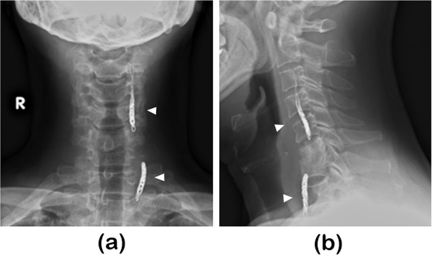
Furukawa et al. BMC Musculoskeletal Disorders       (2021) 22:22                                                                         Page 3 of 7

 Fig. 2 Follow-up magnetic resonance imaging performed at referring hospitala T2 weighted parasagittal image at left side revealed a cyst at C6/
 C7 level (arrow)b T2-weighted axial image revealed a cyst at the retropharyngeal space, adjacent to the infected vertebral body (arrow). Dural sac
 compression by the epidural abscess remained (arrowhead)

examination revealed muscle weakness at the bilateral                       cyst (Fig. 3a). Color Doppler ultrasonography showed
finger extension. There was no evidence of pyramidal                        blood flow inside the cyst, which was linked to the verte-
signs or sensory disturbance.                                               bral artery (Fig. 3b and c). Therefore, we suspected the
  An ultrasonographic examination of the neck was per-                      cyst to be a vertebral aneurysm and performed contrast-
formed in order to evaluate the properties of the neck                      enhanced computed tomography (CT) for confirmation.
cyst (which was thought to be an abscess). Ultrasono-                       The contrast-enhanced CT scans showed blood flow to
graphic examination showed pulsatile turbulence in the                      the cyst from the vertebral artery; therefore, the cyst,

 Fig. 3 Ultrasonographic examination of the necka Imaging with normal parameters showed pulsatile turbulence in the cyst (arrowhead)b Color
 Doppler imaging showed heterogeneous blood flow inside the cyst (arrowhead)c Color Doppler imaging showed the vertebral artery
 (arrowhead) linked to the cyst
Furukawa et al. BMC Musculoskeletal Disorders       (2021) 22:22                                                                        Page 4 of 7

 Fig. 4 Contrast-enhanced computed tomography.a The arterial phase showed slight contrast-enhancement of the left vertebral artery (arrowhead)
 b The delayed phase showed a contrast-enhanced lesion in the cyst (arrowhead). The lesion was linked to the left vertebral artery

initially thought to be an abscess, was diagnosed as an                    was 4600/µl in a blood examination performed on day 91.
infected aneurysm of the vertebral artery (Fig. 4). The                    When the antibiotic therapy was finished, his muscle
patient underwent endovascular treatment (parent artery                    weakness recovered and his VAS for neck pain remained
occlusion; PAO) at our department of Neurosurgery                          0. The C5-C6 vertebral body was completely fused. At the
(Fig. 5) and his neck pain subsequently disappeared                        2-year follow-up, there were no new lesions or recurrence
completely (VAS was 0).                                                    of the pyogenic spondylitis or aneurysm.
  He was transferred back to the referring hospital on day
47 without complications resulting from the treatment.                     Discussion
Cefazolin was administered to the patient until day 58,                    Most incidences of pyogenic spondylitis are reported as
after which cefaclor was orally administered until day 91                  bloodstream infections that often infect intervertebral
(as an outpatient). CRP was 0.02 mg/dl and WBC count                       discs [7]. Our case did not result from iatrogenic

 Fig. 5 X-rays obtained after endovascular treatmentBoth the anteroposterior (a) and mediolateral views (b) showed the coiled left vertebral
 artery (arrowhead)
Furukawa et al. BMC Musculoskeletal Disorders         (2021) 22:22                                                                     Page 5 of 7

conditions or an underlying disease, thus, the route of                       generally considered the most sensitive method for
infection was unknown. For pyogenic spondylitis, a bi-                        examination of pyogenic spondylitis [18]. In our case,
opsy is recommended to determine the responsible bac-                         during the follow-up consultation, monitoring of the
teria [8]. Initially, biopsy and drainage were                                pyogenic spondylitis was performed by MRI and X-ray
recommended because of the presence of a cyst in our                          photography. Since the spine is adjacent to blood vessels,
case, which thought to be an abscess. Generally, CT-                          it is difficult to distinguish whether a paraspinal cyst is
guided or fluoroscopy-guided biopsy is performed for                          an abscess or aneurysm. Therefore, it is necessary to ex-
pyogenic spondylitis [9]. In our case, we planned                             clude aneurysms by ultrasonographic examination or
ultrasound-guided biopsy because the cyst and vessels                         contrast-enhanced CT when a paraspinal cyst is ob-
were not deeply located and could be easily detected by                       served, even if the cyst is thought to be an abscess.
ultrasonography in real time. Ultrasonographic findings                          The treatment for infected aneurysms of the vertebral
revealed that the cyst (initially thought to be an abscess)                   arteries, includes surgical treatments for ruptured cases
was actually an aneurysm of the vertebral artery.                             and large aneurysms [10, 11]. In addition, endovascular
  Although there are reports of infected aneurysms, a                         treatments have been reported for saccular aneurysms
limited number of reports have described infected aneu-                       [13], and conservative treatment has been reported for
rysms of the vertebral arteries as indicated in Table 1                       asymptomatic fusiform aneurysms [12]. According to
[10–13]. Moreover, to our knowledge, this is the first re-                    the literature, surgery is recommended for ruptured
port describing an infected aneurysm of the vertebral ar-                     cases and large aneurysms of vertebral artery. When de-
tery due to cervical pyogenic spondylitis. The first                          ciding on the management strategy of unruptured aneu-
infected aneurysm was reported by Osler [14], which                           rysms of the vertebral artery, adopting a treatment
had been caused by invasion of the arterial wall by bac-                      strategy similar to that of cerebral aneurysms is desir-
teria and its destruction by neutrophils.                                     able. Conservative treatment is indicated for aneurysms
                                                                              with a diameter ≤ 5 mm or ≤ 7 mm, while surgery is rec-
  Infected aneurysms are broadly classified into those                        ommended for larger aneurysms [19, 20]. Infected aneu-
arising from infection of arterial walls and the infection                    rysms are particularly more likely to expand or rupture
of existing aneurysms. The routes of infections of arterial                   and require aggressive surgery [21]. In terms of
walls are further classified into four categories: (1) in-                    aneurysm morphology, it has been reported that saccular
fected endocarditis [14], (2) adjacent infected lesions                       aneurysms have a higher risk of rupture than fusiform
[15], (3) bloodstream infection due to a damaged vascu-                       aneurysms [22], and high-risk saccular aneurysms can be
lar intima [16], (4) infection due to trauma [17]. The                        detected by determining their aspect ratio [23, 24]. In
route of infection in our case was considered to be the                       addition, if clinical symptoms suggestive of imminent
adjacent infected lesion.                                                     rupture are present, such as pain, or if the diameter of
  In previous reports, the diagnosis of an infected                           the aneurysm increases rapidly, urgent surgery is re-
aneurysm of the vertebral artery was confirmed by con-                        quired. In summary, we believe that safe, conservative
ventional and contrast-enhanced CT. An aneurysm may                           treatment of infected aneurysms of vertebral artery are
be diagnosed by palpation, but this was difficult in our                      only possible for small, asymptomatic, fusiform aneu-
case because the common carotid artery was in close                           rysms. Aneurysms with a diameter of 5 mm or less are
proximity to the aneurysm. Therefore, we confirmed                            particularly good candidates for conservative treatment.
that the cyst (which was initially thought to be an ab-                       Even when electing conservative treatment, it is neces-
scess) was an aneurysm by ultrasonographic examin-                            sary to perform CT or ultrasonographic follow-up with
ation, which is a non-invasive and convenient procedure.                      strict antihypertensive control and be aware of whether
Furthermore, contrast-enhanced CT scans showed that                           the aneurysm diameter has expanded. Before surgery, it
the cyst was an aneurysm of the vertebral artery. MRI is                      is necessary to check the dominant side of the vertebral

Table 1 Past reports of infected aneurysms of the vertebral arteries: a systematic review
Reference                          Age (years), sex      Cause                      Responsible bacteria                     Treatment
Flye et al. 1971 [10]              42, Male              Iatrogenic                 S. aureus                                Surgery (rupture)
Singh et al. 2005 [11]             7, Male               Infective endocarditis     Unknown                                  Surgery
Gupta et al. 2014 [12]             25, Male              Lemierre’s disease         Methicillin-resistant S. aureus (MRSA)   Antibiotics
Hashimoto et al. 2015 [13]         75, Male              Cholangitis                S. aureus                                Endovascular Surgery
Current report                     59, Male              Pyogenic spondylitis       S. aureus                                Endovascular Surgery
S. aureus: Staphylococcus aureus
Furukawa et al. BMC Musculoskeletal Disorders (2021) 22:22 Page 6 of 7
artery and determine the presence of anomalies such as Consent for publication
posterior inferior cerebellar artery (PICA) termination, Written informed consent was obtained from the patient for publication of
this Case report and any accompanying images. A copy of the written
which is reportedly found in 0.04–1.3% of cases [25–27]. consent is available for review by the Editor of this journal.
If there is a risk of cerebral infarction due to
embolization of the vertebral artery on the side of the Competing interests
aneurysm, occipital artery-PICA bypass or endovascular The authors declare that they have no competing interests.
treatment with a stent should be performed. Since endo- Author details
vascular modalities are developing rapidly, even ruptured 1
Department of Orthopedic Surgery, Nara Medical University, 840 Shijo-cho,
aneurysms and aneurysms that require revascularization Kashihara City 6348522, Nara, Japan. 2Department of Emergency and Critical
Care Medicine, Nara Medical University, 840 Shijo-cho, Kashihara City
may be managed endovascularly, except in cases where 6348522, Nara, Japan.
endovascular treatment is technically difficult or when
there are concerns about placing a prosthetic device at Received: 18 June 2020 Accepted: 14 December 2020
the infected site. In the current case, the patient suffered
from severe neck pain, and we suspected that rupture of References
a saccular aneurysm was impending. Minimally invasive 1. Issa K, Diebo BG, Faloon M, Naziri Q, Pourtaheri S, Paulino CB, et al. The
endovascular treatment was thus performed. Generally, epidemiology of vertebral osteomyelitis in the United States from 1998 to
2013. Clin Spine Surg. 2018;31:E102-8.
PAO or stent implantation are performed as the endo- 2. Sugawa M, Tanaka R, Nakamura M, Isaka N, Nishimura J, Kimura M, et al. A
vascular treatment. We selected PAO because of the suf- case of infectious pseudoaneurysm of the abdominal aorta associated with
ficient blood flow from the contralateral vertebral artery infectious spondylitis due to Klebsiella pneumoniae. Jpn J Med. 1989;28:402–
5.
and concerns about placing an artificial object at the in- 3. Reddy DJ, Shepard AD, Evans JR, Wright DJ, Smith RF, Ernst CB.
fected site. As a result, cervical pain disappeared and a Management of infected aortoiliac aneurysms. Arch Surg (Chicago, Ill: 1960).
good outcome was achieved. 1991;126:873-8; discussion 8–9.
4. Hagino RT, Clagett GP, Valentine RJ. A case of Pott’s disease of the spine
In conclusion, we described the treatment of an in- eroding into the suprarenal aorta. J Vasc Surg. 1996;24:482–6.
fected aneurysm of the vertebral artery following cervical 5. Doita M, Marui T, Kurosaka M, Yoshiya S, Tsuji Y, Okita Y, et al. Contained
pyogenic spondylitis. This report highlights the need to rupture of the aneurysm of common iliac artery associated with pyogenic
vertebral spondylitis. Spine (Phila Pa 1976). 2001;26:E303-7.
evaluate paraspinal cysts located around the vertebral ar- 6. Tsuji Y, Okita Y, Niwaya K, Tsukube T, Doita M, Marui T, et al. Allograft
tery using not only X-ray, conventional CT, and MRI, replacement of common iliac artery mycotic aneurysm caused by
but also ultrasonographic examination and contrast- Bacteroides fragilis vertebral spondylitis–a case report. Vasc Endovascular
Surg. 2003;37:441–4.
enhanced CT considering the possibility of an aneurysm 7. Berbari EF, Kanj SS, Kowalski TJ, Darouiche RO, Widmer AF, Schmitt SK, et al.
being present. Moreover, we recommend that the Infectious iseases Society of America (IDSA) clinical practice guidelines for
morphology of the aneurysm be evaluated to determine the diagnosis and treatment of native vertebral osteomyelitis in adults. Clin
Infect Dis. 2015;61:e26–46.
the most effective treatment required. The cervical pain 8. Lew DP, Waldvogel FA. Osteomyelitis. Lancet. 2004;364:369–79.
of the patient in the current study disappeared after 9. Pola E, Taccari F. Multidisciplinary management of pyogenic
endovascular treatment. spondylodiscitis: epidemiological and clinical features, prognostic factors
and long-term outcomes in 207 patients. Eur Spine J. 2018;27:229–36.
10. Flye MW, Wolkoff JS. Mycotic aneurysm of the left subclavian and vertebral
Abbreviations
arteries. A complication of cervicothoracic sympathectomy. Am J Surg.
CRP: C-reactive protein; CT: Computed tomography; MRI: Magnetic
1971;122:427–9.
resonance imaging; MRSA: Methicillin-resistant Staphylococcus aureus;
11. Singh D, Pinjala RK, Purohit AK, Reddy LR, Bhattacharjee S. Giant mycotic
PAO: Parent artery occlusion; PICA: Posterior inferior cerebellar artery;
aneurysm of the vertebral artery: a case report. J Vasc Surg. 2005;42:348–51.
VAS: Visual analogue scale; WBC: White blood cell
12. Gupta T, Parikh K, Puri S, Agrawal S, Agrawal N, Sharma D, et al. The
forgotten disease: Bilateral lemierre’s disease with mycotic aneurysm of the
Acknowledgements
vertebral artery. Am J Med Case Rep. 2014;15:230–4.
Not applicable.
13. Hashimoto K, Isaka F, Yamashita K. An infected aneurysm of the vertebral
artery treated with a stent-graft: a case report. Neurol Med Chir (Tokyo).
Authors’ contributions 2015;55:852–5.
TF was a major contributor in writing the manuscript. MK conceived the 14. Osler W. The Gulstonian lectures, on malignant endocarditis. BMJ. 1885;1:
report and helped to draft the manuscript. MK, HS, MT, SK, and YY examined 467–70.
and treated the patient. AO, YS and YY collected and organized the patient’s 15. Hsu RB, Lin FY. Psoas abscess in patients with an infected aortic aneurysm. J
data. HS and YT supervised to wrote the paper. All authors read and Vasc Surg. 2007;46:230–5.
approved the final manuscript. 16. Itatani K, Miyata T, Komiyama T, Shigematsu K, Nagawa H. An ex-situ arterial
reconstruction for the treatment of an infected suprarenal abdominal aortic
Funding aneurysm involving visceral vessels. Ann Vasc Surg. 2007;21:380–3.
None. 17. Samore MH, Wessolossky MA, Lewis SM, Shubrooks SJ Jr, Karchmer AW.
Frequency, risk factors, and outcome for bacteremia after percutaneous
Availability of data and materials transluminal coronary angioplasty. Am J Cardiol. 1997;79:873–7.
Not applicable. 18. An HS, Seldomridge JA. Spinal infections: diagnostic tests and imaging
studies. Clin Orthop Relat Res. 2006;444:27–33.
Ethics approval and consent to participate 19. Wermer MJ, van der Schaaf IC, Algra A, Rinkel GJ. Risk of rupture of
Formal approval is not required for this type of study. Moreover, the need unruptured intracranial aneurysms in relation to patient and aneurysm
for informed consent to participate was waived. characteristics: an updated meta-analysis. Stroke. 2007;38:1404–10.Furukawa et al. BMC Musculoskeletal Disorders (2021) 22:22 Page 7 of 7
20. Wiebers DO, Whisnant JP, Huston J 3rd, Meissner I, Brown RD Jr, Piepgras
DG, et al. Unruptured intracranial aneurysms: natural history, clinical
outcome, and risks of surgical and endovascular treatment. Lancet. 2003;
362:103–10.
21. Rakita D, Newatia A, Hines JJ, Siegel DN, Friedman B. Spectrum of CT
findings in rupture and impending rupture of abdominal aortic aneurysms.
Radiographics. 2007;27:497–507.
22. Nathan DP, Xu C, Pouch AM, Chandran KB, Desjardins B, Gorman JH 3rd,
et al. Increased wall stress of saccular versus fusiform aneurysms of the
descending thoracic aorta. Ann Vasc Surg. 2011;25:1129–37.
23. Akai T, Hoshina K, Yamamoto S, Takeuchi H, Nemoto Y, Ohshima M, et al.
Biomechanical analysis of an aortic aneurysm model and its clinical
application to thoracic aortic aneurysms for defining “saccular” aneurysms. J
Am Heart Assoc. 2015;4:e001547.
24. Natsume K, Shiiya N, Takehara Y, Sugiyama M, Satoh H, Yamashita K, et al.
Characterizing saccular aortic arch aneurysms from the geometry-flow
dynamics relationship. J Thorac Cardiovasc Surg. 2017;153:1413–120.
25. OʼDonnell CM, Child ZA, Nguyen Q, Anderson PA, Lee MJ. Vertebral artery
anomalies at the craniovertebral junction in the US population. Spine (Phila
Pa 1976). 2014;39:E1053-7.
26. Fortuniak J, Bobeff E, Polguj M, Kośla K, Stefańczyk L, Jaskólski DJ.
Anatomical anomalies of the V3 segment of the vertebral artery in the
Polish population. Eur Spine J. 2016;25:4164–70.
27. Wakao N, Takeuchi M, Nishimura M, Riew KD, Kamiya M, Hirasawa A, et al.
Vertebral artery variations and osseous anomaly at the C1-2 level diagnosed
by 3D CT angiography in normal subjects. Neuroradiology. 2014;56:843–9.
Publisher’s Note
Springer Nature remains neutral with regard to jurisdictional claims in
published maps and institutional affiliations.You can also read