Blind Exchange With Mini-Pinning Technique Using the Tron Stent Retriever for Middle Cerebral Artery M2 Occlusion Thrombectomy in Acute Ischemic ...
←
→
Page content transcription
If your browser does not render page correctly, please read the page content below
ORIGINAL RESEARCH
                                                                                                                                                  published: 19 May 2021
                                                                                                                                         doi: 10.3389/fneur.2021.667835

                                               Blind Exchange With Mini-Pinning
                                               Technique Using the Tron Stent
                                               Retriever for Middle Cerebral Artery
                                               M2 Occlusion Thrombectomy in
                                               Acute Ischemic Stroke
                                               Takeshi Yoshimoto 1*, Kanta Tanaka 2,3 , Junpei Koge 3 , Masayuki Shiozawa 3 ,
                                               Hiroshi Yamagami 4 , Manabu Inoue 2,3 , Naruhiko Kamogawa 1,3 , Tetsu Satow 5 ,
                                               Hiroharu Kataoka 5 , Kazunori Toyoda 3 , Masafumi Ihara 1 and Masatoshi Koga 3
                                               1
                                                Department of Neurology, National Cerebral and Cardiovascular Center, Suita, Japan, 2 Division of Stroke Care Unit,
                                               National Cerebral and Cardiovascular Center, Suita, Japan, 3 Department of Cerebrovascular Medicine, National Cerebral
                                               and Cardiovascular Center, Suita, Japan, 4 Department of Stroke Neurology, National Hospital Organization Osaka National
                           Edited by:          Hospital, Osaka, Japan, 5 Department of Neurosurgery, National Cerebral and Cardiovascular Center, Suita, Japan
                  Diogo C. Haussen,
       Emory University, United States
                                               Introduction: The usefulness of the blind exchange with mini-pinning (BEMP) technique
                        Reviewed by:
                  Carlos Pérez García,
                                               has recently been reported for mechanical thrombectomy in patients with stroke owing to
         San Carlos University Clinical        medium vessel occlusion (MeVO). The Tron stent retriever can be delivered and deployed
                       Hospital, Spain
                                               through a 0.0165-inch microcatheter. This retriever has potential as an effective and safe
                      Cynthia Kenmuir,
        UPMC Altoona, United States            treatment for acute ischemic stroke (AIS) due to occlusion of the M2 segment of the
                       Francisco Dias,         middle cerebral artery (MCA). Here, we report the outcomes of the BEMP technique
        University of São Paulo, Brazil
                                               using Tron stent retrievers for M2 occlusion thrombectomy.
                *Correspondence:
                 Takeshi Yoshimoto             Methods: Consecutive patients with AIS owing to M2 occlusion who underwent the
   yoshimototakeshi1982@ncvc.go.jp
                                               BEMP technique using 2 × 15-mm or 4 × 20-mm Tron stent retrievers were included. The
                  Specialty section:
                                               technique involves deploying a Tron stent retriever through a 0.0165-inch microcatheter,
        This article was submitted to          followed by microcatheter removal and blind navigation of a 3MAX or 4MAX aspiration
      Endovascular and Interventional          catheter over the bare Tron delivery wire until the aspiration catheter reaches the clot. A
                           Neurology,
              a section of the journal         Tron stent retriever is inserted into the aspiration catheter like a cork and subsequently
               Frontiers in Neurology          pulled as a unit. We assessed procedural outcomes [first-pass expanded thrombolysis
         Received: 19 February 2021            in cerebral infarction (eTICI) score 2c/3 and 2b/2c/3], safety outcomes [symptomatic
            Accepted: 07 April 2021
                                               intracranial hemorrhage (sICH)], and clinical outcomes (good outcome rate defined as
            Published: 19 May 2021
                                               modified Rankin Scale score 0–2 at 90 days and mortality at 90 days).
                              Citation:
      Yoshimoto T, Tanaka K, Koge J,           Results: Eighteen M2 vessels were treated in 15 patients (six female, median
  Shiozawa M, Yamagami H, Inoue M,
   Kamogawa N, Satow T, Kataoka H,
                                               age: 80 years, and median National Institutes of Health Stroke Scale score:
Toyoda K, Ihara M and Koga M (2021)            18). The BEMP technique was performed successfully in all cases. Whether to
    Blind Exchange With Mini-Pinning
                                               use a 3MAX or 4MAX catheter was determined by considering one of the
       Technique Using the Tron Stent
   Retriever for Middle Cerebral Artery        following target vessels: dominant, non-dominant, or co-dominant M2 (3MAX, n
M2 Occlusion Thrombectomy in Acute             = 9; 4MAX, n = 9). The first-pass eTICI 2c/3 and 2b/2c/3 rates were 47
                      Ischemic Stroke.
             Front. Neurol. 12:667835.
                                               (7/15) and 60% (9/15), respectively; sICH was not observed. Seven patients
     doi: 10.3389/fneur.2021.667835            (47%) achieved good outcomes, and one patient (7%) died within 90 days.

Frontiers in Neurology | www.frontiersin.org                                         1                                             May 2021 | Volume 12 | Article 667835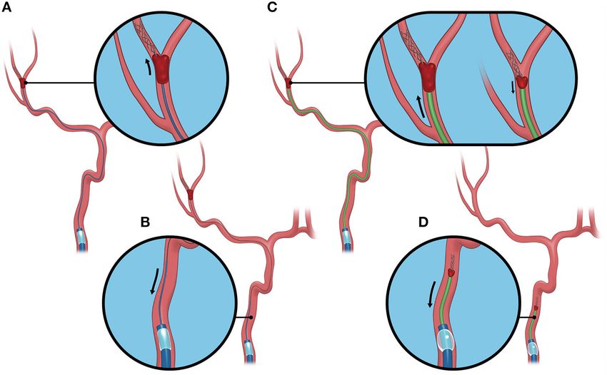
Yoshimoto et al.                                                                                                    M2O Thrombectomy With BEMP Technique

                                               Conclusions: The Tron stent retriever was safely and effectively used in the BEMP
                                               technique for acute MCA M2 occlusion and can be combined with a 0.0165-inch
                                               microcatheter, which may be useful for treating MeVO, in general.

                                               Keywords: BEMP technique, M2 occlusion, Tron stent retriever, thrombectomy, acute ischemic stroke

INTRODUCTION                                                                        the BEMP technique using Tron stent retriever for MCA M2
                                                                                    occlusion thrombectomy.
The usefulness of the blind exchange with mini-pinning
(BEMP) technique has recently been reported for mechanical                          MATERIALS AND METHODS
thrombectomy in patients with acute ischemic stroke (AIS)
owing to medium vessel occlusion (MeVO), i.e., occlusions of the                    Study Design
M2/M3 middle cerebral artery (MCA), A2/A3 anterior cerebral                         All AIS patients admitted to our institute within 7 days from
artery, and P2/P3 posterior cerebral artery segments (1, 2).                        symptom onset or last-known-well date were prospectively
    The BEMP technique, which is the blind catheter exchange                        registered in the National Cerebral and Cardiovascular Center
technique and originally devised using a Trevo 3 × 20-mm                            (NCVC) Stroke Registry (8–13). Data for AIS patients with MCA
retriever (Stryker, Fremont, CA, USA) delivery wire to advance                      M2 occlusion undergoing the BEMP technique were collected
the thromboaspiration catheter and the pinning technique with                       retrospectively from the NCVC Stroke Registry from March
small devices (“mini-pinning”) for thrombectomy of intracranial                     2019 to June 2020. The decision to proceed with endovascular
distal occlusions, has been reported to be helpful and safe for                     therapy (EVT), including the BEMP technique, was made at
optimizing procedural performance when treating MeVO (1, 2).                        the discretion of the investigators in a nonrandomized fashion.
Although original mini-pinning was required to pull the stent                       This study was approved by the institutional review board of
retriever slightly and lock the stent retriever and aspiration                      the NCVC (approval number: M23-073-4). The NCVC Stroke
catheter (1), performing this operation for MCA distal M2                           Registry is registered at ClinicalTrials.gov (NCT02251665).
occlusion is challenging. The Tron stent retriever is a new stent
retriever for AIS owing to not only large intracranial vessel                       Tron Stent Retriever
occlusion but also MeVO (3). As the most significant feature, the                   The Tron stent retriever is a self-expanding stent retriever made
Tron stent retriever can be deployed via a relatively small 0.0165-                 of a nickel–titanium alloy that consists of two types of cells with
inch (0.42-mm) microcatheter, similar to the Aperio (Acandis,                       different shapes (3). The large cell captures the thrombus, and the
Pforzheim, Germany) (2 × 16 mm and 3.5 × 28 mm) (4, 5)                              small cell reduces stent extension at the flexion. When deploying
or Catch View mini (Catch View; Balt, Montmorency, France)                          and retracting from an elongated target vessel, the stent retriever
(3.5 × 20 mm), (6) and has a stent retriever diameter profile                       stretches against the flexion force and passes through the lesser
of 2 mm (3). The Trevo stent retriever, which was used in the                       curvature side of an elongated target vessel. However, the unique
first reported BEMP technique (1), has a minimum low profile                        cell structure of the Tron stent retriever allows the stent structure
of 3 mm, and a microcatheter can be used only up to 0.0175                          to stretch at the bend; therefore, this stent retriever is less likely
in. (0.44 mm), whereas, the Tron stent retriever has a minimum                      to miss a blood clot. The details of the Tron stent retriever are
profile of 2 mm, and a catheter can be used up to 0.0165 in. These                  illustrated in Figure 1.
differences provide the advantage that the Tron stent retriever
can be deployed distally, and that less friction between the stent                  Interventional Protocol With the BEMP
retriever and vessel wall is also expected to reduce hemorrhagic                    Technique Using the Tron Stent Retriever
events (3). Furthermore, the Tron stent retriever is more useful                    All procedures in the BEMP technique using the Tron
for MCA M2 occlusion thrombectomy in the BEMP technique.                            stent retriever were performed by four experienced
Although the traditional combination technique using a Tron                         neurointerventionalists (TY, KTa, JK, and HY) as a front-line
stent retriever with a 3MAX or 4MAX aspiration catheter may                         thrombectomy with local anesthesia or conscious sedation.
be effective in enhancing the first-pass effect (FPE) (7) in MCA                    The BEMP technique was performed through a right femoral
M2 occlusion thrombectomy, we occasionally experience that a                        artery approach, and the occluded-side cervical internal carotid
4MAX aspiration catheter is often outsized for the target vessel,                   artery (ICA) was catheterized using a balloon guide catheter
and that a 3MAX aspiration catheter for the target thrombus                         (BGC). The proximal BGC used in all procedures was a 9-Fr
volume is insufficient when trying to engage these aspiration                       Optimo BGC (Tokai Medical Products Inc., Aichi, Japan). The
catheters proximal to the thrombus. In the BEMP technique, the                      BEMP technique using the Tron stent retriever is performed
aspiration catheter (3MAX or 4MAX aspiration catheter) can be                       initially by advancing a 0.0165-inch microcatheter (Excelsior
determined after confirming the target vessel diameter (1, 2).                      SL-10 microcatheter; Target Therapeutics/Stryker, Fremont, CA,
Therefore, the BEMP technique using Tron stent retriever has                        USA) distal to the occlusion site. Next, a 2 × 15-mm or 4 ×
potential as an effective and safe treatment for MeVO, including                    20-mm Tron stent retriever (JIMRO; Terumo, Takasaki, Gunma,
occlusion of the MCA M2. Here, we report the outcomes of                            Japan) is deployed via a 0.0165-inch microcatheter, followed

Frontiers in Neurology | www.frontiersin.org                                    2                                       May 2021 | Volume 12 | Article 667835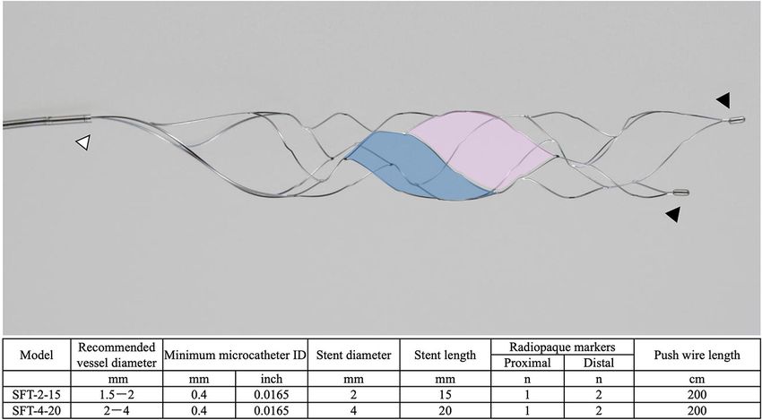
Yoshimoto et al.                                                                                                              M2O Thrombectomy With BEMP Technique

 FIGURE 1 | The details of the Tron stent retriever. Photograph of the Tron stent retriever, which is constructed of two types of cells. There are a radiopaque marker in
 the proximal end (white arrowhead) and two markers attached to the distal end (black arrowheads). The large cell (pink) captures the thrombus, and the small cell
 (blue) reduces stent extension at the flexion.

by microcatheter removal and immediate blind navigation of a                            in cerebral blood flow of 6 s (16, 17). Occlusion sites were
retriever is pulled into the DAC like placing a bottle cork, and                        determined using digital subtraction angiography. The stroke
the pinned clot with the stent retriever and the DAC then forms                         causative mechanism was determined according to the Trial of
a single unit. The Tron stent retriever and DAC are subsequently                        ORG 10172 in Acute Stroke Treatment criteria by board-certified
retracted as a unit to retrieve the clot (1). Figure 2 shows the                        stroke neurologists.
BEMP technique performed as the primary maneuver. Figure 3
illustrates an example of the BEMP technique using the Tron                             M2 Definition
stent retriever for MCA M2 occlusion.                                                   The MCA M2 segment was defined as the vessels from the
                                                                                        main MCA bifurcation/trifurcation to the circular sulcus of the
Clinical Data Collection and Definitions                                                insula (18, 19). M2 occlusions in which the occluded MCA
The baseline clinical characteristics for the following variables                       bifurcated after the horizontal segment were considered proximal
were collected: sex, age, prestroke modified Rankin Scale                               occlusions. Proximal M2 was defined as the horizontal M2
(mRS) score, medical history (hypertension, dyslipidemia,                               segment from the main MCA bifurcation/trifurcation within
diabetes mellitus, current smoking, ischemic stroke, and atrial                         1 cm from the MCA bifurcation and distal M2 as the Sylvian
fibrillation), systolic blood pressure at admission, baseline                           M2 segment, starting 1 cm from the bifurcation/trifurcation to
National Institutes of Health Stroke Scale (NIHSS) score,                               the circular sulcus of the insula (19–21). M2 caliber dominance
baseline Alberta Stroke Program Early Computed Tomographic                              was considered present if the M2 branch had a larger diameter
Score (ASPECTS) on diffusion-weighted imaging (DWI) or                                  than the other branches on digital subtraction angiography or
computed tomography (CT), baseline ischemic core volume,                                if the perfusion defect associated with the occluded M2 branch
time logistics (onset-to-groin puncture time and groin puncture-                        was larger than 50% of the MCA territory. Only when the
to-revascularization time), treatment profile (intravenous                              diameters of both the inferior and superior branches were equal
thrombolysis), type of presentation by vessel occlusion (isolated                       and the associated perfusion defect was 6 s) to determine M2 caliber dominance.
of
Yoshimoto et al. M2O Thrombectomy With BEMP Technique
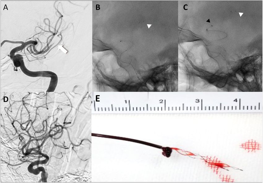
FIGURE 2 | Illustrative cases of the blind exchange and mini-pinning technique using a Tron stent retriever to remove a clot. Illustration of blind exchange and
mini-pinning using the Tron stent retriever in a dominant and proximal middle cerebral artery M2 occlusion. (white arrow: proximal end of the clot) (A) Lateral
angiogram showing an M2 occlusion. (B) A 0.0165-inch microcatheter in place after deployment (white arrowhead: distal end of the retriever). (C) The 3MAX
aspiration catheter is advanced into the face of the clot (black arrowhead) over the bare retriever delivery wire. (D) Final angiogram showing successful reperfusion. (E)
Mini-pinning (2-mm Tron stent retriever and 3MAX aspiration catheter) with “corking” of the thrombus.
and occlusions were classified according to their clinical scenario: (SAH) (diffuse or focal within the territory of the artery treated
isolated M2 occlusion, tandem occlusion, or multi-vessel M2 for distal occlusion), arterial spasm (defined as any degree of
occlusion. Tandem occlusion was defined as an M2 occlusion spasm in treated vessels), emboli to the same or different territory
with a concomitant larger vessel/proximal occlusion. Multi- after thrombectomy of a proximal occlusion, active extravasation,
vessel M2 occlusion was defined as ipsilateral occlusions of and mortality at 90 days. Efficacy outcome was defined as the rate
the M2 superior trunk of the MCA and M2 inferior trunk of of good outcome (mRS score of 0–2 at 90 days).
the MCA.
Statistical Analysis
Continuous variables are reported as median [interquartile range
Outcomes
(IQR)], and categorical variables are reported as proportions.
The procedural outcomes were defined as a first-pass expanded
Analyses were performed using Stata 15.1 (StataCorp, College
thrombolysis in cerebral infarction (eTICI) score of 2c/3 and
Station, TX, USA).
2b/2c/3 for the targeted M2 (23). Safety outcomes were defined as
the presence of any parenchymal hematoma (PH) [hemorrhagic
infarction according to the European Cooperative Acute Stroke RESULTS
Study (ECASS); PH type 1 or 2], any intracranial hemorrhage
(ICH), symptomatic ICH (sICH), which was defined according The study flowchart is provided in Figure 4. A total of 774
to the ECASS II criteria (any ICH with a ≥4-point increase in patients had a diagnosis of AIS within 24 h of onset between
the NIHSS score from baseline) (24), subarachnoid hemorrhage March 2019 and June 2020 in the NCVC Stroke Registry, and of
Frontiers in Neurology | www.frontiersin.org 4 May 2021 | Volume 12 | Article 667835Yoshimoto et al. M2O Thrombectomy With BEMP Technique
FIGURE 3 | Blind exchange and mini-pinning using a Tron stent retriever to retrieve a clot. Illustrative example of the blind exchange and mini-pinning technique using
a Tron stent retriever to treat a middle cerebral artery M2 occlusion. We routinely place a large-bore balloon guide catheter in the cervical internal carotid artery. (A) The
biaxial system, including the microcatheter and microguidewire, is advanced as a unit, with the microcatheter and guidewire traversing the occluded segment. The
Tron stent retriever is deployed via a 0.0165-inch microcatheter distal to the clot. (B) In the next step, the microcatheter is removed from the body. (C) A 3MAX or
4MAX aspiration catheter, as a distal aspiration catheter, is blindly navigated over the bare Tron delivery wire (“blind exchange”) until the aspiration catheter reaches the
clot. (C, left) The Tron stent retriever is pulled into the distal aspiration catheter like placing a bottle cork, and the pinned clot with the stent retriever and the distal
aspiration catheters then forms a single unit. (C, right). (D) The Tron stent retriever and aspiration catheter are subsequently retracted as a unit to retrieve the clot.
these, 62 patients had MCA M2 occlusion, including 25 patients proximal M2 occlusion, and 7/18 vessels had distal M2 occlusion.
who underwent EVT. Of the 25 M2 occlusion patients, 18 MCA Dominance of either superior or inferior division was observed
M2 occlusion patients were treated with the BEMP technique. Of in 12/15 (80%) patients, and co-dominance of M2 branches was
the seven patients who did not undergo the BEMP technique, observed in 3 (20%) patients with M2 occlusion. Dominance
two patients were enrolled in another device clinical trial, and of a branch was more commonly seen in the inferior division
in one patient, we were unable to advance the BGC into the (7/10). A first-pass eTICI score of 2c/3 was achieved in 7/15 (47%)
ICA; this patient underwent a simple stent retrieval technique. vessels, and a first-pass eTICI score of 2b/2c/3 was achieved in
The remaining four patients underwent another EVT technique 9/15 (60%) vessels. A final eTICI score of 2c/3 was obtained
(Aspiration-Retriever Technique for Stroke technique (25) using in nine patients (60%), and a final eTICI score of 2b/2c/3 was
the EmboTrap II device [Neuravi/Cerenovus, Miami, FL, USA] obtained in 14/15 patients (93%). The median (IQR) number of
or a Trevo stent retriever and a large-bore aspiration catheter) at passes was 1 (1–3).
the investigator’s discretion. The BEMP technique using a Trevo Concerning the safety and efficacy outcomes, PH, SAH,
stent retriever was performed in three patients before the use of arterial spasm, and emboli to the same territory after
the Tron stent retriever was approved. thrombectomy of a proximal occlusion were seen in one
Six patients (40%) were female; the median (IQR) age was patient each, respectively. ICH was seen in 6 patients (40%); no
80 (75–83) years; the median NIHSS score was 18 (11–25); sICH, active extravasation, or emboli to the different territory
baseline ASPECTS on DWI or CT: 7 (6–10); onset-to-groin after thrombectomy of a proximal occlusion were observed.
puncture time: 141 (101–170) min. The median groin puncture- The rate of good outcome was 47% (7/15), and 1 patient died
to-revascularization time was 55 (45–75) min, of which the with a history of severe heart failure. The patients’ baseline
median groin puncture-to-revascularization time in isolated M2 characteristics and outcomes are summarized in Table 1, and
occlusion was 35 (27–41) min, and that for tandem occlusion the cohort clinical outcomes and the outcomes in the M2
or multi-vessel M2 occlusion was 82 (58–112) min. Intravenous occlusion patients undergoing BEMP using the Tron stent
alteplase was used in 9 (60%) patients, and a BGC was used in retriever and other stent retrievers (Trevo stent retriever) are
15 (100%) patients; eight patients had isolated M2 occlusions. summarized in Table 2.
Tandem occlusion included the M2 plus the intracranial ICA In seven patients with MCA M2 occlusion who did not
(n = 3) or MCA M1 (n = 1). Three patients had multi-vessel undergo the BEMP technique, the rate of a first-pass eTICI score
M2 occlusion. At the M2 occlusion site, 11/18 (61%) vessels had of 2c/3 was 29% (2/7), and the rate of a first-pass eTICI score
Frontiers in Neurology | www.frontiersin.org 5 May 2021 | Volume 12 | Article 667835Yoshimoto et al. M2O Thrombectomy With BEMP Technique
FIGURE 4 | Study flowchart. ACA, anterior cerebral artery; BA, basilar artery; BEMP, blind exchange with mini-pinning; BGC, balloon guide catheter; CCA, common
carotid artery; EVT, endovascular therapy; ICA, internal carotid artery; MCA, middle cerebral artery; NCVC, National Cerebral and Cardiovascular Center; PCA,
posterior cerebral artery; VA, vertebral artery.
of 2b/2c/3 was 43% (3/7). ICH was seen in four patients (57%); this limitation. Furthermore, the stiffness of the retriever delivery
no sICH or active extravasation was observed. The rate of good wire, distal anchoring by the deployed Tron stent retriever, which
outcome was 43% (3/7). has less friction on M2 vessels, and coverage of the wire by the
BGC eliminate the risk of pushing/losing the delivery wire in the
DISCUSSION patient. The subsequent straightening of the vessels that can be
achieved by gently pulling the retriever wire greatly facilitates
We describe the BEMP technique using the Tron stent retriever, overcoming the barriers to M2 navigation of DACs. Pérez-García
which appears to be helpful and safe in optimizing the procedural et al. reported that the BEMP technique using 3 × 15-mm, 3
performance in the treatment of MCA M2 occlusion. In the × 20-mm, or 3.5 × 28-mm stent retrievers led to higher the
traditional combination technique, we sometimes experience that first-pass eTICI 2c/3 recanalization rates (57.1%) and a lower
a 4MAX aspiration catheter (1.42-mm distal outer diameter) is incidence of sICH (1.9%) than that using mini-stent retrievers
outsized for the target vessel, and a 3MAX aspiration catheter alone in thrombectomy for intracranial MeVOs (2). In our BEMP
(1.27-mm distal outer diameter) is insufficient for the target technique using 2 × 15-mm or 4 × 20-mm Tron stent retrievers,
thrombus volumes. Therefore, before navigating these catheters the first-pass eTICI 2c/3 recanalization was observed in 47% of
in a coaxial manner, it is necessary to thoroughly judge what types the M2 vessels, and no sICH was seen for MCA M2 occlusion.
of aspiration catheters are required in advance concerning the These results may indicate clinical benefit in the BEMP technique
diameter and sites of the occluded MCA M2 vessels. The blind using Tron stent retrievers for MCA M2 occlusion. Additionally,
exchange technique was derived from the need to circumvent the minimum sizes of low-profile stent retrievers reported in the
Frontiers in Neurology | www.frontiersin.org 6 May 2021 | Volume 12 | Article 667835Yoshimoto et al. M2O Thrombectomy With BEMP Technique
TABLE 1 | Baseline characteristics and outcomes. TABLE 1 | Continued
BEMP technique BEMP technique
N = 15 patients N = 15 patients
(18 vessels) (18 vessels)
Baseline characteristics Any ICH 6/15 (40)
Female 6/15 (40) Symptomatic ICH 0
Age, years 80 (75–83) Subarachnoid hemorrhage 1/15 (7)
Prestroke mRS score 0 (0–2) Arterial spasm 1/15 (7)
Hypertension 12/15 (80) Emboli to the same territory after 1/15 (7)
Dyslipidemia 7/15 (47) thrombectomy of a proximal occlusion
Diabetes mellitus 4/15 (27) Emboli to the different territory after 0
thrombectomy of a proximal occlusion
Current smoking 4/15 (27)
Active extravasation 0
Ischemic stroke 2/15 (13)
Mortality at 90 days 1/15 (7)
Atrial fibrillation 10/15 (67)
Efficacy outcomes
Systolic blood pressure at admission, mmHg 152 (143–169)
mRS score of 0–2 at 90 days 7/15 (47)
Baseline NIHSS score 18 (11–25)
Baseline ASPECTS on DWI or CT 7 (6–10) Data are presented as number (%) or median (interquartile range).
Baseline ischemic core volume, mL 31 (8–67) ASPECTS, Alberta Stroke Program Early Computed Tomographic Score; BEMP, blind
exchange with mini-pinning; CT, computed tomography; DWI, diffusion-weighted imaging;
Intravenous alteplase 9/15 (60)
eTICI, expanded Thrombolysis in Cerebral Infarction; ICH, intracranial hemorrhage; mRS,
Time logistics modified Rankin Scale; NIHSS, National Institutes of Health Stroke Scale.
Onset-to-groin puncture time, min 141 (101–170)
Groin puncture-to-revascularization time, min 55 (45–75)
Groin puncture-to-revascularization time in 35 (27–41)
the isolated M2 occlusion, min (n = 8)
BEMP technique were in the 3-mm range (Aperio: 3.5 mm and
Type of presentation (by vessel)
Catch Mini: 3 mm). Few reports of the BEMP technique used
Isolated M2 occlusion 8/15 (53)
tiny, 2-mm stent retrievers. By combining a stent retriever with
a 2-mm profile and the effective BEMP technique, it may be
Tandem occlusion 4/15 (27)
possible to more effectively treat MeVO, such as M2 distal and
Multi-vessel M2 occlusion 3/15 (20)
M3 occlusions, which have been somewhat challenging.
M2 occlusion site (N = 18)
The concept of the BEMP technique using the Tron stent
Proximal M2 11/18 (61)
retriever for MCA M2 occlusion is to deploy the retriever and
Distal M2 7/18 (39)
advance the DAC until it reaches the clot. Relatively similar
M2 type (N = 18)
techniques for proximal occlusions have been described, mostly
Dominant M2 10/18 (56)
consisting of single-arm studies (25–31) and some comparative
Co-dominant M2 3/18 (17)
studies (26, 30–32) showing that the combined use of contact
Non-dominant M2 5/18 (28)
aspiration and the stent retriever technique may be superior
M2 division (N = 18)
to either retriever or aspiration thrombectomy alone. However,
Superior M2 8/18 (44)
there are few reports of techniques specifically analyzing MCA
Inferior M2 10/18 (56) M2 occlusions. The critical aim of the BEMP technique using the
Aspiration catheter (N = 18) Tron stent retriever is to push the DAC slightly past the proximal
3MAX 9/18 (50) end of the clot in order to “cork” (partially insert) the thrombus
4MAX 9/18 (50) into the catheter and to pull the Tron stent retriever a little into
Stroke causative mechanism the DAC to “plug” the DAC with the thrombus. This increases
Large artery atherosclerosis 2/15 (13) the retrieval force by combining the retriever’s traction with
Cardioembolic 9/15 (60)
aspiration, which may explain the relatively higher rates of the
Undetermined 4/15 (27)
first-pass eTICI 2b/2c/3 and the trend toward increased the first-
Outcomes
pass eTICI 2c/3 in our results. Moreover, the friction for retrieval
Procedural outcomes
is attenuated because less retriever is exposed, consequently
First-pass eTICI score 2c/3 7/15 (47)
First-pass eTICI score 2b/2c/3 9/15 (60)
decreasing stretching and torsion in tortuous and small distal
Final eTICI score 2c/3 9/15 (60) vessels (32).
Final eTICI score 2b/2c/3 14/15 (93) It has been reported that the outer diameters of MCA M2
The number of passes 1 (1–2)
and M3 segments are 1.4–2.3 and 0.8–1.5 mm, respectively (33).
Safety outcomes The diameters of previous reported mini-stent retrievers are 3–
Parenchymal hematoma 1/15 (7) 3.5 mm (1, 2, 6), which is considered slightly large for MCA
M2 or M3 segments (34). Since the Tron stent retriever has a
(Continued) 2-mm profile, it can be safely deployed even in non-dominant
Frontiers in Neurology | www.frontiersin.org 7 May 2021 | Volume 12 | Article 667835Frontiers in Neurology | www.frontiersin.org
Yoshimoto et al.
TABLE 2 | The clinical characteristics and outcomes in MCA M2 occlusion patients undergoing the BEMP technique using the Tron vs. other stent retrievers.
Stent Profile Patient Vessel no. Age, Core Tmax >6 s Site Proximal/ Dominant Divisions DAC First-pass First-pass Final eTICI Any ICH PH 3M-
retriever no. years volume, mL volume, mL distal eTICI score eTICI score score mRS
2c/3 2b/2c/3
Tron 2 mm 1 1 79 0 62 L Proximal Dominant Inferior 4MAX N N 2b N N 4
Tron 2 mm 2 2 78 8 NA R Distal Non- Superior 3MAX Y Y 2b N N 1
dominant
Tron 4 mm 3 3 75 85 NA R Proximal Dominant Inferior 4MAX N N 2a Y Y 5
Tron 4 mm 4 4 83 17 NA R Proximal Dominant Inferior 4MAX N Y 3 N N 4
Tron 2 mm 4 5 – – – R Proximal Non- Superior 3MAX N Y – – – –
dominant
Tron 4 mm 5 6 81 0 NA L Distal Dominant Inferior 4MAX Y Y 2c N N 3
Tron 2 mm 6 7 81 0 NA L Distal Non- Superior 3MAX N N 3 Y N 1
dominant
Tron 2 mm 6 8 – – – L Proximal Dominant Inferior 3MAX N N – – – –
Tron 2 mm 7 9 80 0 NA L Distal Co-dominant Superior 3MAX Y Y 2c N N 0
8
Tron 4 mm 8 10 81 64 185 L Proximal Dominant Superior 4MAX N N 3 Y N 4
Tron 4 mm 9 11 83 67 94 L Proximal Dominant Inferior 4MAX Y Y 2c Y N 6
Tron 2 mm 10 12 91 29 NA R Distal Dominant inferior 4MAX N Y 2b Y N 5
Tron 2 mm 11 13 71 8 61 L Distal Co-dominant Inferior 3MAX N N 2b N N 1
Tron 2 mm 12 14 52 32 47 R Proximal Co-dominant Inferior 4MAX Y Y 2b N N 0
Tron 2 mm 13 15 75 5 71 L Distal Dominant Superior 4MAX Y Y 2c Y N 1
Tron 4 mm 14 16 71 32 NA L Proximal Non- Superior 3MAX Y Y 2c N N 0
dominant
Tron 2 mm 15 17 85 74 NA L Distal Dominant Superior 3MAX N N 3 N N 5
Tron 2 mm 15 18 – – – L Proximal Non- Inferior 3MAX N N – – – –
dominant
M2O Thrombectomy With BEMP Technique
The other stent retriever
Trevo 4 mm – – – 12 114 L Proximal Dominant Inferior 4MAX N N 0 Y N 4
May 2021 | Volume 12 | Article 667835
Trevo 3 mm – – – 8 58 R Proximal Co-dominant Superior 3MAX N N 2b Y N 3
Trevo 3 mm – – – 21 74 L Proximal Dominant Inferior 3MAX N N 2b Y N 3
BEMP, blind exchange with mini-pinning; DAC, distal aspiration catheter; eTICI, expanded Thrombolysis in Cerebral Infarction; ICH, intracranial hemorrhage; MCA, middle cerebral artery; mRS, modified Rankin Scale; NA, not applicable;
PH, parenchymal hematoma; Tmax, time-to-maximum.Yoshimoto et al. M2O Thrombectomy With BEMP Technique
distal M2 occlusions with a small diameter. Finally, by adding the followed, unmeasured variables could have introduced bias.
distal support in the clot’s vicinity, the force vector is optimized Finally, the costs related to the use of an additional device must be
as it approaches the angle of movement. The frequency of carefully considered. However, our results are encouraging, and
hemorrhagic complications was relatively small, in our study, further studies are justified.
and only one PH, which was caused by recanalization, could In conclusion, the Tron stent retriever was safely and
be directly attributed to the BEMP technique for MCA M2 effectively used in the BEMP technique for acute MCA M2
occlusion. Although the number was as small as three, any ICH occlusion, which was originally devised using the Trevo 3 × 20-
frequency was almost the same as that of the BEMP technique mm retriever (1). The features of the Tron stent retriever allow it
using the Tron stent retriever compared with other stent to be combined with 0.0165-inch microcatheters, and this device
retrievers. Additionally, although the effect of clot composition may be useful for treating MeVO. Our results might indicate an
on the performance of the different techniques is not well- extended indication and emerging thrombectomy technique in
defined, this appears to play an integral role in procedural AIS to M2 and M3 MeVO using the Tron stent retriever.
performance (31, 32).
One of the present technique’s strengths is that an optimal DATA AVAILABILITY STATEMENT
DAC (4MAX or 3MAX aspiration catheter) can be determined
after deploying a stent retriever, confirming intermediate flow The original contributions generated for this study are included
restoration, and determining the target vessel diameter if the in the article/supplementary material, further inquiries can be
combined use of contact aspiration and the stent retriever directed to the corresponding author/s.
technique is considered effective (1, 26). By capturing part of
the Tron stent retriever by the low-profile DAC (mini-pinning), ETHICS STATEMENT
there is less contact surface between the stent retriever and the
arterial wall, reducing the radial and tractional force exerted by The studies involving human participants were reviewed and
the stent retriever on medium-sized branches and reducing the approved by this study was approved by the institutional
risk of vessel laceration in MCA M2 thrombectomy. However, the review board of the NCVC (approval number: M23-073-4).
following should be noted when performing the BEMP technique The NCVC Stroke Registry is registered at ClinicalTrials.gov
with the Tron stent retriever: if the inside of the aspiration (NCT02251665). The patients/participants provided their
catheter is not coated, such as with the SOFIA (MicroVention, written informed consent to participate in this study. Written
Tustin, CA, USA), the inside of the aspiration catheter may be informed consent was obtained from the individual(s) for
damaged when blindly guiding the aspiration catheter to the the publication of any potentially identifiable images or data
proximal thrombus. Moreover, several pitfalls with the BEMP included in this article.
technique should be noted. When the ICA bifurcation was
strongly angled or when the BGC was placed in the common AUTHOR CONTRIBUTIONS
carotid artery owing to difficulty in access, the bare Tron delivery
wire flexed more strongly, and the DAC entered the external TY, KTa, JK, and HY: study conception. TY, KTa, and JK:
carotid artery. The key to addressing this pitfall is to follow the acquisition of data. TY: analysis and interpretation of the data
bare Tron delivery wire by advancing the DAC while removing and drafting the manuscript. TY, KTa, JK, TS, KTo, and MK:
the deflection by pulling the bare Tron delivery wire slightly, editing the manuscript for intellectual content. All authors
without forcing the DAC to advance. In our study, the BEMP revising the manuscript critically for important intellectual
technique with the Tron stent retriever caused this pitfall in content, final approval of the version to be published, and
3/15 patients, but the above method solved this problem in all agreement to be accountable for all aspects of the work and
three patients. ensuring that questions related to the accuracy or integrity of any
This study has several limitations, and the first limitation part of the work are appropriately investigated and resolved.
was the small sample size. Second, the BEMP technique using
the Tron stent retriever cannot be compared with the BEMP ACKNOWLEDGMENTS
technique using other devices or another technique, including
a simple stent retrieval technique, contact aspiration, or the We thank Jane Charbonneau, DVM, from Edanz Group (https://
combined use of contact aspiration the stent retriever technique. en-author-services.edanzgroup.com/ac) for editing a draft of
Third, because no formal protocol for device selection was this manuscript.
REFERENCES 2. Pérez-García C, Moreu M, Rosati S, Simal P, Egido JA, Gomez-Escalonilla C,
et al. Mechanical thrombectomy in medium vessel occlusions: blind exchange
1. Haussen DC, Al-Bayati AR, Eby B, Ravindran K, Rodrigues GM, with mini-pinning technique versus mini stent retriever alone. Stroke. (2020)
Frankel MR, et al. Blind exchange with mini-pinning technique for 51:3224–31. doi: 10.1161/STROKEAHA.120.030815
distal occlusion thrombectomy. J Neurointervent Surg. (2019) 12:392– 3. Imamura H, Sakai N, Yamagami H, Satow T, Matsumoto Y, Imai K, et al.
5. doi: 10.1136/neurintsurg-2019-015205 Clinical trial of the new stent retriever Tron FX for both proximal and
Frontiers in Neurology | www.frontiersin.org 9 May 2021 | Volume 12 | Article 667835Yoshimoto et al. M2O Thrombectomy With BEMP Technique
distal intracranial large vessel occlusions. J Stroke Cerebrovasc Dis. (2021) M2 segment middle cerebral artery occlusions: meta-analysis of data
30:105585. doi: 10.1016/j.jstrokecerebrovasdis.2020.105585 from the HERMES Collaboration. J Neurointerv Surg. (2019) 11:1065–
4. Kaschner M, Lichtenstein T, Weiss D, Turowski B, Goertz L, Kluner C, 9. doi: 10.1136/heartjnl-2014-307109.258
et al. The new fully radiopaque Aperio hybrid stent retriever: efficient and 22. Compagne KCJ, Van Der Sluijs PM, Van Den Wijngaard IR, Roozenbeek B,
safe? An early multicenter experience. World Neurosurg. (2020) 141:e278– Mulder MJHL, van Zwam HM, et al. Endovascular treatment: the role of
88. doi: 10.1016/j.wneu.2020.05.104 dominant caliber M2 segment occlusion in ischemic stroke. Stroke. (2019)
5. Kallenberg K, Solymosi L, Taschner CA, Berkefeld J, Schlamann M, Jansen 50:419–27. doi: 10.1161/STR.0000000000000185
O, et al. Endovascular stroke therapy with the Aperio thrombectomy device. 23. Liebeskind DS, Bracard S, Guillemin F, Jahan R, Jovin TG, Majoie
J Neurointerv Surg. (2016) 8:834–9. doi: 10.1136/neurintsurg-2015-011678 CB, et al. eTICI reperfusion: defining success in endovascular stroke
6. Onal Y, Velioglu M, Demir U, Celikoglu E, Karakas HM. Feasibility of therapy. J Neurointerv Surg. (2019) 11:433–8. doi: 10.1136/neurintsurg-2018-
distal mechanical thrombectomy in M3, A3 and P3 segments via a 0.013- 014127
inch delivery system: preliminary experience. Turk Neurosurg. (2020) 30:614– 24. Hacke W, Kaste M, Fieschi C, von Kummer R, Davalos A, Meier D, et al.
20. doi: 10.5137/1019-5149.JTN.30083-20.2 Randomised double-blind placebo-controlled trial of thrombolytic therapy
7. Zaidat OO, Castonguay AC, Linfante I, Gupta R, Martin CO, Holloway WE, with intravenous alteplase in acute ischaemic stroke (ECASS II). Second
et al. First pass effect. A new measure for stroke thrombectomy devices. Stroke. European-Australasian Acute Stroke Study Investigators. Lancet. (1998)
(2018) 49:660–6. doi: 10.1161/STROKEAHA.117.020315 352:1245–51. doi: 10.1016/S0140-6736(98)08020-9
8. Sato S, Uehara T, Ohara T, Suzuki R, Toyoda K, Minematsu, et al. Factors 25. Massari F, Henninger N, Lozano JD, Patel A, Kuhn AL, Howk M, et al. ARTS
associated with unfavorable outcome in minor ischemic stroke. Neurology. (Aspiration-Retriever Technique for Stroke): initial clinical experience. Interv
(2014) 8:174–81. doi: 10.1212/WNL.0000000000000572 Neuroradiol. (2016) 22:325–32. doi: 10.1177/1591019916632369
9. Hashimoto T, Hayakawa M, Funatsu N, Yamagami H, Satow T, Takahashi 26. Haussen DC, Lima A, Nogueira R. The Trevo XP 320 mm retriever (’Baby
JC, et al. Histopathologic analysis of retrieved thrombi associated with Trevo’) for the treatment of distal intracranial occlusions. J Neurointerv Surg.
successful reperfusion after acute stroke thrombectomy. Stroke. (2017) (2016) 8:295–9. doi: 10.1136/neurintsurg-2014-011613
47:3035–7. doi: 10.1161/STROKEAHA.116.015228 27. Jadhav AP, Aghaebrahim A, Horev A, Giurgiutiu DV, Ducruet AF, Jankowitz
10. Takasugi J, Yamagami H, Noguchi T, Morita Y, Tanaka T, Okuno B, et al. Stent retriever-mediated manual aspiration thrombectomy for acute
Y, et al. Detection of left ventricular thrombus by cardiac magnetic ischemic stroke. Interv Neurol. (2017) 6:16–24. doi: 10.1159/000449321
resonance in embolic stroke of undetermined source. Stroke. (2017) 48:2434– 28. Maus V, Behme D, Kabbasch C, Borggrefe J, Tsogkas I, Nikoubashmanet
40. doi: 10.1161/STROKEAHA.117.018263 O, et al. Maximizing first-pass complete reperfusion with SAVE. Clin
11. Toyoda K, Koga M, Yamagami H, Yokota C, Sato S, Inoue M, et al. Neuroradiol. (2018) 28:327–38. doi: 10.1007/s00062-017-0566-z
Seasonal variations in neurological severity and outcomes of ischemic 29. Hesse AC, Behme D, Kemmling A, Zapf A, Hokamp NG, Frischmuthet I,
stroke: 5-year single-center observational study. Circ J. (2018) 82:1443– et al. Comparing different thrombectomy techniques in five large-volume
50. doi: 10.1253/circj.CJ-17-1310 centers: a ’real world’ observational study. J Neurointerv Surg. (2018) 10:525–
12. Fujita K, Tanaka K, Yamagami H, Ide T, Ishiyama H, Sonoda K, 9. doi: 10.1136/neurintsurg-2017-013394
et al. Detrimental effect of chronic hypertension on leptomeningeal 30. Delgado Almandoz JE, Kayan Y, Young ML, Fease JL, Scholz JM,
collateral flow in acute ischemic stroke. Stroke. (2019) 50:1751– Milner AM, et al. Comparison of clinical outcomes in patients with
7. doi: 10.1161/STROKEAHA.119.025142 acute ischemic strokes treated with mechanical thrombectomy using either
13. Yoshimoto T, Inoue M, Tanaka K, Kanemaru K, Koge J, Shiozawa M, Solumbra or ADAPT techniques. J Neurointerv Surg. (2016) 8:1123–
et al. Identifying large ischemic core volume ranges in acute stroke that 8. doi: 10.1136/neurintsurg-2015-012122
can benefit from mechanical thrombectomy. J NeuroInterv Surg. (2020). 31. Bourcier R, Mazighi M, Labreuche J, Fahed R, Blanc R, Gory B, et al.
doi: 10.1136/neurintsurg-2020-016934. [Epub ahead of print]. Susceptibility vessel sign in the ASTER trial: higher recanalization rate and
14. Purushotham A, Campbell BCV, Straka M, Mlynash M, Olivot JM, Bammer more favourable clinical outcome after first line stent retriever compared to
R, et al. Apparent diffusion coefficient threshold for delineation of ischemic contact aspiration. J Stroke. (2018) 20:268–76. doi: 10.5853/jos.2018.00192
core. Int J Stroke. (2015) 10:348–53. doi: 10.1111/ijs.12068 32. Gunning GM, McArdle K, Mirza M, Duffy S, Gilvarry M, Brouwer
15. Campbell BCV, Christensen S, Levi CR, Desmond PM, Donnan PA, et al. Clot friction variation with fibrin content; implications
GA, Davis SM, et al. Cerebral blood flow is the optimal CT for resistance to thrombectomy. J Neurointerv Surg. (2018) 10:34–
perfusion parameter for assessing infarct core. Stroke. (2011) 8. doi: 10.1136/neurintsurg-2016-012721
42:3435–40. doi: 10.1161/STROKEAHA.111.618355 33. Umansky F, Juarez SM, Dujovny M, Ausman JI, Diaz FG, Gomes F, et al.
16. Olivot JM, Mlynash M, Thijs VN, Kemp S, Lansberg MG, Wechsler L, et al. Microsurgical anatomy of the proximal segment of the middle cerebral artery.
Optimal Tmax threshold for predicting penumbral tissue in acute stroke. J Neurosurg. (1984) 61:458–67. doi: 10.3171/jns.1984.61.3.0458
Stroke. (2009) 40:469–75. doi: 10.1161/STROKEAHA.108.526954 34. Machi P, Jourdan F, Ambard D, Reynaud C, Lobotesis K,
17. Lansberg MG, Lee J, Christensen S, Straka M, De Silva DA, Mlynashet Sanchezet M, et al. Experimental evaluation of stent retrievers’
M, et al. RAPID automated patient selection for reperfusion therapy: mechanical properties and effectiveness. J Neurointerv Surg. (2017)
a pooled analysis of the Echoplanar Imaging Thrombolytic Evaluation 9:257–63. doi: 10.1136/neurintsurg-2015-012213
Trial (EPITHET) and the Diffusion and Perfusion Imaging Evaluation for
Understanding Stroke Evolution (DEFUSE) study. Stroke. (2011) 42:1608– Conflict of Interest: The authors declare that the research was conducted in the
14. doi: 10.1161/STROKEAHA.110.609008 absence of any commercial or financial relationships that could be construed as a
18. Fischer E. Die Lageabweichungen der vorderen Hirnarterieim Gefassbild. potential conflict of interest.
Zentralblatt Fur Neurochirurgie. (1938) 3:300–12.
19. Goyal M, Ospel JM, Menon BK, Hill, M.D. MeVO: the next frontier? Copyright © 2021 Yoshimoto, Tanaka, Koge, Shiozawa, Yamagami, Inoue,
J Neurointerv Surg. (2020) 12:545–7. doi: 10.1136/neurintsurg-2020-015807 Kamogawa, Satow, Kataoka, Toyoda, Ihara and Koga. This is an open-access article
20. Ospel JM, Menon BK, Demchuk AM, Almekhlafi MA, Kashani N, Mayank distributed under the terms of the Creative Commons Attribution License (CC BY).
A, et al. Clinical course of acute ischemic stroke due to medium vessel The use, distribution or reproduction in other forums is permitted, provided the
occlusion with and without intravenous alteplase treatment. Stroke. (2020) original author(s) and the copyright owner(s) are credited and that the original
51:3232–40. doi: 10.1161/STROKEAHA.120.030227 publication in this journal is cited, in accordance with accepted academic practice.
21. Menon BK, Hill MD, Davalos A, Roos YBWEM, Campbell BCV, Dippel No use, distribution or reproduction is permitted which does not comply with these
DWJ, et al. Efficacy of endovascular thrombectomy in patients with terms.
Frontiers in Neurology | www.frontiersin.org 10 May 2021 | Volume 12 | Article 667835You can also read


