High-dose pre-operative helical tomotherapy (54 Gy) for retroperitoneal liposarcoma
←
→
Page content transcription
If your browser does not render page correctly, please read the page content below
Sargos et al. Radiation Oncology 2012, 7:214
http://www.ro-journal.com/content/7/1/214

 RESEARCH                                                                                                                                       Open Access

High-dose pre-operative helical tomotherapy
(54 Gy) for retroperitoneal liposarcoma
Paul Sargos1,2, Catherine Dejean1, Bénédicte Henriques de Figueiredo1,2, Véronique Brouste3, Binh Nguyen Bui4,
Antoine Italiano4,2, Eberhard Stoeckle5 and Guy Kantor1,2*

  Abstract
  Purpose: To evaluate the feasibility of pre-operative radiotherapy (54 Gy) with Helical Tomotherapy (HT) followed
  by surgery.
  Methods and materials: Ten patients with non-metastatic resectable retroperitoneal liposarcomas were treated by
  pre-operative tomotherapy (54 Gy) and surgery. Clinical and biological toxicities were evaluated on the CTCAEV3.0
  scale. For nine patients, delivered tomotherapy plans were compared with retrospectively-planned dynamic
  intensity-modulated radiotherapy (IMRT) dosimetric studies.
  Results: No immediate or late Grade>2 toxicities were observed after radiotherapy. Post-operatively, one patient
  died and three patients experienced Grade 3 toxicity (two digestive and one metabolic). These toxicities
  disappeared and only two patients presented persistent Grade 1 paresthesia. R0 resection was obtained for four
  patients, R1 for four, and R2 resection for two. With a median follow-up of 26 months, no local or metastatic
  relapse was observed. Dosimetric comparisons between HT and retrospectively-planned IMRT demonstrate
  adequate target volume coverage for both techniques. Gastrointestinal sparing is higher with HT with a D200cc
  reduced by 5 Gy. Integral dose (ID) was increased in HT.
  Conclusions: High dose pre-operative radiotherapy (54 Gy) for retroperitoneal liposarcoma is feasible and mostly
  well tolerated. Cumulative toxicity and tolerance depend mainly on patient’s general status. Image-guided radiation
  therapy (IGRT) is essential, irrespective of the IMRT technique used. Furthermore, HT offers the possibility of sparing
  selected areas in such complex volumes.
  Keywords: Retroperitoneal sarcoma, Liposarcoma, Pre-operative radiotherapy, Helical tomotherapy, Intensity
  modulated radiotherapy, Surgery

Background                                                                           the adjacent viscera, however R0 resections are only
Retroperitoneal sarcomas represent 12-15% of soft-tissue                             achieved in 60-85% of cases [3-5]. To improve local con-
sarcomas and liposarcomas are the most common histo-                                 trol in retroperitoneal sarcomas, combination treatments
logical sub-type. They remain asymptomatic for an                                    including both surgery and radiotherapy have been
extended period and diagnosis is often made only when                                developed (from strategies used for soft tissue sarcoma
considerable growth becomes evident, involving or com-                               of the extremities).
pressing the surrounding organs. With 5-year local re-                                 One of the major obstacles for radiotherapy (pre-,
currence rates varying from 35-85% according to                                      intra- or post-operative) is the proximity of adjacent
different publications, local control is the principal aim                           organs. Reports from studies delivering 45 to 50 Gy have
for managing these tumours [1,2]. Treatment usually                                  not confirmed the efficacy of radiotherapy in the treat-
involves an en bloc surgery to remove the tumour and                                 ment of these tumours, nor defined the best therapeutic
                                                                                     sequence [6,7].
* Correspondence: g.kantor@bordeaux.unicancer.fr                                       In this study we examined high-dose pre-operative ir-
1
 Department of Radiation Oncology, Institut Bergonié, Bordeaux, France
2
 Université Bordeaux Segalen, 229 cours de l’Argonne, Bordeaux 33076,
                                                                                     radiation of retroperitoneal liposarcoma. The principal
France                                                                               objective was to evaluate the feasibility of high-dose
Full list of author information is available at the end of the article

                                       © 2012 Sargos et al.; licensee BioMed Central Ltd. This is an Open Access article distributed under the terms of the Creative
                                       Commons Attribution License (http://creativecommons.org/licenses/by/2.0), which permits unrestricted use, distribution, and
                                       reproduction in any medium, provided the original work is properly cited.
Sargos et al. Radiation Oncology 2012, 7:214 Page 2 of 7
http://www.ro-journal.com/content/7/1/214
pre-operative irradiation by tomotherapy at 54 Gy by gross tumoral volume (GTV) represented by a high
analysing immediate and late clinical tolerance after density zone on the CT scan related to a dedifferentiated
radiotherapy and after surgery. Secondary objectives component. This CTV also encompasses the adjacent
were to evaluate the quality of the surgery, local con- fatty structure assumed to be involved. Contact areas
trol and overall survival (OS) rates. Finally, dosimetric such as posterior wall, adjacent viscera (especially kid-
comparisons were made between the helical plan by ney, large bowel and small bowel) were included, as were
HT used for treatment with retrospectively-planned the zones of contact between the lesion and the directly
conformational sliding window intensity-modulated adjacent organs (zones at high risk of R1 resection).
radiotherapy (IMRT). Limits between normal fat and pathologic fat were occa-
sionally difficult to define. The planning target volume
Methods and materials (PTV) was obtained by a 5mm 3D geometric expansion
Ten patients with newly diagnosed retroperitoneal lipo- from the CTV (except for one patient). This margin was
sarcoma treated consecutively with pre-operative radio- determined after observation of interfraction tumor mo-
therapy (HT) and surgery at our Institution between tion of retroperitoneal tumour patients treated by
August 2007 and September 2008 were included in this tomotherapy in the department. This small margin is
pilot, prospective study. All patients were treated by a justified by the limited mobility of these tumours that
unique team consisting of a radiation oncologist and a are restricted within a confined space as well as by the
surgeon. No chemotherapy was delivered. Patients were daily position monitoring in tomotherapy. The specific
receiving first-line treatment, did not have any metasta- organs at risk (OAR) and constraints are described
ses, and presented with tumours that the surgeon judged Table 2. Small bowel loops were excluded from the PTV
to be resectable. Three patients with multiple focality and a virtual space corresponding to the small bowel
comprising of sarcomatosis, or history of complicated without PTV was created.
prior laparotomies with extensive adhesions precluding Treatment planning was carried out with the
extended field radiotherapy, were excluded. The anato- Tomotherapy HiArt dedicated inverse planning system.
mopathologic characteristics are consistent with the For each patient this planning was undertaken using a
French National Comprehensive Cancer Care Centre pitch of 0.3 and a field width of 2.5cm, except for one
(FFCLCC) classification [8]. Patient characteristics are patient where a field of 5cm was necessary to minimise
described in Table 1. Institutional review board approval treatment duration. On average, the modulation factor
was obtained for this pilot study. was 1.62. The dose, prescribed at the median PTV (as
recommended for the ICRU report n°83 dedicated to
Treatment IMRT), was 54 Gy, delivered in 30 fractions of 1.8Gy,
Pre-operative radiotherapy by HT five days weekly. The review of the dose volume histo-
Computed tomography (CT) images were acquired in grams and of different dosimetric indices (Table 2) also
two series, before and after intravenous injection to enabled a quantitative evaluation of treatment plans
identify the vascular axis. Contours of the clinical target [10].
volume (CTV) were defined jointly by the surgeon and Before each irradiation session, a MegaVoltage scan
the radiation oncologist. The CTV corresponded to the was carried out for each patient with the compact HT
Table 1 Patients and Tumour characteristics for retroperitoneal liposarcoma patients treated with pre-operative helical
tomotherapy
Patient Gender Age (yrs) ECOG* ASA** Tumour size (cm) Side Histology Grade
1 F 39 1 1 20 Right Dedifferentiated liposarcoma 3
2 F 62 1 1 26 Left Dedifferentiated liposarcoma 3
3 M 69 1 1 11 Left Well-differentiated liposarcoma 1
4 M 48 1 2 29 Right Dedifferentiated liposarcoma 3
5 M 73 1 2 19.5 Left Dedifferentiated liposarcoma 3
6 F 78 1 2 30 Left Dedifferentiated liposarcoma 3
7 F 51 1 1 25 Left Well differentiated liposarcoma 1
8 F 50 1 1 22 Right Dedifferentiated liposarcoma 1
9 M 79 1 2 30 Right Dedifferentiated liposarcoma 3
10 F 52 1 1 40 Right and left Well-differentiated liposarcoma 1
*Eastern Cooperative Oncology group.
**American Society of Anesthesiologists.Sargos et al. Radiation Oncology 2012, 7:214 Page 3 of 7
http://www.ro-journal.com/content/7/1/214
Table 2 Dosimetric constraints utilized for tomotherapy planning for pre-operative helical tomotherapy
Index Expected value Definition
Target volume
-Planning Target Volume (PTV) V95 >95% Volume receiving at least 95% of prescribed dose
D95 >95% (51.3 Gy) Dose received by 95% of the volume
D98 >90% (48.6 Gy) Dose received by 98% of the volume
D2Sargos et al. Radiation Oncology 2012, 7:214 Page 4 of 7
http://www.ro-journal.com/content/7/1/214
Table 3 Results of the dosimetric comparison between weight loss was 2 kg after surgery (but the weight of
tomotherapy and intensity-modulated radiotherapy the surgical specimen removed must be taken into ac-
(IMRT) count). Two patients experienced pain and Grade 1
Volume Indices Tomotherapy IMRT neuropathies in a form of dermatomal paresthesia after
Planning Target Volume V95 (%) 97 96.3 resection of nerves in the surgical area. This symptom-
(PTV) atology was a consequence of tumour involvement of
D95 (Gy) 52.4 51.7
D98(Gy) 50.6 50.9
the psoas muscle that was manipulated during surgery.
Finally, one patient presented with a Grade 3 renal fail-
D2 (Gy) 55.2 54.6
ure (creatinine clearance = 28ml/min). This patient was
D_mean (Gy) 53.8 53.4 77-years-old and had received surgery that was macro-
SD* 1.06 0.92 scopically complete but had a total unilateral nephrec-
DSC† 0.88 0.91 tomy. This renal failure was probably linked to a
HI†† 0.086 0.068 multifactor kidney malfunction (age, hypovolemia and
Gastrointestinal D2 (Gy) 50.8 51.8
nephrectomy).
V45 (%) 7.3 10.3
Tumour control and late toxicity
V20 (%) 45.6 45.3 No patient was lost to follow-up. With a median follow-
D200cc (Gy) 41.3 46.5 up of 26 months (range: 12–36), no local or systematic
Contralateral kidney D2 (Gy) 8.3 8.9 recurrence was observed and all nine surviving patients
EUD§ (Gy) 3.9 3.5 were in a good general state. No R2 patients presented
Spinal canal D2 32.5 39.1
local relapses. In terms of delayed toxicities, only two
patients presented persistent and non invalidating pain
Healthy tissue Integral dose (Joules) 354.1 294.7
and paresthesia in manipulated and irradiated zones.
Integral dose RVRII 321.5 259.6 The other toxicities described during the immediate
(Joules)
postoperative period were all resolved. At the moment
* SD=Standard Deviation, †DSC=Dice Similarity Coefficient, ††
HI=Homogeneity Index, § EUD= Equivalent Uniform Dose, II RVR= Remaining of the last evaluation, patients had gained an average of
Volume at Risk. three kilos.
(Results in bold indicate statistical significance at the pSargos et al. Radiation Oncology 2012, 7:214 Page 5 of 7
http://www.ro-journal.com/content/7/1/214
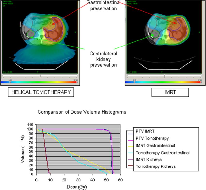
Figure 1 Dosimetric comparison between Tomotherapy and intensity-modulated radiotherapy (IMRT) for a left retroperitoneal
liposarcoma (for the same prescribed dose of 54 Gy, note the greater gastrointestinal preservation with helical tomotherapy and the
equivalent controlateral kidney preservation).
IMRT regardless of the calculation method utilised Radiotherapy schedule
(D’souza et al. [12] or ICRU N°83). Dynamic IMRT with No randomised study has yet compared results from
equally-distributed fields of 18MV photons enables the radiotherapy administered at different stages of treat-
reduction of the ID to healthy tissues by 60 joules on ment (pre-, intra- or post-operative) [15]. Further, recur-
average (p=0.008) compared to HT (Figure 1). rence tends to occur at distance after pre-operative
radiation, whereas it is more likely to be local after post-
operative radiation [16]. Pre-operative radiation appears
Discussion to offer several advantages compared to post-operative
The mainstay of treatment of retroperitoneal liposarco- radiation. The OAR (intestines, liver), repressed by the
mas is surgery [13,14]. Due to rarity of these tumours, tumour are more easily preserved and toxicities, particu-
data on radiotherapy are limited and many questions re- larly digestive, are reduced. Bossi et al.’s series evaluating
main unanswered. In the current series focussing on pre-operative voluntarily partial radiation at 50 Gy
retroperitoneal liposarcoma treated by high-dose pre- shows good tolerance because less than 10% of patients
operative Helical Tomotherapy, toxicity was low to presented with Grade 3 digestive toxicity [17]. In con-
moderate and mostly grade2 toxicity was reports 42% of Grade 2 digestive toxicity [18].
observed nor any delayed digestive toxicity. No occlu-
sion or perforation occurred. Furthermore, R0 surgery Dose level
was achieved in 40% of cases, R1 for 40% and R2 in the Results concerning local control in our series may indi-
remaining 20%. With a median follow-up of 26 months, cate the superiority of a high dose (54 Gy), but this
no local recurrence occurred. would need to be confirmed in a larger prospective trial.Sargos et al. Radiation Oncology 2012, 7:214 Page 6 of 7
http://www.ro-journal.com/content/7/1/214
Data from the French multicentre TOMOREP protocol the constraints and conformational quality, the conform-
are expected shortly. Local control appears dose- ity index approaches the ideal value of 1 in tomotherapy,
dependent [19]. For 104 patients receiving radiation after whereas in IMRT it moves away from 1 up until the iso-
complete surgery, Catton et al. showed that time to local dose of 98% (p=0.051). In terms of OAR-sparing, digest-
recurrence was 30 months after low dose radiation ive organs are spared more in tomotherapy (D200cc
(under 35 Gy) vs. 103 months after radiation at doses lowered by 5 Gy). Given the short duration of follow-up,
higher than 35 Gy (p=0.06) [1]. it is however impossible to know the clinical conse-
The increase in pre-operative or post-operative radi- quences of this dosimetric gain.
ation doses requires the use of new radiation techniques It is also particularly important to evaluate the “low
(intensity modulation) to allow sufficient sparing of doses” delivered during the intensity modulation treat-
healthy surrounding tissues. Theoretical and clinical ment [22]. Using a biological model, Hall et al. estimate
dosimetric studies have already shown that the doses that the risk of a second cancer at 10 years is potentially
delivered can be increased with IMRT. In a previous doubled when an IMRT technique is used compared to
study [6], the post-operative use of IMRT enabled the a 3D-CRT across all pathology types [23]. Our compari-
dose to be raised to 54 Gy instead of 45 Gy in 3- son indicates that the integral dose delivered is weaker
dimensional conformational radiotherapy (3D-CRT) (on average 60 Joules) in dynamic IMRT than in
while improving the protection factor for surrounding tomotherapy, irrespective of the calculation method used
organs by 20%. The dose reductions outside the PTV (D’Souza or Remaining Volume at Risk, RVR). The inte-
enabled this prescribed dose increase of 9 Gy to reach gral dose seems to increase with tomotherapy when
54 Gy [6]. Similar results were found by Koshy et al. [20] similar energy is used. The larger field length (in a longi-
or in Bossi et al.’s series with increased gastrointestinal tudinal direction) of the tomotherapy plans relative to
and renal sparing on dosimetric comparisons between retrospectively-planned IMRT could be one reason for
3D-CRT and IMRT for 10 patients (dose of 50.4 Gy). these higher integral dose values [22].
With the same dosimetric limitations used for 3D-CRT However, intensity modulation treatments, given the
planning and in IMRT, the average dose to the small in- high gradients, must be accompanied by daily position
testine decreased from 36 Gy in 3D-CRT to 27 Gy in monitoring to ensure accurate target positioning. In
IMRT. Tumoral coverage (V95) is improved with IMRT, tomotherapy this is ensured by daily megavoltage CT
increasing from 95.3% to 98.6%. Pre-operatively, Bossi scans and during arc therapy by daily cone beam CT
et al. also found greater renal sparing with IMRT (kidney scans (CBCT)[24]. Overall, the evaluation of the integral
volume receiving more the 15 Gy reduced from 19.8cc dose delivered to healthy tissues by a treatment must
in RTC3D to 3.2 cc with IMRT) [17]. take into account both the energy emitted by the radi-
Similarly, Tzeng et al. report the results of 16 patients ation itself but also that emitted by the repositioning im-
receiving radiation at 57.5 Gy. Local control was 80% agery [25].
with only 25% of patients experiencing Grade 2 digestive
toxicity probably due to the fact that the high-dose PTV Conclusions
was smaller than the standard PTV, and intentionally far The low toxicities in our series, both during and imme-
from the small bowel [7]. diately after radiotherapy and after surgery, show the
technical and clinical feasibility of a pre-operative radi-
IMRT techniques ation strategy at a high dose of 54 Gy for patients with
Technological improvements in radiotherapy are ever- retroperitoneal liposarcoma. Clinical evaluation of the
increasing and intensity modulated treatment can be age and general status of patients is essential to judge
administered by various means (tomotherapy, dynamic feasibility. This irradiation must be delivered with inten-
IMRT, arc therapy, etc.) [21].. The comparative dosim- sity modulation to allow for sufficient sparing of sur-
etry study shows very good tumoral coverage for the two rounding at-risk organs. Tomotherapy, including image-
techniques examined, with all dosimetric evaluation con- guided radiation therapy (IGRT) systems, appears to be
straints defined by the ICRU N°83 respected. In particu- an attractive option with regard to gastrointestinal pres-
lar, homogeneity of coverage, evaluated through HI, is ervation, especially in large and convex volumes. This
similar across IMRT and tomotherapy. In terms of con- technique enables us to treat safely a larger number of
formity, the DSC (calculated with the 95% isodose) is patients who would not otherwise have been able to re-
better after IMRT. The use of the 95% isodose for this ceive radiation. With regard to the delayed effects of in-
comparison is, however, questionable given the possibil- tegral dose, only long-term and prospective data will
ities of optimisation offered by these modern techniques. answer this specific question. In terms of the cancer out-
When evaluation isodoses are increased progressively comes in this series, no local or distant recurrence was
from 95 to 100% to calculate the DSC, thus increasing observed with a median follow-up of 26 months, butSargos et al. Radiation Oncology 2012, 7:214 Page 7 of 7
http://www.ro-journal.com/content/7/1/214
longer follow-up needs to be obtained to confirm these 9. Martin E, Pointreau Y, Roche-Forestier S, Barillot I: Normal tissue tolerance
preliminary results. to external beam radiation therapy: small bowel. Cancer Radiother 2010,
14:350–353.
10. Kantor G, Mahe MA, Giraud P, Alapetite C, Durdux C, Fourquet A, Gardner
Abbreviations M, Le Prise E, Maire JP, Richaud P, et al: French national evaluation for
CTCAE: Common terminology criteria for adverse events; ICRU: International helicoidal tomotherapy: description of indications, dose constraints and
Commission on Radiation units; IMRT: Intensity-modulated radiotherapy; set-up margins. Cancer Radiother 2007, 11:331–337.
MV: Mega volt; ID: Integral dose; HT: Helical Tomotherapy; Gy: Grays; 11. Kantor G, Mahe MA, Giraud P, Lisbona A, Caron J, Mazal A: [Helical
IGRT: Image-guided radiation therapy; OS: Overall survival; CT: Computed tomotherapy: general methodology for clinical and dosimetric
tomotherapy; RVR: Remaining risk volume; DSC: Dice similarity coefficient; 3D evaluation (national French project)]. Cancer Radiother 2006, 10:488–491.
-CRT: Three dimensional conformational radiotherapy; PTV: Planned target 12. D’Souza WD, Rosen II: Nontumor integral dose variation in conventional
volume; OAR: Organs at risk; GTV: Growth tumoral volume; CTV: Clinical radiotherapy treatment planning. Med Phys 2003, 30:2065–2071.
target volume; NCI: National Cancer Institute; EUD: Equivalent Uniform Dose; 13. Youssef E, Fontanesi J, Mott M, Kraut M, Lucas D, Mekhael H, Ben-Josef E:
WHO: World Health Organisation; ASA: American Society of Long-term outcome of combined modality therapy in retroperitoneal
Anesthesiologists; HI: Homogeneity Index. and deep-trunk soft-tissue sarcoma: analysis of prognostic factors. Int J
Radiat Oncol Biol Phys 2002, 54:514–519.
Competing interests 14. Stoeckle E, Coindre JM, Bonvalot S, Kantor G, Terrier P, Bonichon F, Nguyen
The author’s declare that they have no competing interests. Bui B: Prognostic factors in retroperitoneal sarcoma: a multivariate
analysis of a series of 165 patients of the French Cancer Center
Authors’ contributions Federation Sarcoma Group. Cancer 2001, 92:359–368.
PS conceived of the study, participated in its design and coordination and 15. Pawlik TM, Pisters PW, Mikula L, Feig BW, Hunt KK, Cormier JN, Ballo MT,
helped to draft the manuscript. CD supervised the technical and theoretical Catton CN, Jones JJ, O’Sullivan B, et al: Long-term results of two
technique of the physical data. BHdF participated in the design of the study prospective trials of preoperative external beam radiotherapy for
and helped to review the manuscript. VB participated in the design of the localized intermediate- or high-grade retroperitoneal soft tissue
study and performed the statistical analysis. BNB, AI, ES and GK conceived of sarcoma. Ann Surg Oncol 2006, 13:508–517.
the study, participated in its design and coordination and helped to draft 16. Zlotecki RA, Katz TS, Morris CG, Lind DS, Hochwald SN: Adjuvant radiation
the manuscript. All authors read and approved the final manuscript. therapy for resectable retroperitoneal soft tissue sarcoma: the University
of Florida experience. Am J Clin Oncol 2005, 28:310–316.
Acknowledgements 17. Bossi A, De Wever I, Van Limbergen E, Vanstraelen B: Intensity modulated
We thank Pippa McKelvie-Sebileau of Institut Bergonié for English medical radiation-therapy for preoperative posterior abdominal wall irradiation
editorial assistance. of retroperitoneal liposarcomas. Int J Radiat Oncol Biol Phys 2007,
67:164–170.
Author details 18. Gilbeau L, Kantor G, Stoeckle E, Lagarde P, Thomas L, Kind M, Richaud P,
1
Department of Radiation Oncology, Institut Bergonié, Bordeaux, France. Coindre JM, Bonichon F, Bui BN: Surgical resection and radiotherapy for
2
Université Bordeaux Segalen, 229 cours de l’Argonne, Bordeaux 33076, primary retroperitoneal soft tissue sarcoma. Radiother Oncol 2002,
France. 3Clinical and Epidemiological Research Unit, Institut Bergonié, 65:137–143.
Bordeaux, France. 4Department of Medical Oncology, Institut Bergonié, 19. Tepper JE, Suit HD, Wood WC, Proppe KH, Harmon D, McNulty P: Radiation
Bordeaux, France. 5Department of Surgery, Institut Bergonié, Bordeaux, therapy of retroperitoneal soft tissue sarcomas. Int J Radiat Oncol Biol
France. Phys 1984, 10:825–830.
20. Koshy M, Landry JC, Lawson JD, Staley CA, Esiashvili N, Howell R, Ghavidel S,
Received: 17 July 2012 Accepted: 12 December 2012 Davis LW: Intensity modulated radiation therapy for retroperitoneal
Published: 17 December 2012 sarcoma: a case for dose escalation and organ at risk toxicity reduction.
Sarcoma 2003, 7:137–148.
21. Paumier A, Le Pechoux C, Beaudre A, Negretti L, Ferreira I, Roberti E, Brahim
References J, Lefkopoulos D, Daly-Schweitzer N, Bourhis J, Bonvalot S: IMRT or
1. Catton CN, O’Sullivan B, Kotwall C, Cummings B, Hao Y, Fornasier V: conformal radiotherapy for adjuvant treatment of retroperitoneal
Outcome and prognosis in retroperitoneal soft tissue sarcoma. sarcoma? Radiother Oncol 2011, 99:73–78.
Int J Radiat Oncol Biol Phys 1994, 29:1005–1010. 22. Wiezorek T, Schwahofer A, Schubert K: The Influence of Different IMRT
2. Lewis JJ, Leung D, Woodruff JM, Brennan MF: Retroperitoneal soft-tissue Techniques on the Peripheral Dose. Strahlenther Onkol 2009, 185:696–702.
sarcoma: analysis of 500 patients treated and followed at a single 23. Hall EJ, Wuu CS: Radiation-induced second cancers: the impact of 3D-CRT
institution. Ann Surg 1998, 228:355–365. and IMRT. Int J Radiat Oncol Biol Phys 2003, 56:83–88.
3. Bonvalot S, Vanel D, Le Cesne A, Terrier P, Le Pechoux C: Surgery of 24. Boda-Heggemann J, Lohr F, Wenz F, Flentje M, Guckenberger M:
retroperitoneal sarcomas. Cancer Radiother 2006, 10:41–49. kV Cone-Beam CT-Based IGRT. Strahlenther Onkol 2011, 187:284–291.
4. Bonvalot S, Rivoire M, Castaing M, Stoeckle E, Le Cesne A, Blay JY, 25. Lisbona A, Averbeck D, Supiot S, Delpon G, Ali D, Vinas F, Diana C, Murariu
Laplanche A: Primary retroperitoneal sarcomas: a multivariate analysis of C, Lagrange JL: IMRT combined to IGRT: increase of the irradiated
surgical factors associated with local control. J Clin Oncol 2009, 27:31–37. volume. Consequences? Cancer Radiother 2010, 14:563–570.
5. Gronchi A, Lo Vullo S, Fiore M, Mussi C, Stacchiotti S, Collini P, Lozza L,
Pennacchioli E, Mariani L, Casali PG: Aggressive surgical policies in a
doi:10.1186/1748-717X-7-214
retrospectively reviewed single-institution case series of retroperitoneal
Cite this article as: Sargos et al.: High-dose pre-operative helical
soft tissue sarcoma patients. J Clin Oncol 2009, 27:24–30. tomotherapy (54 Gy) for retroperitoneal liposarcoma. Radiation Oncology
6. Musat E, Kantor G, Caron J, Lagarde P, Laharie H, Stoeckle E, Angles J, 2012 7:214.
Gilbeau L, Bui BN: Comparison of intensity-modulated postoperative
radiotherapy with conventional postoperative conformal radiotherapy
for retroperitoneal sarcoma. Cancer Radiother 2004, 8:255–261.
7. Tzeng CW, Fiveash JB, Popple RA, Arnoletti JP, Russo SM, Urist MM,
Bland KI, Heslin MJ: Preoperative radiation therapy with selective dose
escalation to the margin at risk for retroperitoneal sarcoma. Cancer 2006,
107:371–379.
8. Coindre JM, Trojani M, Contesso G, David M, Rouesse J, Bui NB, Bodaert A,
De Mascarel I, De Mascarel A, Goussot JF: Reproducibility of a
histopathologic grading system for adult soft tissue sarcoma.
Cancer 1986, 58:306–309.You can also read