Clinical diagnosis, treatment and screening of the VHL gene in three von Hippel Lindau disease pedigrees
←
→
Page content transcription
If your browser does not render page correctly, please read the page content below
EXPERIMENTAL AND THERAPEUTIC MEDICINE 20: 1237-1244, 2020

      Clinical diagnosis, treatment and screening of the VHL gene
             in three von Hippel‑Lindau disease pedigrees
                         GUOBING LIN1, YIHUA ZHAO2, ZHEWEI ZHANG3 and HUIJIANG ZHANG4

                   1
                   Department of Urology, The First People's Hospital of Wenling, Wenling, Zhejiang 317500;
            2
            Department of Urology, Yueqing People's Hospital, Yueqing, Zhejiang 325600; 3Department of Urology,
            The Second Affiliated Hospital of Zhejiang University School of Medicine, Hangzhou, Zhejiang 310009;
                  4
                   Department of Urology, Lishui City People's Hospital, Lishui, Zhejiang 323000 P.R. China

                                     Received September 1, 2018; Accepted August 23, 2019

                                                    DOI: 10.3892/etm.2020.8829

Abstract. The present study aimed to investigate the clinical        2018 (2‑4). Currently, >40 lesions for VHL disease have
characteristics of von Hippel‑Lindau (VHL) disease and the           been primarily observed in the central nervous system (CNS)
clinical significance of VHL gene detection. The clinical            and in 14 different organs, including hemangioblastomas
materials of patients with VHL disease were collected from           (HB; 44‑72% of cases), renal cell carcinoma (RCC; 25‑45%
3 different families between May 1985 and October 2017.              of cases), pheochromocytoma (PHEO)/paragangliomas (PGL;
A systematic pedigree study and VHL gene detection at the            10‑30% of cases), endolymphatic sac tumors (ELSTs; 6‑15% of
germline level were performed together with a literature             cases), renal cysts (60% of cases), pancreatic neuroendocrine
review. Of the 22 patients from 3 VHL pedigrees, 10 exhib-           tumors (PNETs; 5‑10% of cases), pancreatic serous cystade-
ited VHL gene mutations (3 genotypes) at the germline level.         noma/cysts (72‑90% of cases), and papillary cystadenoma (PC)
The genotypes of pedigree were VHL‑p.R161Q (c.482G>A),               of the epididymis and the broad ligament of the uterus (2‑15).
VHL‑p.N78S (c.233A>G), and VHL‑p.R167Q (c.500G>A).                   VHL disease is primarily caused by inactivation of the VHL
During the follow‑up period, the symptoms were stable in 10          tumor‑suppressive protein. The VHL gene (OMIM, 608537)
patients, including 2 cases of central nervous system heman-         is located on human chromosome 3p25.3 and encodes the
gioblastomas (CNS‑HB), 3 cases of bilateral multiple renal           VHL protein (pVHL), which forms a complex with elongation
cell carcinoma (RCC) and 5 cases of adrenal pheochromocy-            factor and is critical for pVHL to function as an E3 ligase (16).
toma without local recurrence or distant metastasis. Patients        VHL germline mutations result in the dysfunction of E3 ligase
with p.R161Q and p.N78S were not associated with CNS‑HB,             and the accumulation of hypoxia‑inducible factor‑α (HIF‑α;
which was different from the clinical phenotype of previously        VHL‑Elongin‑HIF‑α complex), leading to decreased ubiqui-
reported families. RCC were Fuhrman II grade, which was              tylation and proteasomal degradation of HIFs, namely HIF‑1α
consistent with the previous study. The results of the present       and HIF‑2α. Elevated levels of HIFs subsequently regulate
study indicated that the standardization of early diagnosis and      overactivation of the downstream pathways in which vascular
the improvement of long‑term efficacy may be achieved by             endothelial growth factor (VEGF), platelet‑derived growth
combining clinical screening and VHL gene detection.                 factor‑β (PDGF‑β) and transforming growth factor‑α (TGF‑α)
                                                                     are involved, which accelerates tumorigenesis (14). The present
Introduction                                                         study analyzed the clinical data of 9 patients from 3 families
                                                                     with VHL disease. The mutations in the VHL gene of patients
Von Hippel‑Lindau (VHL) disease (1) is a rare autosomal              and their family members were determined.
dominant inherited disease that predisposes the affected
individual to various benign or malignant tumors with an             Materials and methods
incidence rate of 1 in every 36,000‑50,000 worldwide in
                                                                     Patients. Between May 1985 and October 2017, three Han pedi-
                                                                     grees with VHL disease were recruited separately and followed up
                                                                     by Lishui People's Hospital (Lishui, China), the Second Affiliated
                                                                     Hospital of Zhejiang University School of Medicine (Hangzhou,
Correspondence to: Dr Huijiang Zhang, Department of
                                                                     China) and the First People's Hospital of Wenling City (Wenling,
Urology, Lishui City People's Hospital, 15 Dazhong Street, Lishui,
Zhejiang 323000, P.R. China                                          China), respectively. The patients underwent collection of their
E‑mail: iszhanghuijiang@163.com                                      medical history, family survey (pedigree analysis) and a detailed
                                                                     physical examination and associated auxiliary examination by a
Key words: von Hippel‑Lindau disease, VHL gene, molecular            specialist (GL, ZZ and HZ, respectively) from each of the three
diagnosis, case report                                               hospitals aforementioned in a blinded manner. There were 21
                                                                     members in the pedigrees, including 15 males and 6 females,
                                                                     aged 9‑66 years (Fig. 1 and Table I). The present study was
1238 LIN et al: IDENTIFICATION OF VON HIPPEL-LINDAU DISEASE
Figure 1. Pedigree tree of three families with VHL disease. F1, family 1; F2, family 2; F3, family 3; Squares, male; circles, female. VHL, von Hippel‑Lindau;
HB, hemangioblastoma; RCC, renal cell carcinoma; PHEO, pheochromocytoma.
approved by the Ethics Committee of Lishui People's Hospital Follow‑up. All patients were followed up by telephone or
and written informed consent was obtained from all patients. hospital visits for 5 years. The follow‑up interval was every
2 months in the first year, then every 6 months in the second
Detection of VHL gene mutation. A total of 5 ml peripheral year and once every year thereafter. During follow‑up, general
blood (EDTA anticoagulant) was collected from each of the conditions, including blood pressure, blood biochemical
21 family members from the 3 families, including the 3 probands indices, recurrence or metastasis of tumor were assessed for
(F1‑II6, F2‑II1 and F3‑III2). Extraction of genomic DNA from every patient.
peripheral blood leukocytes was performed using a QIAamp
Blood kit (Qiagen GmbH) according to the manufacturer's Results
protocol. The primer sequences were designed based on the
VHL gene sequence found in GeneBank using the Primer‑Blast Family 1 (F1) investigation and general clinical material.
online tool (https://omictools.com/primer‑blast‑tool) and Proband (F1‑II6) was female and aged 50 years. The patient
synthesized by Sangon Biotech Co., Ltd. The primer sequences was admitted to Lishui People's Hospital (Lishui, China) in
were forward, 5'‑ACCGGTGTGGCTCTTTAACA‑3' and May 1985 due to paroxysmal headache, palpitation and hyper-
reverse, 5'‑TCCTGTACTTACCACAACAACCTT‑3'. The 20 µl hidrosis that had occurred for 3 months. The results of physical
solution used for the PCR amplification consisted of 17 µl examinations revealed that patient heart rate was 110 bpm and
KAPA2G Robust HotStart ReadyMix (cat. no. KK5701; Beijing blood pressure was 210/120 mmHg. Ultrasound and CT results
Huaruikang Technology Co., Ltd.), 1 µl each of the aforemen- revealed a mass sized 5.5x4.5x2.8 cm on the right side of the
tioned upstream and downstream primers (10 µM) and 1 µl of adrenal gland and it was considered as right PHEO. Retinal
genomic DNA (200 ng) from both the patients and family hemangioblastoma was excluded by an ophthalmologist. After
members. PCR was performed as follows: Pre‑denaturation at blood pressure and volume expansion were adjusted with
94˚C for 5 min, followed by 30 cycles of denaturation at 94˚C for phenylbenzylamine hydrochloride tablets, right adrenal tumor
30 sec, annealing at 65˚C for 30 sec, 72˚C for 1 min; and exten- resection was performed under general anesthesia. The post-
sion at 72˚C for 5 min. The 2 µl PCR amplified product was used operative pathology results confirmed that the patient suffered
for subsequent electrophoresis with 1% agarose gel for identifi- from right PHEO.
cation and purification. The gel was stained with ethidium The proband's son (F1‑III6) was 25 years old and was
bromide (cat. no. E7637; Sigma‑Aldrich; Merck KGaA) for admitted to Lishui People's Hospital (Lishui, China) in
visualization. The purified product was sent to Shanghai Xiang March 2002 due to paroxysmal headache and hypertension
Yin Biotechnology Co., Ltd. for sequencing with an ABI 3730XL (140‑160/100‑110 mmHg). The results of the physical exami-
sequencer. All mutations were confirmed by bi‑directional nation revealed that his levels of serum catecholamine were
sequencing. increased, including dopamine (88.64 ng/l; normal reference
value,Table I. Clinical phenotype and genotype characteristics of patients with VHL syndrome in 3 families.
Age at VHL CNS-HB
diagnosis gene tumor RCC PHEO Operative Pathological Final
Patients Sex (years) mutation size (cm) size (cm) size (cm) type diagnosis diagnosis VHL type
F1-II6 Female 18 p.R161Q - - Right, 5.5x4.5x2.8 Right PHEO Right PHEO Right PHEO VHL-IIB
resection
F1-III2 Male 9 (right)/ p.R161Q - - Left, 7.0x5.5x3.4; Left and right Bilateral PHEO Bilateral PHEO VHL-IIB
17 (left) right, 5.6x3.8x3.0 PHEO resection (metachronous) (metachronous)
F1-III6 Female 28 p.R161Q - - Left, 6.0x4.7x3.5; Bilateral PHEO Bilateral PHEO Bilateral PHEO VHL-IIB
right, 3.5x2.8x2.5 resection (synchronous) (synchronous)
F2-II1 Male 24 p.N78S - Left, 1.0x1.0x0.8; Right, 4.1x3.4x3.9 Right PHEO Right PHEO + Right PHEO + VHL-IIB
right, 1.0x1.0x1.0 resection + right right RCC + right PHEOa +
RCC resection + bilateral bilateral epididymal
resection epididymal cust + multiple
of bilateral cyst pancreatic cysts
epididymal cyst
F2-II2 Male 27 p.N78S - Right, 4.0x3.0x2.5 - Right radical Right RCC Right PHEOa + VHL-IIB
nephrectomy multiple cysts of kidney
and pancreas + bilateral
epididymal nodules
F2-III1 Female 5 p.N78S - - - - - VHL gene VHL-IIB
mutation carrier
F3-II1 Male 66 p.R167Q - Left, 1.0x1.0x0.8; Right, 4.5x3.5x2.6 Right PHEO Right PHEO + Right PHEO + VHL-IIB
right, 1.0x1.0x0.9 resection + right RCC bilateral RCCa +
right RCC multiple cysts of kidney
resection and pancreas + PNETs
EXPERIMENTAL AND THERAPEUTIC MEDICINE 20: 1237-1244, 2020
F3-III1 Female 45 p.R167Q 3.2x2.1x1.0 - - Cerebellar HB HB Cerebellar HB VHL-IIIB
resection
F3-III2 Male 34 p.R167Q Right 2.1x1.0x0.9; - - Gamma knife - Multiple CNS-HBb VHL-IIIB
right, 0.6x0.5x0.5 radiosurgery
for CNS-HB
F3-IV1 Male 21 p.R167Q - - - - - VHL variant carrier VHL-IIIB
a
Bilateral multiple RCC; bclinical diagnosis. VHL, von Hippel-Lindau; CNS, central nervous system; HB, hemangioblastomas; RCC, renal cell carcinoma; PHEO, pheochromocytoma; PNETs, pancreatic
neuroendocrine tumors.
12391240 LIN et al: IDENTIFICATION OF VON HIPPEL-LINDAU DISEASE
Figure 2. Imaging examination results. (A) Coronal and (B) axial abdominal MRI revealed right PHEO, multiple renal RCC and multiple pancreatic cysts
in F2‑II1. Arrows indicate cysts. (C) Coronal and (D) axial abdominal CT revealed multiple RCC of the right kidney with cystic degeneration and multiple
pancreatic cysts in F2‑II2. Arrows indicate cysts. (E) Axial abdominal CT revealed right PHEO, left RCC and neuroendocrine tumors of pancreatic head
in F3‑II1. (F) Axial cranial MRI revealed hemangioblastoma with a clear boundary. The right margin of the sellar region and vermis of cerebellum were
significantly enhanced in F3‑III2. PHEO, pheochromocytoma; RCC, renal cell carcinoma; F, family.
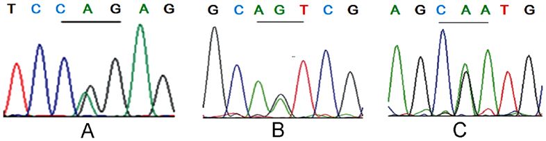
Figure 3. Results of VHL gene detection. (A) A G to A heterozygous mutation (p.R161Q) was presented in the 482nd codon of proband F1‑II11. (B) An A to G
missense mutation (p.N78S) in the 233rd codon of the proband F2‑II1 was identified and (C) a G to A heterozygous mutation (p.R167Q) in the 500th codon
was exhibited in the proband F3‑III1. VHL, von Hippel‑Lindau; F, family.
patient suffered from right PHEO. In 2010, this patient suffered under general anesthesia. The postoperative pathology results
from paroxysmal hypertension and headache for a second time confirmed that the patient suffered from bilateral PHEO. The
and his levels of serum catecholamine were also increased, father of this patient (F1‑II2, the proband's brother) died from
including dopamine (600.36 ng/l), adrenaline (297.94 ng/l) hypertensive intracerebral hemorrhage 26 years ago, aged
and norepinephrine (1,093.84 ng/l). The ultrasound and CT 38 years.
results revealed a mass sized 7.0x5.5x3.4 cm on the left side of
the adrenal gland. After blood pressure and volume expansion Family 2 (F2) investigation and general clinical material.
were adjusted with phenylbenzylamine hydrochloride tablets, The proband (F2‑II1) of F2 was male and aged 33 years. The
left adrenal tumor resection was performed under general patient was admitted to the Second Affiliated Hospital of
anesthesia. The postoperative pathology results confirmed that Zhejiang University School of Medicine (Hangzhou, China)
the patient suffered from left PHEO. The final diagnosis of this in September 2009 due to paroxysmal headache, dizziness
patient was bilateral PHEO. and palpitation with hyperhidrosis. Physical examination
The proband's niece (F1‑III2) was 38 years old and was results revealed that the patients' heart rate was 100 bpm
admitted to Lishui People's Hospital in October 2002 due to and blood pressure was 105/78 mmHg. MRI results revealed
persistent headache and dizziness that had lasted for 3 days. a mass sized 4.1x3.4x3.9 cm at the right adrenal gland and
The results of physical examination demonstrated that the multiple renal tumors (the largest diameter of the tumors,
patient's blood pressure was 160/100 mmHg and their serum 1.0x1.0x1.0 cm on the left; 1.0x1.0x0.8 cm on the right) and
catecholamine levels were increased. Ultrasound/CT revealed multiple cysts in the pancreas (Fig. 2A and B). An ophthal-
a bilateral mass on the adrenal gland. The left mass was mologist (from the Second Affiliated Hospital of Zhejiang
6.0x4.7x3.5 cm in size and the right mass was 3.5x2.8x2.5 cm University School of Medicine) excluded the existence of
in size. After blood pressure and volume expansion were retinal hemangioblastoma preoperatively. The concentration
adjusted with phenylbenzylamine hydrochloride tablets, of urine vanillicmandelic acid at 24 h was 138.3 µmol/24 h
bilateral adrenal tumor laparoscopic resection was performed (normal reference value,EXPERIMENTAL AND THERAPEUTIC MEDICINE 20: 1237-1244, 2020 1241
manifestations, the patient was considered as right PHEO, with infection and the adjustment of blood pressure and volume
double renal masses and multiple pancreatic cysts. After the expansion with phenylbenzylamine hydrochloride tablets,
patient's blood pressure and volume expansion was adjusted resection of the right adrenal tumor and right kidney tumor
with phenylbenzylamine hydrochloride tablets, resection of were performed under general anesthesia. The postoperative
the right adrenal tumor and the upper pole of the right kidney pathology results revealed that the patient suffered from right
were performed under general anesthesia. The postoperative PHEO and right RCC (Fuhrman II grade).
pathology results confirmed that the patient had right PHEO
and right RCC (Fuhrman II grade) (17). In April 2012, the VHL gene detection results. Of the 22 patients from 3 VHL
patient underwent bilateral epididymal nodule excision due to families, 10 had three VHL germline missense mutations
the presence of multiple nodules in the bilateral epididymal within coding regions, respectively (Fig. 3). In the F1 family,
head. Postoperative pathology results confirmed that the 3 cases harbored the p.R161Q (c.482G>A) mutation at
patient suffered from papillary cystadenoma of the bilateral exon 3 (Fig. 3A); 3 cases in the F2 family harbored the p.N78S
epididymal head. (c.233A>G) mutation at exon 1 (Fig. 3B); and 4 cases in the
The proband's younger brother (F2‑II2) was aged 32 years. F4 family harbored the p.R167Q (c.500G>A) mutation at
In 2012, the patient presented with multiple masses in exon 3 (Fig. 3C). Apart from 8 patients with the VHL clinical
both kidneys (left, 1.0x1.0x1.0 cm; right, 4.0x3.5x2.6 cm), phenotype who had been surgically and pathologically diag-
multiple cysts in both kidneys, and pancreas and epididymal nosed, 2 new cases (F2‑III1 and F3‑IV2) were revealed to be
nodules (Fig. 2C and D). The patient underwent laparoscopic asymptomatic carriers of VHL gene mutations. The remaining
right radical nephrectomy under general anesthesia. The post- 12 family members had no VHL gene mutations and no
operative pathology results confirmed that the patient suffered clinical manifestations or abnormalities following imaging
from multiple RCC in the right kidney (Fuhrman II grade). and serological tests associated with VHL disease. The
IFN and IL‑2 were used for postoperative treatment. clinical phenotype and genotype characteristics are presented
in Table I.
Family 3 (F3) investigation and general clinical material.
Proband (F3‑III2) was male and aged 39 years. In April 2013, Discussion
the patient was admitted to the First People's Hospital of
Wenling City (Wenling, China) having suffered with dizzi- VHL disease, also known as VHL syndrome, is characterized
ness and headaches for 1 month. A cranial MRI examination by a variety of benign, malignant tumors and multiple organ
revealed that there was a mass sized 2.1x1.0x0.9 cm at the right cysts (18). VHL patients usually develop clinical symptoms
margin of the sellar region and a mass sized 0.6x0.5x0.5 cm at after reaching 20 years of age, with 90‑100% penetrance of
the cerebellar vermis. The masses were markedly enhanced clinical symptoms between 65 and 70 years old (19). Retinal
with clear margins. The patient was considered to suffer from and cerebellar HB are typically the most common and earliest
CNS‑HB. An ophthalmologist (The First People's Hospital presenting forms of VHL disease (4,7,9). VHL syndrome can be
of Wenling City) excluded retinal hemangioblastoma. As divided into two types: Type I and type II. Type I syndrome has
the patient refused surgical treatment, cranial gamma knife CNS‑HB and/or RCC among other tumors (except PHEO) and
radiotherapy (peripheral dose 5 Gy, central dose 10 Gy) was can also be divided into type IA (high‑risk RCC) and type IB
performed 6 times with a 5 Gy dose at the peripheral and (without RCC). The patients with type II must have PHEO and
10 Gy dose at the central positions. CNS‑HB of the F3‑III2 can be divided into type IIA (includes PHEO but not RCC),
was stable after 5 years of follow‑up. type IIB (RCC + PHEO) and type IIC (only PHEO) (2,3,14).
The proband's elder sister was aged 45 years (F3‑III1). The clinical diagnosis of this disease should be based on VHL
In May 2015, the patient was admitted to the First People's symptoms, heredity and family factors. If a patient possesses
Hospital of Wenling City due to dizziness lasting 3 months. A one of the following three conditions, they may be diagnosed
brain MRI revealed that there was a mass sized 3.2x2.1x1.0 cm with VHL disease: i) At least two positions where CNS‑HB
at the vermis of the cerebellum, with clear margins. The mass exists; ii) CNS‑HB at one position and one other organ tumor
was excised via surgery. The postoperative pathology results (RCC, PHEO/PGL, PNETs or ELSTs); iii) at least one visceral
confirmed that the patient suffered from cerebellar hemangio- tumor (RCC, PHEO/PGL, PNETs or ELSTs) associated with
blastoma. the VHL gene mutation at the pathogenic germline level, or
The proband's father (F3‑II1) was aged 66 years. The a parent has been diagnosed with VHL (3). Despite the high
patient was hospitalized in the First People's Hospital of heterogeneity of clinical phenotypes, VHL gene mutations
Wenling City presenting with cough and expectoration with could be detected at the germline level in almost all patients
low fever for 2 months in February 2014. Physical examination with VHL disease (2). The patients in the three families
results revealed that the patient's temperature was 37.8˚C and included in the present study were diagnosed as VHL type II.
his blood pressure fluctuated between 104‑123/66‑96 mmHg. The patients in F2 and F3 were VHL type IIB and the patients
CT examination results revealed that the patient suffered in F1 were VHL type IIC (Table I).
from chronic obstructive pulmonary disease with pulmo- The VHL gene contains three exons and encodes a poly-
nary infection. Ultrasound and CT results revealed a mass saccharide anchored membrane protein containing 213 amino
sized 4.5x3.5x2.6 cm on the right side of the adrenal gland, acids (20). The N‑terminal region of the protein contains α
multiple solid lesions of both kidneys (left, 1.0x1.0x0.8 cm; and β domains (20). The α domain binds to the elongation
right, 1.0x1.0x0.9 cm) with multiple small cysts and pancreatic factor C/B to form a complex and the β domain binds to the
head lesions (2.6x2.0x2.0 cm). Following treatment of a lung HIF‑ α gene (21). pVHL downregulates the expression of1242 LIN et al: IDENTIFICATION OF VON HIPPEL-LINDAU DISEASE HIF‑α, activates the ubiquitination of epidermal growth factor VHL‑associated lesions (22). Patients with VHL that present receptor (EGFR), regulates glucose metabolism‑associated with missense mutations in the HIF‑α binding site (HM) are genes [glucose transporter 1 (GLUT1) and posphofructoki- associated with a lower risk of PHEO and higher risks of CHB nase 1], growth factors (TGF, PDGF and VEGF) and the cascade and pancreatic tumors or cysts, while missense mutations reaction of nerve growth factor/JunB/Egl nine homolog 3‑asso- located at sites other than that of the HIF‑α binding site (nHM) ciated apoptotic pathway, ultimately leading to the formation of are associated with a higher risk of PHEO, which results in better benign and malignant multiple organ diseases (21). Emerging survival time (24). Furthermore, patients with truncating muta- evidence has indicated that HIF‑1α serves an important role tions are more likely to develop RCC than those with HM (26). during clear cell (cc)RCC and HB development (21). Previously, In contrast to the present study, it has been indicated that p.N78S almost 400 VHL gene mutants have been revealed to cause (HM)‑associated PHEO alone, p.R161Q (nHM)‑associated VHL disease (22). The majority of mutations are missense RCC/PHEO and p.R167Q (nHM)‑associated CNS/RCC/PHEO (27‑38%), nonsense (13‑27%), large fragment deletions (9‑20%), exhibit diverse phenotypes (16). Of note, the pathogenesis of microdeletions (10%), truncation and rearrangement (25%) CHB and RCC, upregulated HIF‑α expression and consequent mutations (22). Splicing site mutations are rare. Of all the muta- overexpression of VEGF and other HIF‑associated genes are tions, ~20% of the patients exhibited new mutations (de novo) the primary causative factors of tumor progression (14). In or their parents are chimeras (22). VHL type I disease is caused PHEO, HIF‑α (particularly HIF‑2α) dysregulation results in primarily by a large fragment deletion of the VHL gene, the overexpression of various HIF‑inducible genes, including including C3orf10 gene inactivation, truncation and missense GLUT1 and VEGF, which themselves serve important roles in mutations, which lead to the loss of pVHL function or struc- tumor development (27). However, the development of PHEO tural changes in the protein (3). VHL type II disease associated appears to be HIF‑independent. In patients with the typical IIC with PHEO is mostly caused by missense mutations (78‑96%), phenotype, as exhibited by the F1 family of the current study, which often lead to partial functional defects of the pVHL. The mutant pVHL is able to degrade HIFs, and it has been hypoth- most frequent mutations of VHL type II disease are p.R167W esized that mutations associated with PHEO may induce gain and p.R167Q mutations at position 167 of exon 3 in the VHL of function through an intact but altered pVHL (28). In addi- gene (2,3). Notably, although the stabilization of HIF‑α is tion, Gossage et al (14) reported that mutations in the elongin closely associated with the occurrence of RCC, both IIA and C binding domain of pVHL are associated with PHEO. These IIB antimutagenesis can inactivate VHL function, which in binding sites are implicated in the p53‑mediated apoptosis of turn inhibits the regulation of HIF‑α activation. However, only sympathetic neuronal precursor cells, which then go on to form patients in stage IIB exhibit ccRCC (23). Currently, certain PHEO (14). It is speculated that such large phenotypic varia- hypotheses suggest that the occurrence of ccRCC is associated tion may result from numerous other factors that are potentially with the expression of HIF‑2α (20,21). However, IIA mutations environmental and may affect the phenotypes induced by do not form enough HIF‑2α to form HB, despite producing the specific mutations (7). appropriate quantity of HIF‑α. This explains why HB is the The current clinical treatment strategy of VHL disease most common clinical manifestation of VHL (21). Liu et al (15) focuses primarily on the treatment of VHL disease‑associated demonstrated that patients with non‑HIF‑ α binding sites CNS‑HB, RCC, PHEO and PNETs (19). The average age of demonstrated an improved survival rate when compared with CNS‑HB at diagnosis is 33 years (range, 7‑78 years) (8,19). those that exhibited HIF‑α binding sites and truncated muta- For CNS‑HB, 90% presented in multiple forms and the tions (15). All patients in the three families of the present study majority occurred in the cerebellum (45‑50%) and spinal were VHL type II with missense mutations in the VHL gene cord (40‑45%) (8,20). Complete excision of CNS‑HB based and the patients of the F3 family harbored p.R167Q (α) muta- on microsurgical treatment was rare (
EXPERIMENTAL AND THERAPEUTIC MEDICINE 20: 1237-1244, 2020                                      1243

The majority of peripheral retinal HB cases can be treated with      VHL disease were not associated with ELSTs and it was spec-
laser photocoagulation (small peripheral HB), cryotherapy            ulated that the phenomenon may be associated with the small
(large HB) or photodynamic therapy (31). However, these              sample size of the family and patients. In addition, two asymp-
treatments cannot be used when the tumor is near the optic           tomatic VHL gene carriers (F2‑III1, 5 years of age, p.N78S;
nerve, in which case the therapeutic approach is only surveil-       F3‑IV2, 21 years of age, p.R167Q) were identified via gene
lance, resulting in a high risk of damage to the optic nerve (32).   testing. Early diagnosis and genetic counseling, regular cancer
The average age at diagnosis of VHL‑RCC is 39 years (range,          screening and surveillance are therefore helpful for disease
13‑70 years) (13,20). VHL‑RCC is characterized by simulta-           management and for the improvement of prognosis (40).
neous or heterogeneous bilateral tumors that are multifocal              In conclusion, VHL is a complex disease that can be
and low grade with a slow progression (13,20). The metastasis        easily misdiagnosed. Future studies should aim to improve
rate of VHL‑RCC is very low when tumors are 3 cm, which should effectively decrease renal insufficiency         the decrease in clinical risk of VHL disease.
despite the high recurrence rate of localized neoplasms (13).
    In the present study, the diagnostic ages of 3 patients with     Acknowledgements
VHL‑RCC (F2‑II1, F2‑II2 and F3‑II1) were 33, 32 and 66
years, respectively. F2‑II2 underwent radical resection due to       Not applicable.
multiple and large tumors of the right kidney. The other two
cases underwent unilateral single tumor enucleation (similar         Funding
to biopsy for definite pathology), all of which were Fuhrman
grade II and RCC was relatively stable after follow‑up. The          No funding was received.
average age at diagnosis of VHL‑PHEO was 27 years (range,
2.75‑58.00 years) (33,34). Of the VHL‑PHEO cases, 90% of             Availability of data and materials
tumors were located in the adrenal gland, and 20‑50% of
VHL‑PHEO were bilateral (33). Malignant VHL‑PHEO was                 The datasets used and/or analysed during the current study are
rare (1‑5%) (33). VHL‑PHEO can be asymptomatic at an early           available from the corresponding author on reasonable request.
stage and often secretes large quantities of norepinephrine,
which differs from MEN2‑PHEO, where large quantities of              Authors' contributions
adrenaline are secreted (2,33‑35). PHEO excision or partial
adrenalectomy decreases the risk of or avoids adrenal cortical       HZ designed the study. GL collected the clinical data,
insufficiency or crisis (36). It is worth noting that, 50% of        performed the experiments and wrote the manuscript. YZ and
patients developed a second PHEO within 30 years after initial       ZZ collected clinical data. All authors read and approved the
diagnosis (2,36,37). In the present study, the average diagnostic    final manuscript.
age of 5 patients with VHL‑PHEO was 34.6 years. There were
3 patients (F1‑II6, F2‑II1 and F3‑II1) with unilateral PHEO          Ethics approval and consent to participate
and two patients (F1‑III2 and F1‑III6) with bilateral PHEO,
including one patient (F1‑III2) with first‑time diagnosis of         The present study was approved by the Ethics Committee of
unilateral PHEO and contralateral PHEO occurring 8 years             Lishui People's Hospital, The Second Affiliated Hospital of
after surgery. Patients received PHEO resection, after which         Zhejiang University School of Medicine and the First People's
adrenal function was normal. A previous study determined             Hospital of Wenling City. Written informed consent was
that the average diagnostic age of PNETs was 35 years (range,        obtained from all participants.
10‑75 years). PNETs were asymptomatic and grew slowly. Of
the PNETs, 80‑93% were 3.0 cm, doubling time
1244 LIN et al: IDENTIFICATION OF VON HIPPEL-LINDAU DISEASE
3. Chittiboina P and Lonser RR: von Hippel-Lindau disease. In: 24. Shuin T, Ashida S, Yao M and Kanno H: Von Hippel‑Lindau
Handbook of Clinical Neurology. Elsevier, pp139‑156, 2015. disease. Nihon Rinsho 58: 1448‑1454, 2000 (In Japanese).
4. Aronoff L, Malkin D, van Engelen K, Gallinger B, Wasserman J, 25. Nordstrom‑O'Brien M, van der Luijt RB, van Rooijen E,
Kim RH, Villani A, Meyn MS and Druker H: Evidence for van den Ouweland AM, Majoor‑Krakauer DF, Lolkema MP,
genetic anticipation in vonHippel‑Lindau syndrome. J Med van Brussel A, Voest EE and Giles RH: Genetic analysis of von
Genet 55: 395‑402, 2018. Hippel‑Lindau disease. Hum Mutat 31: 521‑537, 2010.
5. Kruizinga RC, Sluiter WJ, de Vries EG, Zonnenberg BA, 26. Liu SJ, Wang JY, Peng SH, Li T, Ning XH, Hong BA, Liu JY,
Lips CJ, van der Horst‑Schrivers AN, Walenkamp AM and Wu PJ, Zhou BW, Zhou JC, et al: Genotype and phenotype
Links TP: Calculating optimal surveillance for detection of von correlation in von Hippel‑Lindau disease based on alteration
Hippel‑Lindau‑related manifestations. Endocr Relat Cancer 21: of the HIF‑alpha binding site in VHL protein. Genet Med 20:
63‑71, 2013. 1266‑1273, 2018.
6. Palmer LS and Linehan WM: Editorial comment. J Urol 183: 27. Young RM and Simon MC: Untuning the tumor metabolic machine:
2351‑2351, 2010. HIF‑α: pro‑ and antitumorigenic? Nat Med 18: 1024‑1025, 2012.
7. Iida K, Okimura Y, Takahashi K, Inomata S, Iguchi G, Kaji H 28. Hoffman MA, Ohh M, Yang H, Klco JM, Ivan M and
and Chihara K: A variety of phenotype with R161Q germline Kaelin WG Jr: von Hippel‑Lindau protein mutants linked to
mutation of the von Hippel‑Lindau tumor suppressor gene in type 2C VHL disease preserve the ability to downregulate HIF.
Japanese kindred. Int J Mol Med 13: 401‑404, 2004. Hum Mol Genet 10: 1019‑1027, 2001.
8. Gong K, Zhang N, Zhang K and Na Y: The relationship of 29. Niemelä M, Lemeta S, Sainio M, Rauma S, Pukkala E, Kere J,
erythropoietin overexpression with von Hippel‑Lindau tumour Böhling T, Laatikainen L, Jääskeläinen J and Summanen P:
suppressor gene mutations between hypoxia‑inducible factor‑1α Hemangioblastomas of the retina: Impact of von Hippel‑Lindau
and ‑2α in sporadic clear cell renal carcinoma. Int J Mol Med 26: disease. Invest Ophthalmol Vis Sci 41: 1909‑1915, 2000.
907‑912, 2010. 30. González Escobar AB, Morillo Sánchez MJ and García‑Campos JM:
9. David Dornbos 3III, Kim HJ, Butman JA and Lonser RR: Review Von Hippel‑Lindau disease: Family study. Arch Soc Esp
of the neurological implications of von Hippel‑Lindau disease. Oftalmol 87: 368‑372, 2012 (In Spanish).
JAMA Neurol 75: 620-627, 2018. 31. Richard S, Gardie B, Couvé S and Gad S: Von Hippel‑Lindau:
10. Asthagiri AR, Mehta GU, Zach L, Li X, Butman JA, How a rare disease illuminates cancer biology. Semin Cancer
Camphausen KA and Lonser RR: Prospective evaluation of Biol 23: 26‑37, 2013.
radiosurgery for hemangioblastomas in von Hippel‑Lindau 32. Lefevre A, Mathis T, Denis P and Kodjikian L: Retinal heman-
disease. Neuro Oncol 12: 80‑86, 2010. gioblastoma: Treatment strategy and long‑term follow‑up in a
11. Lonser RR, Butman JA, Huntoon K, Asthagiri AR, Wu T, retrospective cohort. J Fr Ophtalmol 41: 164‑169, 2018 (In French).
Bakhtian KD, Chew EY, Zhuang Z, Linehan WM and 33. Woodward ER and Maher ER: Von Hippel‑Lindau disease
Oldfield EH: Prospective natural history study of central nervous and endocrine tumour susceptibility. Endocr Relat Cancer 13:
system hemangioblastomas in von Hippel‑Lindau disease. 415‑425, 2006.
J Neurosurg 120: 1055‑1062, 2014. 34. Sovinz P, Urban C, Uhrig S, Stepan V, Lackner H, Schwinger W,
12. Nambu S, Otani R, Higuchi F, Uzuka T, Matsuda H, Kim P and Benesch M, Moser A, Spuller E and Speicher MR:
Ueki K: Histology of hemangioblastoma treated with stereotactic Pheochromocytoma in a 2.75‑year‑old‑girl with a germline von
radiosurgery confirms its effectiveness. J Clin Neurosci 51: Hippel‑Lindau mutation Q164R. Am J Med Genet A 152A:
43‑45, 2018. 1752‑1755, 2010.
13. Kim E and Zschiedrich S: Renal cell carcinoma in von 35. Colvin A, Saltzman AF, Walker J, Bruny J and Cost NG:
Hippel‑Lindau disease‑from tumor genetics to novel therapeutic Metastatic pheochromocytoma in an asymptomatic 12‑Year‑Old
strategies. Front Pediatr 6: 16, 2018. with von Hippel‑Lindau disease. Urology 119: 140‑142, 2018.
14. Gossage L, Eisen T and Maher ER: VHL, the story of a tumour 36. Petr EJ and Else T: Genetic predisposition to endocrine tumors:
suppressor gene. Nat Rev Cancer 15: 55‑64, 2015. diagnosis, surveillance and challenges in care. Semin Oncol 43:
15. Liu SJ, Wang JY, Peng SH, Li T, Ning XH, Hong BA, Liu JY, 582‑590, 2016.
Wu PJ, Zhou BW, Zhou JC, et al: Genotype and phenotype 37. Lenders JW, Duh Q‑Y, Eisenhofer G, Gimenez‑Roqueplo AP,
correlation in von Hippel‑Lindau disease based on alteration of Grebe SK, Murad MH, Naruse M, Pacak K and Young WF Jr;
the HIF‑ α binding site in VHL protein. Genetics in Medicine Endocrine Society: Pheochromocytoma and paraganglioma: An
Official Journal of the American College of Medical Genetics, endocrine society clinical practice guideline. J Clin Endocrinol
2018. Metab 99: 1915‑1942, 2014.
16. Liu Q, Yuan G, Tong D, Liu G, Yi Y, Zhang J, Zhang Y, Wang LA, 38. Tirosh A, Sadowski SM, Linehan WM, Libutti SK, Patel D,
Wang L, Zhang D, et al: Novel genotype‑phenotype correlations Nilubol N and Kebebew E: Association of VHL genotype with
in five Chinese families with Von Hippel‑Lindau disease. Endocr pancreatic neuroendocrine tumor phenotype in patients with von
Connect 7: 870‑878, 2018. Hippel-Lindau disease. JAMA Oncol 4: 124‑126, 2018.
17. Smith ZL, Pietzak EJ, Meise CK, Van Arsdalen K, Wein AJ, 39. Krauss T, Ferrara AM, Links TP, Wellner U, Bancos I,
Malkowicz SB and Guzzo TJ: Simplification of the Fuhrman Kvachenyuk A, Villar Gómez de Las Heras K, Yukina MY,
grading system for renal cell carcinoma. Can J Urol 22: 8069‑8073, Petrov R, Bullivant G, et al: Preventive medicine of von
2015. Hippel‑Lindau disease‑associated pancreatic neuroendocrine
18. Ben‑Skowronek I and Kozaczuk S: Von Hippel‑Lindau tumors. Endocr Relat Cancer 25: 783‑793, 2018.
Syndrome. Horm Res Paediatr 84: 145‑152, 2015. 40. Wang J‑Y, Peng S‑H, Li T, Ning XH, Liu SJ, Hong BA, Liu JY,
19. Chou A, Toon C, Pickett J and Gill AJ: von Hippel‑Lindau Wu PJ, Zhou BW, Zhou JC, et al: Risk factors for survival in
syndrome. Front Horm Res 41: 30‑49, 2013. patients with von Hippel‑Lindau disease. J Med Genet 55:
20. Arjumand W and Sultana S: Role of VHL gene mutation in 322‑328, 2018.
human renal cell carcinoma. Tumour Biol 33: 9‑16, 2012.
21. Tarade D and Ohh M: The HIF and other quandaries in VHL This work is licensed under a Creative Commons
disease. Oncogene 37: 139‑147, 2018. Attribution-NonCommercial-NoDerivatives 4.0
22. Ordóñez‑Navadijo Á, Fuertes‑Yebra E, Acosta‑Iborra B, Balsa E, International (CC BY-NC-ND 4.0) License.
Elorza A, Aragonés J and Landazuri MO: Mutant versions of von
Hippel‑Lindau (VHL) can protect HIF1α from SART1‑mediated
degradation in clear‑cell renal cell carcinoma. Oncogene 35:
587‑594, 2016.
23. Clifford SC, Cockman ME, Smallwood AC, Mole DR,
Woodward ER, Maxwell PH, Ratcliffe PJ and Maher ER:
Contrasting effects on HIF‑1alpha regulation by disease‑causing
pVHL mutations correlate with patterns of tumourigenesis in von
Hippel‑Lindau disease. Hum Mol Genet 10: 1029‑1038, 2001.You can also read