Cerebral Amyloid Angiopathy: CT and MR Imaging Findings1 - Neurotalk
←
→
Page content transcription
If your browser does not render page correctly, please read the page content below
Note: This copy is for your personal non-commercial use only. To order presentation-ready
                          copies for distribution to your colleagues or clients, contact us at www.rsna.org/rsnarights.

                EDUCATION EXHIBIT                                                                                                                             1517

                                            Cerebral Amyloid
RadioGraphics

                                            Angiopathy: CT and
                                            MR Imaging Findings1
                                            Christine P. Chao, MD ● Amy L. Kotsenas, MD ● Daniel F. Broderick, MD
                    TEACHING
                    POINTS
                    See last page           Cerebral amyloid angiopathy (CAA) is an important but underrecog-
                                            nized cause of cerebrovascular disorders that predominantly affect el-
                                            derly patients. CAA results from deposition of !-amyloid protein in
                                            cortical, subcortical, and leptomeningeal vessels. This deposition is
                                            responsible for the wide spectrum of clinical symptoms and neuroim-
                                            aging findings. Many cases of CAA are asymptomatic. However, when
                                            cases are symptomatic, patients can present with transient neurologic
                                            events, progressive cognitive decline, or potentially devastating intra-
                                            cranial hemorrhage. Computed tomography is the imaging study of
                                            choice for evaluation of suspected acute cortical hemorrhage, which
                                            may be accompanied by subarachnoid, subdural, or intraventricular
                                            hemorrhage. Magnetic resonance imaging is best suited for identifica-
                                            tion of small or chronic cortical hemorrhages and ischemic sequelae of
                                            this disease, exclusion of other causes of acute cortical-subcortical
                                            hemorrhage, and assessment of disease progression. Accurate recogni-
                                            tion of imaging findings is important in guiding clinical decision mak-
                                            ing in patients with CAA.
                                            ©
                                                RSNA, 2006

                Abbreviations: CAA " cerebral amyloid angiopathy, FLAIR " fluid-attenuated inversion recovery, GRE " gradient echo, ICH " intracranial hem-
                orrhage, TIA " transient ischemic attack

                RadioGraphics 2006; 26:1517–1531 ● Published online 10.1148/rg.265055090 ● Content Codes:
                1From   the Department of Radiology, Mayo Clinic, 4500 San Pablo Rd, Jacksonville, FL 32224. Recipient of a Certificate of Merit award for an educa-
                tion exhibit at the 2004 RSNA Annual Meeting. Received April 17, 2005; revision requested July 12 and received September 8; accepted September
                14. All authors have no financial relationships to disclose. Address correspondence to A.L.K. (e-mail: kotsenas.amy@mayo.edu).
                ©
                    RSNA, 2006
1518 September-October 2006 RG f Volume 26 ● Number 5
RadioGraphics
Introduction features, and review management and prognosis
Teaching Cerebral amyloid angiopathy (CAA) is an impor- and the differential diagnosis.
Point tant cause of spontaneous cortical-subcortical
intracranial hemorrhage (ICH) in the normoten- Histopathologic Features
sive elderly. CAA is a cerebrovascular disorder CAA is characterized by the deposition of !-amy-
characterized by the deposition of !-amyloid pro- loid protein in the media and adventitia of small
tein in the media and adventitia of small and me- and medium-sized vessels of the cerebral cortex,
dium-sized vessels of the cerebral cortex, subcor- subcortex, and leptomeninges, with sparing of
tex, and leptomeninges. Both sporadic and he- similarly sized vessels in the deep white matter
reditary forms may occur. Hereditary syndromes (1). CAA is not associated with the presence of
of CAA are rare and generally demonstrate auto- systemic amyloidosis (4). The structural changes
somal dominant transmission. Hereditary forms in the vascular wall related to !-amyloid deposi-
of CAA display a broader range of clinical mani- tion are associated with fibrinoid necrosis, focal
festations than the sporadic form and have been vessel wall fragmentation, and microaneurysms,
seen at a younger age, as early as the third decade which all predispose the patient to repeated epi-
in some variants (1). In contrast, the sporadic sodes of blood vessel leakage or frank hemor-
form is more common in the elderly and increases rhage. Furthermore, at sites of fibrinoid necrosis,
in both prevalence and severity with increasing there may be luminal narrowing, which can lead
age. The focus of this article is the more common to ischemic change (4). Histologically, !-amyloid
sporadic, age-related form of CAA. deposits stained with Congo red show classic yel-
Although found at autopsy in only 33% of low-green birefringence under polarized light
60 –70 year olds, the prevalence of age-related (Fig 1).
CAA increases to 75% of those older than 90
years (2). Despite its high prevalence, CAA re- Clinical Features
mains an underrecognized cause of cerebrovascu- When CAA is symptomatic, there are several
lar disease, clinically as well as at imaging, in part clinical presentations, which include sudden neu-
because many patients are asymptomatic. When rologic deficit (stroke) related to acute ICH,
symptomatic, typical presentations include acute symptoms resembling a TIA, or dementia.
ICH, symptoms resembling a transient ischemic The most common presentation of CAA is the
attack (TIA), or dementia. However, these symp- development of a sudden neurologic deficit sec-
toms are not specific for CAA and are often not ondary to an acute ICH (5). Specific clinical
readily associated with CAA. symptoms and signs depend on both the size and
Teaching CAA manifests radiologically as part or all of location of the ICH. ICH related to CAA can
Point a constellation of findings including acute or have a similar presentation as acute ICH related
chronic ICHs in a distinctive cortical-subcortical to other causes: headache, nausea and vomiting,
distribution, leukoencephalopathy, and atrophy. loss of consciousness, focal neurologic deficits,
Early recognition of such imaging findings is im- and seizures.
portant; not only is the radiologist sometimes the CAA patients may also present with symptoms
first to raise the possibility of CAA, but the diag- resembling a TIA. Greenberg et al (6,7) noted
nosis of CAA most often requires a combination that the TIA-like symptoms associated with CAA
of clinical assessment and radiologic evaluation may be distinguished from a true TIA by the
(3). With continued aging of the population, smooth spread of symptoms from one body part
CAA will become even more prevalent, making to another and may in fact be secondary to sei-
correct characterization of imaging findings im- zures. Distinction between these TIA-like symp-
portant. toms and true TIAs may be difficult but is impor-
In this article, we describe the histopathologic tant, as the treatment may be different.
and clinical features of sporadic CAA, discuss Dementia in CAA may be seen prior to symp-
diagnostic considerations, present the imaging tomatic ICH in 25%– 40% of patients. CAA-re-
lated dementia may be slowly progressive, similar
to that seen in patients with Alzheimer diseaseRG f Volume 26 ● Number 5 Chao et al 1519
RadioGraphics
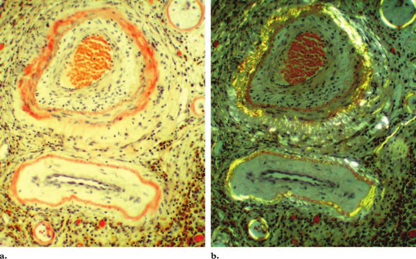
Figure 1. Histologic appearance of !-amyloid deposition in cerebral cortical vessels. (a) Photomicrograph
(original magnification, #100; Congo red stain) shows highlighted !-amyloid deposits along the vessel walls.
(b) Photomicrograph (original magnification, #100; Congo red stain) obtained with polarized light shows the
classic yellow-green birefringence of the !-amyloid deposits.
dementia, with which CAA is frequently associ- overlap in diseases that result in acute neurologic
ated. Forty percent of CAA patients with demen- deficits, TIA-like symptoms, and dementia.
tia show changes of Alzheimer disease at autopsy. The Boston criteria were developed in the mid-
Teaching
Conversely, up to 90% of patients with Alzheimer 1990s as a tool to both improve and standardize
Point
disease have changes of CAA at autopsy (1). How- the diagnosis of CAA (7,12). The criteria specify
ever, dementia may be present in patients with four diagnostic categories: definite CAA, prob-
CAA in the absence of pathologic changes of Alz- able CAA with supporting pathologic evidence,
heimer disease and may in those cases be related probable CAA, and possible CAA, depending on
to small vessel ischemic changes (1,5). Alterna- a combination of clinical, imaging, and histologic
tively, CAA has been seen in patients with sub- data. A “definite” diagnosis of CAA is made with
acute cognitive decline that progresses rapidly a full postmortem examination providing confir-
over the course of a few weeks. These patients mation of lobar, cortical, or corticosubcortical
may present with confusion and disorientation, ICH and severe CAA. Rarely, a biopsy may be
without the focal neurologic deficits that may be performed at the time of hematoma evacuation or
seen in patients with a cerebral infarct or CAA- to exclude other causes of ICH. This pathologic
related acute ICH (8 –11). tissue may reveal CAA, which with the appropri-
ate clinical data, leads to a diagnosis of CAA as
Diagnostic Consider- “probable with supporting pathologic evidence.”
ations: The Boston Criteria CAA is considered “probable” if there is an
The clinical differentiation of CAA-related versus
non–CAA-related symptomatology may be very
difficult and unreliable, as there is significant1520 September-October 2006 RG f Volume 26 ● Number 5
RadioGraphics
The Boston Criteria for Diagnosis of CAA
Clinical Postmortem Pathologic Cortical
Diagnostic Category History Examination Specimen ICH*
Definite CAA Yes Yes Yes ...
Probable CAA with Yes No Yes ...
pathologic evidence
Probable CAA Yes No No $1
Possible CAA Yes No No 1 only
*ICH " intracranial hematoma.
appropriate clinical history as well as imaging
findings of multiple cortical-subcortical hemato-
mas, which may be of varying ages and sizes, in a
patient 55 years old or older, with no other clini-
cal or radiologic cause of hemorrhage. Finally,
clinical data suggesting CAA and the imaging
finding of a single cortical-subcortical hematoma
in a patient older than 55 years, without other
cause of hemorrhage, leads to a “possible” diag-
nosis of CAA (13) (Table).
As histologic analysis is often not practical,
recognition of the imaging findings of CAA is im-
portant for correct diagnosis and proper treat-
ment of patients. Knudsen et al (3) studied 39
cases of cortical-subcortical ICH to validate the
Boston Criteria. Clinical and magnetic resonance
(MR) imaging evidence of CAA was compared
with results from autopsy, biopsy, or surgical
evacuation of hematomas. In those patients diag-
nosed with “probable” CAA by means of the Bos-
ton Criteria, 13 of 13 patients (100%) had a
pathologic diagnosis of CAA. A diagnosis of “pos-
Figure 2. Determination of ICH location in a 74-
sible” CAA was confirmed in 16 of 26 patients year-old man with acute onset of expressive aphasia,
(62%) with pathologic specimens. confusion, and a right-sided facial droop. Axial nonen-
hanced CT scan shows a left-sided frontal cortical
Imaging of CAA ICH, a finding most consistent with CAA-related ICH.
Pathologic tissue obtained at hematoma evacuation
Imaging Evaluation was positive for CAA. The location of an ICH is help-
ful in determining the cause of the ICH in a patient
The clinical presentation of a patient dictates the
with a sudden neurologic deficit.
imaging work-up. A patient presenting with an
acute neurologic deficit or TIA-like symptoms
should undergo nonenhanced computed tomog- lishment of the presence or absence of an ICH
raphy (CT) of the head. CT allows rapid estab- and exclusion of the main clinical differential di-
agnostic consideration of an acute cerebral infarc-RG f Volume 26 ● Number 5 Chao et al 1521
RadioGraphics
Figure 3. Sensitivity of GRE imaging for hemosiderin in an 80-year-old man with dementia that has pro-
gressed over the past 4 years. (a) Axial GRE MR image shows multiple foci of signal loss in cortical-subcortical
locations. In a patient with a diagnosis of probable CAA, these foci are consistent with chronic microhemor-
rhages. (b) Axial T2-weighted fast spin-echo MR image does not show the foci of chronic microhemorrhage.
tion. Nonenhanced head CT is the preferred im- A patient presenting with dementia is usually
aging modality for initial work-up as it provides evaluated initially with brain MR imaging, as the
crucial information regarding the characteristics clinical presentation is often nonspecific and the
of the ICH, including size, location, shape, and causes of dementia are numerous. It is critical to
extension to the extraaxial spaces (Fig 2). maintain a high index of suspicion for CAA, espe-
If an ICH is present in a cortical-subcortical cially in the elderly, and to ensure a thorough
location suspicious for CAA, the patient should evaluation by including a GRE sequence in all
undergo additional evaluation with MR imaging patients who are 70 years old or older (14).
including a gradient-echo (GRE) sequence. GRE In general, angiography does not play a role in
is currently the most sensitive MR imaging se- the evaluation of CAA.
quence for detection of the chronic cortical-sub-
cortical microhemorrhage. Local magnetic field Intracranial Hemorrhage
inhomogeneity related to the presence of hemo- Often the acute presenting finding in CAA-re-
siderin causes a marked loss of signal at T2*- lated cerebrovascular disease, CAA-related ICH
weighted GRE imaging (Fig 3). These chronic represents only 2% of all ICH but is an important
microhemorrhages can be associated with acute cause of hemorrhage in normotensive elderly pa-
CAA-related ICH, and detection of these chronic tients without trauma (1), representing 38%–74%
microhemorrhages with GRE imaging increases
the probability for CAA.1522 September-October 2006 RG f Volume 26 ● Number 5
RadioGraphics
Figure 4. Recurrent CAA-related ICH in a 65-year-old woman with progressive aphasia, right
visual field deficits, and headache. (a) Axial nonenhanced scan from the initial CT study shows a
discrete, ovoid, left-sided occipital ICH. (b) Axial GRE MR image obtained the same day shows
numerous cortical-subcortical microhemorrhages, a finding most compatible with a diagnosis of
probable CAA. One month later, the patient returned to the emergency department with an in-
creasing level of confusion. (c) Axial nonenhanced CT scan obtained at that time shows a larger,
more devastating, left-sided parieto-occipital hemorrhage. Owing to the presence of multiple corti-
cal-subcortical microhemorrhages, which are highly suggestive of CAA, the larger ICH was thought
to represent recurrent hemorrhage rather than a hemorrhagic infarction. The patient was not a sur-
gical candidate and was discharged to a hospice 1 week later, where she died after a few days.RG f Volume 26 ● Number 5 Chao et al 1523
RadioGraphics
Figure 6. CAA-related macrohemorrhage with asso-
ciated subdural hemorrhage in a 77-year-old man with
Figure 5. CAA-related macrohemorrhage with asso- severe headache and difficulty walking. Axial nonen-
ciated subarachnoid hemorrhage in an 81-year-old man hanced CT scan shows a large right-sided posterior
with acute dysphasia and agitation. Axial nonenhanced parietal ICH with irregular borders in a cortical loca-
CT scan shows an irregular, 4 # 5-cm, left-sided fron- tion. There is a small right-sided posterior parafalcine
toparietal cortical ICH. The high attenuation in adja- subdural hemorrhage (arrow). The large hematoma
cent sulci (arrowheads) is consistent with subarachnoid causes marked effacement of right cerebral sulci and
hemorrhage. The patient had a diagnosis of probable approximately 9 mm of subfalcine herniation. The pa-
CAA on the basis of a history of two spontaneous right- tient underwent emergency hematoma evacuation;
sided frontal ICHs. CAA was demonstrated at histologic analysis.
of ICH cases in the elderly (3). Symptomatic ICH Macrohemorrhages.—Large intracerebral
is commonly large ($5 mm), in contrast to mi- hemorrhage ($5 mm in size) is most often acutely
crohemorrhages (!5 mm), which are often clini- symptomatic and may manifest as headaches as-
cally silent. Regardless of the size, CAA-related sociated with emesis, focal neurologic deficits,
ICH exhibits a distinctive cortical-subcortical dis- seizures, coma, or death. Nonenhanced CT is the
tribution that generally spares the deep white imaging study of choice in the initial evaluation of
matter, basal ganglia, and brainstem (Fig 4a). patients with suspected acute ICH, allowing rapid
This cortical-subcortical distribution of ICH in yet precise demonstration of location, size, and
CAA correlates with the anatomic distribution of any other associated hemorrhage.
!-amyloid– containing vessels (15–17). Rarely, CAA-related macrohemorrhages typically ex-
the cerebellum is involved (18). CAA-related hibit irregular borders (15) and may be associated
ICH can involve any lobe of the cerebral hemi- with subarachnoid hemorrhage (Fig 5), subdural
spheres (1,16,19). Other neuroimaging findings hemorrhage (Fig 6), or, less commonly, intraven-
suspicious for CAA-related ICH include multi-
plicity (Fig 4b) and recurrence of ICH (Fig 4c).1524 September-October 2006 RG f Volume 26 ● Number 5
RadioGraphics
Figure 7. CAA-related macrohemorrhage with associ-
ated intraventricular hemorrhage in an obtunded 81-year-
old man. (a) Sagittal nonenhanced T1-weighted MR im-
age shows a large frontal cortical ICH. (b) Axial GRE MR
image shows that the right-sided frontal cortical ICH ex-
tends to the right lateral ventricle. GRE images also re-
vealed multiple cortical-subcortical microhemorrhages, a
finding most consistent with a diagnosis of probable CAA.
(c) Axial fluid-attenuated inversion-recovery (FLAIR)
MR image shows the more rarely associated intraventricu-
lar hemorrhage (arrows) as well as subarachnoid hemor-
rhage (arrowhead).
tricular hemorrhage (Fig 7) (15–17). Subarach-
noid and subdural hemorrhage may be due to
direct extension of the cortical-subcortical hemor-
rhage into the subarachnoid or subdural space
(1,20) or to primary subarachnoid or subdural
hemorrhage resulting from disruption of the lep-
tomeningeal vessels by !-amyloid (21). Intraven-
tricular extension of cortical-subcortical CAA-
related macrohemorrhage may also be seen, de-
pending on its size and location (1). Microhemorrhages.—Petechial hemorrhages
(!5 mm in size) are generally asymptomatic.
Walker et al (14) found evidence of microhemor-RG f Volume 26 ● Number 5 Chao et al 1525
RadioGraphics
ence of these cortical microhemorrhages lends
specificity in patients presenting with acute ICH.
Leukoencephalopathy
Leukoencephalopathy—low attenuation of white
matter at CT or high signal intensity of white
matter at T2-weighted MR imaging—is a nonspe-
cific finding that can be due to demyelination,
ischemia, infarction, or edema. CAA should be
considered in the broad differential diagnosis of
leukoencephalopathy, especially if associated with
cortical-subcortical hemorrhage(s) or progressive
dementia (11). Two imaging patterns of leukoen-
cephalopathy in patients with CAA have been
reported.
Leukoencephalopathy with Sparing of U Fi-
bers.—A symmetric periventricular distribution
of white matter high signal intensity, sparing the
U fibers and associated with atrophy, is seen in
patients with a clinically protracted dementia,
Figure 8. CAA-related microhemorrhage in a 76- similar to that seen in patients with Alzheimer
year-old woman with memory loss, seizures, and head- disease. These white matter lesions are similar to
aches. CAA was diagnosed with biopsy at another insti- those seen in Binswanger subcortical encepha-
tution. Axial GRE MR image shows multiple cortical- lopathy and may have a similar etiology. How-
subcortical microhemorrhages, a finding consistent
ever, in CAA, this ischemic white matter damage
with CAA.
is presumed to be caused by diffuse narrowing of
penetrating cortical vessels resulting from !-amy-
rhage in a characteristic cortical-subcortical distri- loid deposition in the adventitia (10,11). Low
bution in 15.5% of elderly patients more than 70 attenuation at CT and/or high signal intensity at
years of age. CT and conventional or fast spin- T2-weighted MR imaging are most prevalent in
echo T1- and T2-weighted MR imaging se- the centrum semiovale and deep white matter
quences are relatively insensitive for such small with sparing of the U fibers, corpus callosum, and
microhemorrhages. Local magnetic field inhomo- internal capsules (Fig 9). These lesions can be
Teaching geneity related to the presence of hemosiderin in both diffuse and focal and may be severe in pa-
Point foci of petechial hemorrhage causes a marked loss tients with long-standing dementia. In patients
of signal at T2*-weighted GRE imaging, which is with ICH, white matter lesions can be observed in
currently the most sensitive sequence for detec- regions remote from the ICH.
tion of the cortical-subcortical microhemorrhage
associated with CAA (14,22) (Fig 8). The pres-1526 September-October 2006 RG f Volume 26 ● Number 5
RadioGraphics
Figure 9. Leukoencephalopathy in a 79-
year-old woman with slowly progressive
dementia similar to Alzheimer dementia.
(a, b) Axial nonenhanced CT scan (a) and
FLAIR MR image (b) show symmetric
periventricular leukoencephalopathy with
sparing of the U fibers, corpus callosum, and
internal capsules. The FLAIR image also
shows encephalomalacia and hemosiderin
from prior macrohemorrhage in the left fron-
tal lobe. (c) Axial GRE MR image shows
multiple bilateral cortical foci of hemosiderin,
thus increasing the specificity for a diagnosis
of probable CAA. The encephalomalacia and
hemosiderin in the left frontal lobe are also
seen.
Leukoencephalopathy with Involvement of U
Fibers.—Several case reports of patients with
pathologically proved CAA have described sub-
acute cognitive decline associated with leukoen-
cephalopathy that extends to involve U fibers and
is associated with mass effect likely related to
edema (9,11,23,24). At T2-weighted MR imag-
ing, white matter high signal intensity is most opsy. Harkness et al (23) proposed that these
prevalent in the centrum semiovale and deep changes may be secondary to !-amyloid–induced
periventricular regions, sparing the corpus callo- vasculopathy— cerebral amyloid inflammatory
sum and internal capsule (Fig 10). Most cases vasculopathy (CAIV). A few biopsy-proved cases
demonstrated perivascular inflammation at bi- of CAIV have responded to immunosuppressive
therapy, with at least partial resolution of leu-
koencephalopathy at imaging (8,10,24).RG f Volume 26 ● Number 5 Chao et al 1527
RadioGraphics
Figure 10. Leukoencephalopathy in a
61-year-old woman with rapidly progressive
cognitive decline. (a) Axial FLAIR MR
image shows asymmetric lobar leukoen-
cephalopathy extending to involve the U
fibers and exerting mass effect on the adja-
cent sulci, most prominently in the poste-
rior left parietal lobe. The absence of signal
abnormality at diffusion-weighted MR im-
aging made an ischemic process or acute
infarction unlikely. CAA was diagnosed
with biopsy. (b) Axial GRE MR image ob-
tained after biopsy shows a few cortical mi-
crohemorrhages (arrows). The patient was
treated with a short course of prednisone
taper therapy, which started at 40 mg and
produced clinical improvement. (c) Fol-
low-up axial FLAIR MR image obtained 1
year later shows near-complete resolution
of the leukoencephalopathy. CAA patients
with subacute cognitive decline and leu-
koencephalopathy may respond to immu-
nosuppressive therapy.
Atrophy CAA, atrophy is most likely the result of chronic
Prominence of the ventricular system and en- small vessel ischemia related to !-amyloid depo-
largement of the sulci representing generalized sition and is usually seen in association with
cerebral and cerebellar atrophy are nonspecific
imaging findings, especially in the elderly. In1528 September-October 2006 RG f Volume 26 ● Number 5
RadioGraphics
Figure 11. Probable CAA in a 72-year-old woman with speech difficulties and waxing and waning memory
loss. (a) Axial FLAIR MR image shows nonspecific atrophy as well as periventricular leukoencephalopathy and
prominent left-sided parieto-occipital leukoencephalopathy. (b) Axial GRE MR image shows cortical-subcorti-
cal microhemorrhages and a small left-sided parietal cortical-subcortical macrohemorrhage. These findings in-
crease suspicion for probable CAA.
leukoencephalopathy (Fig 11a). When atrophy Currently, there is no treatment to halt or re-
and leukoencephalopathy are seen in conjunction verse !-amyloid deposition. Thus, attention is Teaching
with acute or chronic ICH in a cortical-subcorti- directed instead to the prevention of adverse out- Point
cal location, the diagnostic specificity for CAA is comes associated with the natural history of CAA,
increased (Fig 11b). such as recurrent hemorrhages or progressive de-
mentia. Furthermore, higher numbers of micro-
Management and Prognosis hemorrhages on the baseline GRE MR images are
Although surgical intervention for an acute ICH predictive of a greater risk for recurrent bleeding,
was previously thought to be contraindicated in future cognitive impairment, loss of functional
CAA patients because of fear of rebleeding (1), independence, or death (26).
more recent studies have not shown an increased Patients with a new diagnosis of CAA who re-
frequency of adverse outcome in most patients ceive anticoagulation for other disorders should
with CAA-related ICH. Patients 75 years of age undergo evaluation of the risks and benefits of
or older, those with a hematoma in a parietal lobe continued anticoagulation and antiplatelet
location, or those with associated intraventricular therapy. Administration of anticoagulation
hemorrhage are more likely to have an adverse therapy for presumed TIA or warfarin for atrial
postoperative outcome and should be treated fibrillation and other disorders may potentiate the
nonsurgically (19,25). risk of hemorrhage in a CAA patient. Rosand et al
(27) found that even therapeutic levels of antico-
agulation with warfarin (international normalizedRG f Volume 26 ● Number 5 Chao et al 1529
RadioGraphics
Figures 12, 13. (12) Hypertension-related macrohemorrhage in an 80-year-old woman with right-sided
weakness and a blood pressure of 160/85 mm Hg. Axial nonenhanced CT scan shows an area of increased at-
tenuation in the left thalamus, a finding most consistent with an acute hypertensive ICH. (13) Hypertension-
related microhemorrhages in a 91-year-old woman with hypertension and unsteadiness. Axial GRE MR image
shows multiple small foci of hemosiderin in both basal ganglia and thalami, locations more consistent with a
hypertensive cause.
ratio ! 3) are associated with an increased fre- ICH is most commonly caused by hypertension,
quency of warfarin-associated ICH in CAA pa- trauma, bleeding diatheses, amyloid angiopathy,
tients. Furthermore, while warfarin has decreased illicit drug use (mostly amphetamines and co-
the annual risk of stroke in patients more than 75 caine), and vascular malformations. Infrequent
years of age from 3.5%– 8.1% to less than 2%, causes include hemorrhagic tumors, ruptured
it carries an annual rate of ICH of 1.8%, even aneurysms, and vasculitis (28). The history,
higher in CAA patients, thus potentially offsetting physical examination findings, and laboratory
the benefit of warfarin in stroke prevention (27). results often allow establishment of one of these
Other studies have shown fatal outcomes in CAA diagnoses. However, specific characteristics of the
patients undergoing thrombolytic or antiplatelet ICH are just as important in the identification of
therapy for various clinical indications (16). The CAA-related ICH.
risk-benefit ratio of anticoagulation and thrombo- Hypertension is the most common cause of
lytic therapy in CAA patients should be carefully nontraumatic hemorrhage in adults (29). In con-
considered on an individual basis. trast to the typical cortical-subcortical location of
CAA-related hemorrhage, hypertensive hemor-
Differential Diagnosis rhages, both large and small, most commonly oc-
A single large cortical-subcortical ICH in a pa- cur in the deep gray matter, such as the basal gan-
tient presenting with an acute neurologic deficit is glia or thalami, or the brainstem (Figs 12, 13).
not entirely specific for a diagnosis of CAA (16).1530 September-October 2006 RG f Volume 26 ● Number 5
RadioGraphics
Figure 14. Large macrohemorrhage in a 66-year-old man with biopsy-proved brain metastases
from small cell lung cancer who presented with headache, light-headedness, and difficulty walking.
(a) Axial FLAIR MR image shows a large right-sided frontal cortical hematoma with surrounding
vasogenic edema. A fluid-fluid level is present, as is often seen in patients undergoing anticoagula-
tion therapy. This patient was taking clopidogrel for a coronary stent. (b) Axial contrast-enhanced
T1-weighted MR image shows a second, nonhemorrhagic metastatic lesion in the right temporal
lobe (arrow).
Although a hemorrhagic tumor may exhibit a terns of involvement that are characteristic of
cortical-subcortical location similar to CAA-re- CAA, including cortical-subcortical location of
lated hemorrhage, MR imaging may be helpful in macro- and microhemorrhages, which may be
identifying additional enhancing lesions, leading found concurrently with leukoencephalopathy
to a greater suspicion of metastatic disease (Fig and atrophy. Early recognition of the constella-
14). tion of imaging findings associated with CAA fa-
cilitates a clinical diagnosis of CAA and proper
Conclusions patient treatment.
CAA-related hemorrhage is an important cause of
morbidity and mortality in the normotensive el- Acknowledgment: We thank Murli Krishna, MD, for
contributing the pathologic slides.
derly patient. Patients may present with a spec-
trum of clinical findings such as sudden neuro-
References
logic deficit (stroke), TIA-like symptoms, or de-
1. Vinters HV. Cerebral amyloid angiopathy: a criti-
mentia that can be seen in disorders other than cal review. Stroke 1987;18:311–324.
CAA. However, neuroimaging demonstrates pat- 2. Yamada M, Tsukagoshi H, Otomo E, Hayakawa
M. Cerebral amyloid angiopathy in the aged. J
Neurol 1987;234:371–376.RG f Volume 26 ● Number 5 Chao et al 1531
RadioGraphics
3. Knudsen KA, Rosand J, Karluk D, Greenberg 17. Brown RT, Coates RK, Gilbert JJ. Radiographic-
SM. Clinical diagnosis of cerebral amyloid angi- pathologic correlation in cerebral amyloid angi-
opathy: validation of the Boston Criteria. Neurol- opathy: a review of 12 patients. J Can Assoc Ra-
ogy 2001;56:537–539. diol 1985;36:308 –311.
4. Vonsattel JP, Myers RH, Hedley-Whyte ET, Rop- 18. Cuny E, Loiseau H, Rivel J, Vital C, Castel JP.
per AH, Bird ED, Richardson EP. Cerebral amy- Amyloid angiopathy-related cerebellar hemor-
loid angiopathy without and with cerebral hemor- rhage. Surg Neurol 1996;46:235–239.
rhages: a comparative histological study. Ann 19. Yamada M. Cerebral amyloid angiopathy: an
Neurol 1991;30:637– 649. overview. Neuropathology 2000;20:8 –22.
5. Rosand J, Greenberg SM. Cerebral amyloid angi- 20. Yamada M, Itoh Y, Maeda A, Otomo E, Haya-
opathy. Neurologist 2000;6:315–325. kawa M, Miyatake T. Pathogenesis of cerebral
6. Greenberg SM, Vonsattel JP, Stakes JW, et al. The amyloid angiopathy-related hemorrhage in the
clinical spectrum of cerebral amyloid angiopathy: elderly. In: Kisilevsky R, Benson MD, Fragione B,
presentations without lobar hemorrhage. Neurol- Gauldie J, Muckle TJ, Young ID, eds. Amyloid
ogy 1993;43:2073–2079. and amyloidosis. New York, NY: Parthenon,
7. Greenberg SM, Finklestein SP, Schaefer PW. Pe- 1994; 362–364.
techial hemorrhages accompanying lobar hemor- 21. Takeda S, Yamazaki K, Miyakawa T, et al. Sub-
rhage: detection by gradient-echo MRI. Neurology cortical hematoma caused by cerebral amyloid
1996;46:1751–1754. angiopathy: does the first evidence of hemorrhage
8. Eng JA, Frosch MP, Choi K, et al. Clinical mani- occur in the subarachnoid space? Neuropathology
festations of cerebral amyloid angiopathy-related 2003;23:254 –261.
inflammation. Ann Neurol 2004;55:250 –256. 22. Atlas SW, Mark AS, Grossman RI, Gomori JM.
9. Sarazin M, Amarenco P, Mikol J, Dimitri D, Lot Intracranial hemorrhage: gradient-echo MR imag-
G, Bousser MG. Reversible leukoencephalopathy ing at 1.5 T— comparison with spin-echo imaging
in cerebral amyloid angiopathy presenting as sub- and clinical applications. Radiology 1988;168:
acute dementia. Eur J Neurol 2002;9(4):353–358. 803– 807.
10. Gray F, Dubas F, Roullet E, Escourolle R. Leu- 23. Harkness KA, Coles A, Pohl U, Xuereb JH, Baron
koencephalopathy in diffuse hemorrhagic cerebral JC, Lennox GG. Rapidly reversible dementia in
amyloid angiopathy. Ann Neurol 1985;18:54 –59. cerebral amyloid inflammatory vasculopathy. Eur
11. Loes DJ, Biller J, Yuh WT, et al. Leukoencepha- J Neurol 2004;11:59 – 62.
lopathy in cerebral amyloid angiopathy: MR imag- 24. Oh U, Gupta R, Krakauer JW, et al. Reversible
ing in four cases. AJNR Am J Neuroradiol 1990; leukoencephalopathy associated with cerebral
11:485– 488. amyloid angiopathy. Neurology 2004;62:494 –
12. Greenberg SM, Rebeck GW, Vonsattel JP, Go- 497.
mez-Isla T, Hyman BT. Apolipoprotein E epsilon 25. Izumihara A, Ishihara T, Iwamoto N, Yamashita
4 and cerebral hemorrhage associated with amy- K, Ito H. Postoperative outcome of 37 patients
loid angiopathy. Ann Neurol 1995;38(2):254 – with lobar intracerebral hemorrhage related to ce-
259. rebral amyloid angiopathy. Stroke 1999;30:29 –33.
13. Smith EE, Greenberg SM. Clinical diagnosis of 26. Greenberg SM, Eng JA, Ning M, Smith EE, Ro-
cerebral amyloid angiopathy: validation of the sand J. Hemorrhage burden predicts recurrent in-
Boston Criteria. Curr Atheroscler Rep 2003;5: tracerebral hemorrhage after lobar hemorrhage.
260 –266. Stroke 2004;35:1415–1420.
14. Walker DA, Broderick DF, Kotsenas AL, Rubino 27. Rosand J, Hylek EM, O’Donnell HC, Greenberg
FA. Routine use of gradient-echo MRI to screen SM. Warfarin-associated hemorrhage and cerebral
for cerebral amyloid angiopathy in elderly patients. amyloid angiopathy: a genetic and pathologic
AJR Am J Roentgenol 2004;182:1547–1550. study. Neurology 2000;55:947–951.
15. Wagle WA, Smith TW, Weiner M. Intracerebral 28. Caplan LR. Intracerebral hemorrhage. Lancet
haemorrhage caused by cerebral amyloid angiopa- 1992;339:656 – 658.
thy: radiographic-pathological correlation. AJNR 29. Meyer JR, Gorey MT. Differential diagnosis of
Am J Neuroradiol 1984;5:171–176. nontraumatic intracranial hemorrhage. Neuroim-
16. Miller JH, Wardlaw JM, Lammie GA. Intracere- aging Clin N Am 1998;8(2):263–293.
bral haemorrhage and cerebral amyloid angiopa-
thy: CT features with pathological correlation.
Clin Radiol 1999;54:422– 429.RG Volume 26 • Volume 5 • September-October 2006 Chao et al
RadioGraphics
Cerebral Amyloid Angiopathy: CT and MR Imaging Findings
Christine P. Chao, MD et al
RadioGraphics 2006; 26:1517–1531 ● Published online 10.1148/rg.265055090 ● Content Codes:
Page 1518
Cerebral amyloid angiopathy (CAA) is an important cause of spontaneous cortical-subcortical
intracranial hemorrhage (ICH) in the normotensive elderly.
Page 1518
CAA manifests radiologically as part or all of a constellation of findings including acute or chronic
ICHs in a distinctive cortical-subcortical distribution, leukoencephalopathy, and atrophy.
Page 1519
The Boston criteria were developed in the mid-1990s as a tool to both improve and standardize the
diagnosis of CAA (7,12). The criteria specify four diagnostic categories: definite CAA, probable CAA
with supporting pathologic evidence, probable CAA, and possible CAA, depending on a combination
of clinical, imaging, and histologic data.
Page 1525
Local magnetic field inhomogeneity related to the presence of hemosiderin in foci of petechial
hemorrhage causes a marked loss of signal at T2*-weighted GRE imaging, which is currently the most
sensitive sequence for detection of the cortical-subcortical microhemorrhage associated with CAA
(14,22) (Fig 8).
Page 1528
Currently, there is no treatment to halt or reverse β-amyloid deposition. Thus, attention is directed
instead to the prevention of adverse outcomes associated with the natural history of CAA, such as
recurrent hemorrhages or progressive dementia.You can also read