Case Report A rare presentation of giant pediatric pilomyxoid astrocytoma involved in the sellar region: case report and literature review
←
→
Page content transcription
If your browser does not render page correctly, please read the page content below
Int J Clin Exp Med 2018;11(2):1038-1042
www.ijcem.com /ISSN:1940-5901/IJCEM0065452

Case Report
A rare presentation of giant pediatric
pilomyxoid astrocytoma involved in the
sellar region: case report and literature review
Zhihua Li1, Mengzhao Feng2, Fuyou Guo2, Jing Cui1
1
 Department of Parasitology, Medical College, Zhengzhou University, Zhengzhou 450052, Henan, China;
2
 Department of Neurosurgery, The First Affiliated Hospital, Zhengzhou University, Zhengzhou 450052, Henan,
China
Received September 12, 2017; Accepted January 4, 2018; Epub February 15, 2018; Published February 28,
2018

Abstract: Pilomyxoid astrocytoma (PMA) is a rare malignant tumor with a poor prognosis and a high incidence of
dissemination in children. We present a case of an unusual giant pediatric PMA with unique clinical imaging. A
13-month-old female presented with three months of nystagmus and developmental retardation. Magnetic reso-
nance imaging (MRI) showed the size of the tumor was approximately 4.5×4.2×4.0 cm3, and there was significant
enhancement with an abundant blood supply. Three-dimensional printing (3D-P) of the tumor demonstrated that
the bilateral anterior cerebral arteries and the anterior communicating artery were completely wrapped by this gi-
ant tumor. Subsequently, the patient underwent total resection with good recovery with the assistance of 3D-P. The
presumed diagnosis of the lesion was suprasellar germinoma or craniopharyngioma, however, postoperative histo-
pathological examination identified it as being a rare PMA. Uncommon PMA with unique clinical features should be
emphasized as differential diagnosis in the sellar region. 3D-P is a safe and effective tool for successful treatment
of a rare hypervascular PMA.

Keywords: Pediatric brain tumor, pilomyxoid astrocytoma, 3D printer, surgery

Introduction                                                   Case report

The predominant pediatric glioma located in                    A 13-month-old female was admitted to our
the sellar region is pilocytic astrocytoma. How-               hospital with a three-month history of nystag-
ever, pilomyxoid astrocytoma (PMA) is rare                     mus. Physical examination was positive for de-
based on previous literature. PMA is a rare                    velopmental retardation. There was no neuro-
WHO grade II tumor, accepted as a variant of                   logical deficit. A laboratory evaluation revealed
pilocytic astrocytoma in the World Health                      the levels of AFP and HCG were 2.57 ng/ml
Organization (WHO) classification of central                   (normal range: 0~10) and less than 0.1 mIU/ml
nervous system tumors in 2007 [1]. PMA has                     (normal range: 0~5), respectively. A hormonal
previously been described as a solitary rare                   assay of pituitary function was normal except
case in the previous recent literature [2-10]                  for a slight decrease in growth hormone in the
(see Table 1). Here, we report a rare case of                  serum. Computer tomography (CT) revealed a
PMA with an abundant intratumoral blood sup-                   slightly hypodense lesion in the suprasellar
ply in the hypothalamic/chiasmatic region. The                 region (Figure 1A), Magnetic resonance imag-
total resection of the giant PMA was achieved                  ing (MRI) showed a tumor located in the hypo-
with the assistance of three-dimensional print-                thalamic and chiasmatic region and the size of
ing (3D-P). Uncommon PMA with unique clinical                  the tumor was approximately 4.5×4.2×4.0 cm3.
features should be emphasized as the differen-                 The tumor appeared hypointense and hyperin-
tial diagnosis in the hypothalamic and chias-                  tense to the brain on T1-weighted (Figure 1B,
matic region. 3D-P is a safe and effective tool                1C) and T2-weighted MRI, respectively, with
for successful treatment of a rare hypervascu-                 remarkable homogeneous enhancement after
lar PMA.                                                       administration of gadolinium (Figure 1D-F),
Rare giant pediatric pilomyxoid astrocytoma with unique clinical imaging
Table 1. Pediatric pilomyxoid astrocytoma reported in the recent literature
No Author Year Age/Sex Clinical features Location Treatment Prognosis
1 Ceppa E [2] 2007 6.5 Y/Female Weight gain, irritability, Hypothalamic Biopsy + chemo- Tumor recurrence
abnormal eye movements therapy after one year
2 Alimohamadi 2009 12 Y/Male Visual impairment, cognitive Sellar and supra- Surgery + radiother- No recurrence in
M [3] disturbance sellar apy + chemotherapy follow-up of one year
3 Paraskevopoulos 2011 12 Y/Female Gait disturbance, motor C2-C7 intramedul- Surgery Recurrence after 3 M
D [4] and sensory deficits of the lary and development of
left side GBM; dead at 12 M
4 Terasaki M [5] 2012 5 Y/Male Loss of vision Optic nerve lepto- Biopsy + radiothera- No recurrence in
meninges py + chemotherapy follow-up of 2 Y
5 Chonan M [6] 2013 3 Y/Male Truncal ataxia, drowsy Right cerebellum Surgery Dead
6 Pereira FO [7] 2013 11 Y/Female Diplopia, gait disturbance Brainstem Surgery Recurrence after 9 M
7 Tjahjadi M [8] 2015 7 Y/Female Headache, nausea, vomiting, Supraseller region Surgery + radio- Tumor reduction after
bitemporal hemianopia therapy one year
8 Wang Z [9] 2016 13 Y/Male Headache, vomiting Supraseller region VPS + radiotherapy No tumor recurrence
+ subtotal resection after 10 M
9 Homma T [10] 2017 4 M/Male Vomiting, slow weight gain Right temporal lobe Surgery + chemo- Tumor recurrence
+ suprasellar region therapy after 4 Y
10 Present case 2017 13 M/Female Nystagmus, developmental Hypothalamic + Surgery No recurrence in
retardation chiasmatic region follow-up of 3 M
Abbreviation: Y = Year, M = Month, C = Cervical, NA = Not Available, GBM = Glioblastoma, VPS = Ventriculo-peritoneal shunt.
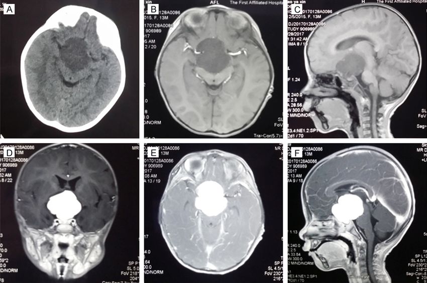
Figure 1. A 13-month-old female with a PMA in the hypothalamic and chiasmatic region shows the tumor compress-
ing the brain stem on CT (A). A preoperative axial and sagittal T1-weighted MRI scan shows a suprasellar lesion with
hypointense signals, respectively (B, C). A coronal axial, sagittal gadolinium-enhanced T1-weighted MRI scan shows
a significant homogeneously enhanced tumor (D-F).
which suggested that the tumor was a hyper- strated the bilateral anterior cerebral arteries
vascular lesion with an abundant intratumoral and anterior communicating artery were com-
blood supply. 3D-P of the tumor was applied in pletely wrapped by this giant tumor (Figure
the present patient (Figure 2A-C). It demon- 3A-D).
1039 Int J Clin Exp Med 2018;11(2):1038-1042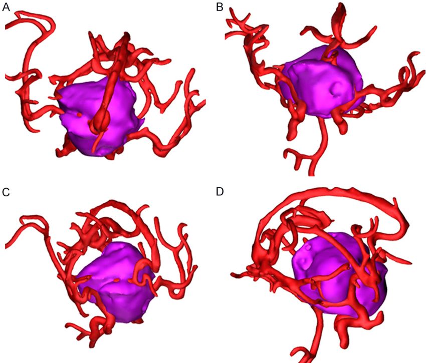
Rare giant pediatric pilomyxoid astrocytoma with unique clinical imaging
Figure 2. Three-dimensional printing of the tumor revealed that the tumor was located at the base of the skull and
surrounding vital structures (A-C).
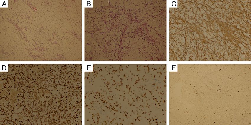
with a loose myxoid back-
ground (Figure 5A, 5B). Imm-
unohistochemical examinati-
on showed the tumor cells
were positive for glial fibrillary
acidic protein (GFAP), S-100,
and Olig2 (Figure 5C-E). Imm-
unohistochemical staining for
Ki-67 was performed, and the
Ki-67 labeling index was app-
roximately 5% (Figure 5F).
Discussion
PMA is usually implicated in
the suprasellar region on the
floor of the third ventricle and
in the thalamic region. It has
been reported that a non-
enhancing solid portion of bo-
th a primary and disseminated
mass have more frequently
Figure 3. Three-dimensional printing of the tumor revealed that bilateral an- been found in PMAs compar-
terior cerebral arteries and anterior communicating artery were completely ed to PAs (100% versus 32%).
wrapped by this giant tumor; the view of anteroposterior position (A, B); and The reason for the lack of en-
the view of lateral position (C, D).
hancement may be the myx-
oid component, based on the
The patient underwent resection of the tumor previous literature. [11] However, the MRI mani-
via a frontal basal interhemispheric approach festation of our case was not consistent with
with assistance of the 3D-P model. The lamina previous reports, and there were several strik-
terminalis was cut for increased exposure dur- ing features obtained from the present patient.
ing the operation, and a gross total tumor res- First, the tumor was very large due to the length
ection was achieved after one week as seen in of the largest diameter surpassing 4 cm.
CT and MRI examinations (Figure 4A-D). No po- Second, the tumor showed remarkably homo-
stoperative radiotherapy or chemotherapy was geneous enhancement after administration of
administered in this patient. During a three- gadolinium. PMA usually exhibit unremarkable
month follow-up, no further tumor progress or enhancement due to low grade glioma, howev-
recurrence was observed. Histological exami- er, hypervascular PMA with significant enhance-
nation revealed monomorphous bipolar cells ment was observed from this patient. The pre-
1040 Int J Clin Exp Med 2018;11(2):1038-1042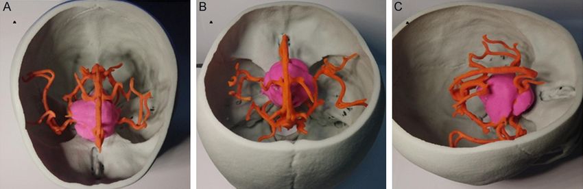
Rare giant pediatric pilomyxoid astrocytoma with unique clinical imaging

                                                                                ic/chiasmatic vital structures
                                                                                and brain stem. Consequently,
                                                                                it was considerably too dan-
                                                                                gerous to totally remove such
                                                                                tumors in infants. In our previ-
                                                                                ous cases, there were occa-
                                                                                sional mortalities with giant
                                                                                suprasellar tumors due to lim-
                                                                                ited exposure and excessive
                                                                                bleeding caused by hypervas-
                                                                                cular lesions as well as inju-
                                                                                ries of the hypothalamus, be-
                                                                                cause patients are easily su-
                                                                                sceptible to shock or even
                                                                                death during surgical inter-
                                                                                vention because of the very
                                                                                limited blood volume in infants
                                                                                or children, especially for hy-
                                                                                pervascular tumors. [12] How
                                                                                should the operational risk
                                                                                of hypervascular tumors be
                                                                                reduced in infants? In addition
                                                                                to the basic skills described in
                                                                                our previous paper, we found
Figure 4. Postoperative CT demonstrated that the tumor was removed fully        that a preoperative 3D-P mo-
and there was excellent decompression of the brain stem (A). A postopera-       del could offer the best visu-
tive sagittal, axial, and coronal gadolinium-enhanced T1-weighted MRI scan      alization in three-dimensional
indicates that the tumor was totally removed at 7 days after the operation
(B-D).
                                                                                space, which not only contrib-
                                                                                uted to the design of the pre-
                                                                                operative approach but also
                                                                                played an important role in de-
                                                                                creasing vascular injury during
                                                                                the operation because bilat-
                                                                                eral anterior cerebral arteries
                                                                                and the anterior communicat-
                                                                                ing artery were completely
                                                                                wrapped by this giant tumor
                                                                                on 3D-P. The use of 3D-P can
                                                                                facilitate the identification of
                                                                                the 3D anatomical relation-
                                                                                ship between the tumor and
                                                                                adjacent artery and it is help-
Figure 5. Hematoxylin-eosin staining demonstrate that the tumor consisted       ful to maximally reduce the
of monomorphous bipolar cells with a loose myxoid background (A: Original       damage to the surrounding
magnification ×100 & B: Original magnification ×200). Immunohistochemi-         artery in deep-seated tumors.
cal examinations for GFAP, S-100, and Olig2, respectively (C-E: Original mag-   Through our experience in this
nification ×400), and Ki-67 (F: Original magnification ×200).
                                                                                case, we found that a 3D-P
                                                                                model is an extremely safe
sumed diagnosis of the lesion was suprasellar                  and useful tool for giant PMAs with hyper-
germinoma or craniopharyngioma and postop-                     vascularity.
erative histopathological examination identi-
fied it as being a rare PMA. This giant tumor                  The clinical features of PMAs are that they
severely compressed the adjacent hypothalam-                   require a more aggressive course, they have a

1041                                                              Int J Clin Exp Med 2018;11(2):1038-1042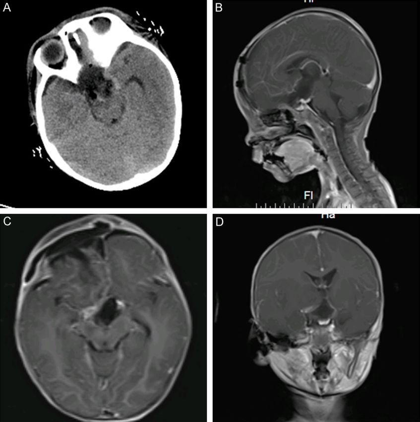
Rare giant pediatric pilomyxoid astrocytoma with unique clinical imaging

higher recurrence rate, and they disseminate           [2]    Ceppa EP, Bouffet E, Griebel R, Robinson C,
along the cerebrospinal fluid pathways. Con-                  Tihan T. The pilomyxoid astrocytoma and its re-
sequently, PMAs are still challenging tumors for              lationship to pilocytic astrocytoma: report of a
neurosurgeons. Moreover, there is no consen-                  case and a critical review of the entity. J
                                                              Neurooncol 2007; 81: 191-196.
sus for standard treatment for PMAs until now.
                                                       [3]    Alimohamadi M, Bidabadi MS, Ayan Z, Ketabchi
Our results show that surgery has a clear role
                                                              E, Amirjamshidi A. Pilomyxoid astrocytoma with
for diagnosis and tumor control as well as relief             involvement of the sella turcica in an adoles-
of the mass effect in optic pathway/hypotha-                  cent. J Clin Neurosci 2009; 16: 1648-1649.
lamic gliomas in children. Simultaneously, the         [4]    Paraskevopoulos D, Patsalas I, Karkavelas G,
authors noted that primary surgical debulking                 Foroglou N, Magras I, Selviaridis P. Pilomyxoid
of the tumor without adjuvant therapy was a                   astrocytoma of the cervical spinal cord in a
safe and effective management. [13] Due to                    child with rapid progression into glioblastoma:
the toxicity of chemotherapy or radiotherapy for              case report and literature review. Childs Nerv
infants, adjuvant therapy was not recommend-                  Syst 2011; 27: 313-321.
ed because this patient underwent total resec-         [5]    Terasaki M, Bouffet E, Maeda M, Sugita Y,
                                                              Sawamura Y, Morioka M. Successful treatment
tion. However, dynamic observation will be very
                                                              of leptomeningeal gliomatosis of pilomyxoid
important for the present patient in the future.
                                                              astrocytoma after failed frontline chemothera-
Conclusion                                                    py. Neurologist 2012; 18: 32-35.
                                                       [6]    Chonan M, Kanamori M, Kumabe T, Saito R,
PMA is a rare malignant tumor with unique                     Watanabe M, Tominaga T. Pilomyxoid astrocy-
manifestation on MRIs. PMA as low grade glio-                 toma of the cerebellum with Williams syn-
                                                              drome: a case report. Childs Nerv Syst 2013;
ma exhibited uncommon remarkable enhance-
                                                              29: 1211-1214.
ment in this patient and 3D-P provided better
                                                       [7]    Pereira FO, Lombardi IA, Mello AY, Romero FR,
visualization for observation of the tumor and                Ducati LG, Gabarra RC, Zanini MA. Pilomyxoid
adjacent vital structures. Total resection of the             astrocytoma of the brainstem. Rare Tumors
PMA without complications was achieved with                   2013; 5: 65-67.
the assistance of 3D-P. Rare PMA with abun-            [8]    Tjahjadi M, Arifin MZ, Sobana M, Avianti A,
dant blood supply should be emphasized as dif-                Caropeboka MS, Eka PA, Aqustina H. Cystic pi-
ferential diagnosis in the hypothalamic and chi-              lomyxoid astrocytoma on suprasellar region in
asmatic region.                                               7-year-old girl: treatment and strategy. Asian J
                                                              Neurosurg 2015; 10: 154-157.
Acknowledgements                                       [9]    Wang Z, Yan HM, Zhou XR, Liu JK, Chang JY,
                                                              Wang YT. Spontaneous intratumoural and in-
This study was supported in part by the Natural               traventricular haemorrhage associated with a
Scientific Foundation of China (No. U1204807                  pilomyxoid astrocytoma in the hypothalamic/
and No. 81172612).                                            chiasmatic region. J Clin Neurosci 2016; 33:
                                                              217-220.
Disclosure of conflict of interest                     [10]   Homma T, Seki T, Suzuki A, Ohta T, Maebayashi
                                                              T, Yoshino A, Kusumi Y, Sugitani M. Cytopath-
None                                                          ological features of pilomyxoid astrocytoma: a
                                                              case report. Cytopathology 2017; 28: 74-77.
Address correspondence to: Jing Cui, Department        [11]   Lee IH, Kim JH, Suh YL, Eo H, Shin HJ, Yoo SY,
of Parasitology, Medical College, Zhengzhou Uni-              Lee KS. Imaging characteristics of pilomyxoid
versity, 40 Daxue Road, Zhengzhou 450052, Henan,              astrocytomas in comparison with pilocytic as-
China. E-mail: cuij@zzu.edu.cn; Fuyou Guo, Depart-            trocytomas. Eur J Radiol 2011; 79: 311-316.
ment of Neurosurgery, The First Affiliated Hospital,   [12]   Guo A, Suresh V, Liu X, Guo F. Clinicopathological
Zhengzhou University, Zhengzhou 450052, Henan,                features and microsurgical outcomes for giant
China. E-mail: chyou666@hotmail.com                           pediatric intracranial tumor in 60 consecutive
                                                              cases. Childs Nerv Syst 2017; 33: 447-455.
References                                             [13]   Goodden J, Pizer B, Pettorini B, Williams D,
                                                              Blair J, Didi M, Thorp N, Mallucci C. The role of
[1]    Louis DN, Ohgaki H, Wiestler OD, Cavenee WK,           surgery in optic pathway/hypothalamic glio-
       Burger PC, Jouvet A, Scheithauer BW, Kleihues          mas in children. J Neurosurg Pediatr 2014; 13:
       P. The 2007 WHO classification of tumours of           1-12.
       the central nervous system. Acta Neuropathol
       2007; 114: 97-109.

1042                                                          Int J Clin Exp Med 2018;11(2):1038-1042
You can also read