A Radiographic Study of Biomechanical Relationship between the Achilles Tendon and Plantar Fascia
←
→
Page content transcription
If your browser does not render page correctly, please read the page content below
Hindawi
BioMed Research International
Volume 2020, Article ID 5319640, 6 pages
https://doi.org/10.1155/2020/5319640

Research Article
A Radiographic Study of Biomechanical Relationship between the
Achilles Tendon and Plantar Fascia

          Genrui Zhu, Zhifeng Wang, Chengjie Yuan, Xiang Geng, Chao Zhang, Jiazhang Huang,
          Xu Wang , and Xin Ma
          Department of Orthopedics, Huashan Hospital, Fudan University, No. 12, Middle Wulumuqi Road, Jingan District, Shanghai,
          China

          Correspondence should be addressed to Xu Wang; wangxuankleii@163.com

          Received 14 September 2019; Revised 12 December 2019; Accepted 25 January 2020; Published 18 February 2020

          Academic Editor: Ali Nokhodchi

          Copyright © 2020 Genrui Zhu et al. This is an open access article distributed under the Creative Commons Attribution License,
          which permits unrestricted use, distribution, and reproduction in any medium, provided the original work is properly cited.
          Background. Previously, scholars have concluded that the Achilles tendon and the plantar fascia were closely biomechanically
          related, although there is little clinical evidence of the relationship between the two. To investigate the biomechanical relationship
          between the Achilles tendon and the plantar fascia, the author used standing lateral ankle radiographs of patients with insertional
          Achilles tendonitis to determine the biomechanical relationship between the Achilles tendon and plantar fascia. Methods. The
          author collected standing lateral ankle radiographs from patients with insertional Achilles tendonitis who accepted surgical
          treatment in the author’s hospital from March 2009 to July 2018. According to whether there were bone spurs on the posterior side
          of the calcaneus, patients were divided into group A (spur present on the posterior side) and group B (spur not present on the
          posterior side). The positive rates of spurs on the plantar side of the calcaneus were determined in group A and group B. The chi-
          square test was used to compare the measurement results between the two groups. Results. In group A, 13 heels were positive for
          calcaneal bone spurs, and the positive rate was 65.0%. In group B, 3 heels were positive for plantar calcaneal spurs, and the positive
          rate was 12%. Among all 16 patients with positive plantar calcaneal spurs, 13 had posterior calcaneal spurs (accounting for 81.3%),
          and 3 had negative results, accounting for 18.7%. There was a significant difference between the results in groups A and B
          (P < 0.05). Conclusion. There is a relationship between posterior calcaneal spurs and plantar calcaneal spurs in patients with
          insertional Achilles tendonitis, which can be inferred as resulting from the increasing tension in the biomechanically complex
          relationship between the Achilles tendon and the plantar fascia.

1. Introduction                                                           the risk of Achilles tendon degeneration and insertional
                                                                          Achilles tendonitis [10]. Further, if the Haglund deformity,
Heel pain includes pain on the plantar side of the calcaneus              insertional Achilles tendonitis and retrocalcaneal bursitis ap-
and in the posterior heel [1]. Plantar heel pain is often caused          pear, then it is known as Haglund syndrome.
by plantar fasciitis, with its typical symptom of pain at the                 In the current clinical work, it was anecdotally found that
medial calcaneal tubercle that is aggravated after standing               patients with plantar heel pain who were diagnosed with
for long periods of time or running [2–5]. Bone spurs at the              plantar fasciitis often presented with posterior heel pain. Due
medial calcaneal tubercle is a typical finding on the X-rays of            to an anatomical connection between the Achilles tendon, the
plantar heel pain patients, and they can be the mechanical                plantar fascia, and the calcaneal, we proposed a hypothesis
factors inducing the occurrence of plantar fasciitis [5–7].               that the Achilles tendon, the plantar fascia, and the calcaneal
    Posterior heel pain is often caused by insertional Achilles           were three parts of a biomechanical complex. And in the
tendonitis, often combined with the Haglund deformity [8, 9].             high-tension state of this complex, like in the insertional
The Haglund deformity is an abnormal cystic projection above              Achilles tendinitis patients, not only the pain appeared on the
the posterior calcaneus, which can cause a high pressure state            both sides of calcaneal, but the compensatory bone spurs
on its rear structure, lead to retrocalcaneal bursitis, and increase      caused by high tension were found bilaterally also [11, 12].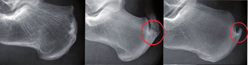
2                                                                                                BioMed Research International

    In the past, the development of bone spurs on both                 Since Haglund deformity was believed to have a rela-
sides of the calcaneus in patients with Achilles tendinop-         tionship with insertional tendinitis, the following various
athy and Haglund syndrome has been observed. Fiamengo              measurements were also made: the Fowler-Philip angle, as
et al. found that the rate of spurs on the posterior side of the   shown in Figure 2, the parallel pitch lines, as shown in
calcaneus in patients with Achilles tendinopathy reached           Figure 3, and the presence of a posterior calcaneal spur, as
44% and the rate of plantar spurs in the same population           shown in Figure 1 [17–19]. There was a flowchart to show
was 6.3% [13]. In a population with Haglund syndrome, the          how the study was conducted, as shown in Figure 4.
rate of posterior calcaneal spurs was 57% and the rate of              The chi-square test was applied to compare the mea-
plantar spurs was 16% [14]. Menz et al. found that 55% of          surements between the two groups, with the P value set at
patients had radiographic evidence of plantar spurs, among         0.05 for statistical significance.
whom 56% had an Achilles tendon spur [15]. To further
clarify the relationship between plantar spurs and Achilles        3. Results
tendinopathy, Vulcano et al. found that 41.9% of the 785
patients diagnosed with Achilles tendinopathy in outpa-            According to the inclusion and exclusion criteria, 43 patients
tient clinics had plantar spurs [16].                              with insertional Achilles tendonitis were enrolled in the
    Although the rates of bone spurs on the posterior and          study group, with 2 having bilateral Achilles tendonitis, so a
plantar sides of the calcaneus were observed in previous           total of 45 cases of insertional Achilles tendonitis were
studies, these studies did not explore the possibility of spurs    collected, as shown in Table 1. Of the 43 patients, 37 were
appeared simultaneously on both sides of calcaneal in the          men (86%), and 6 were women (14%), with an average age of
high-tension state of this biomechanically complex.                40.7 (range 15 to 69) years. There were 15 left heels (34.9%),
    The aim of this study was to demonstrate the biome-            26 right heels (60.5%) and 2 bilateral heels (4.7%).
chanical relationship between the Achilles tendon and the              There were 19 subjects with 20 heels in group A, which
plantar fascia by comparing the probability of developing          consisted of 14 men and 5 women, with an average age of
bilateral calcaneal spurs with that of developing unilateral       46.0 ± 12.4 years (range from 17 to 69 years old). There were
calcaneal spurs in the high tension state. So the author ret-      24 subjects with 25 heels in group B, which consisted of 23
rospectively reviewed patients with insertional Achilles ten-      men and 1 woman, with an average age of 36.9 ± 15.0 years
dinitis who received surgical treatment in the author’s hospital   (range from 15 to 65 years old).
and analyzed the bone structure data from standing lateral             In group A, calcaneal plantar spurs were present in
ankle radiographs.                                                 65.0% (13 out of 20) of the heels with calcaneal posterior
                                                                   spurs, while in group B, calcaneus plantar spurs were present
2. Method                                                          in 12.0% (3 out of 25), as shown in Figure 5. Calcaneal
                                                                   plantar spurs were found in 16 out of 45 heels, of which 13
The study was a retrospective radiographic review of patients      had posterior spurs (81.3%), as shown in Figure 6. However,
with insertional Achilles tendinitis who were admitted for         when the plantar spurs were negative (29 out of 45 heels),
operations at the orthopedic department in Huashan                 there were only 24.1% of heels (7 out of 29) presenting
Hospital of Fudan University (Shanghai, China) from March          posterior spurs, as shown in Table 2. There were significant
2009 to July 2018. The inclusion criteria for the study            differences between the two groups (P < 0.05). The rate of
consisted of patients with a diagnosis of insertional Achilles     spurs presenting on both sides of calcaneus was 28.9% (13
tendinitis. The diagnosis was made by the senior author            out of 45).
based on his clinical examination and radiographic review              In 45 cases of insertional Achilles tendonitis, the positive
and was reassured by surgical pathology. All patients had          rate of parallel pitch lines was 75.6% (34 out of 45). The mean
lateral view standing radiographs taken of the ankle.              Fowler-Philip angle was 51.39 ± 7.85 degrees. There was only
    Patients were excluded from the study group if they had        one heel in which the Fowler angle was more than 75 degrees
previous Achilles tendon surgery or had noninsertional             (88°).
Achilles tendinitis. The present study was approved by the
Institutional Review Board of our institutions, and informed       4. Discussion
consent was obtained from all participants.
    According to whether there were bone spurs on the              Pain in the heel includes pain on the plantar side of the
posterior side of the calcaneus, patients were divided into        calcaneus and posterior heel pain [9, 20, 21]. In the current
group A (spur was present on the posterior side) and               clinical work, it was anecdotally found that patients with
group B (spur was not present on the posterior side), as           plantar heel pain often presented with posterior heel pain
shown in Figure 1. Then, the author evaluated and                  [22]. Calcaneal spurs are outgrowths of bone into tendon
compared the occurrence of plantar calcaneal spurs be-             and ligamentous attachments, appearing mainly at two
tween groups A and B. All lateral radiographs were                 points: one at the posterior aspect of the calcaneus near the
reviewed on a picture archiving and communication                  insertion of the Achilles tendon and the other on the inferior
system by three orthopedic surgeons to identify calcaneal          aspect of the calcaneus, coinciding with the insertion of the
spurs. Patients were considered to have a bone spur only if        posterior fibers of the long plantar ligament.
it was grossly visible without magnification on the stan-               After analyzing imaging studies, the author concluded
dard lateral radiograph.                                           that there was a relationship between plantar calcaneal spurs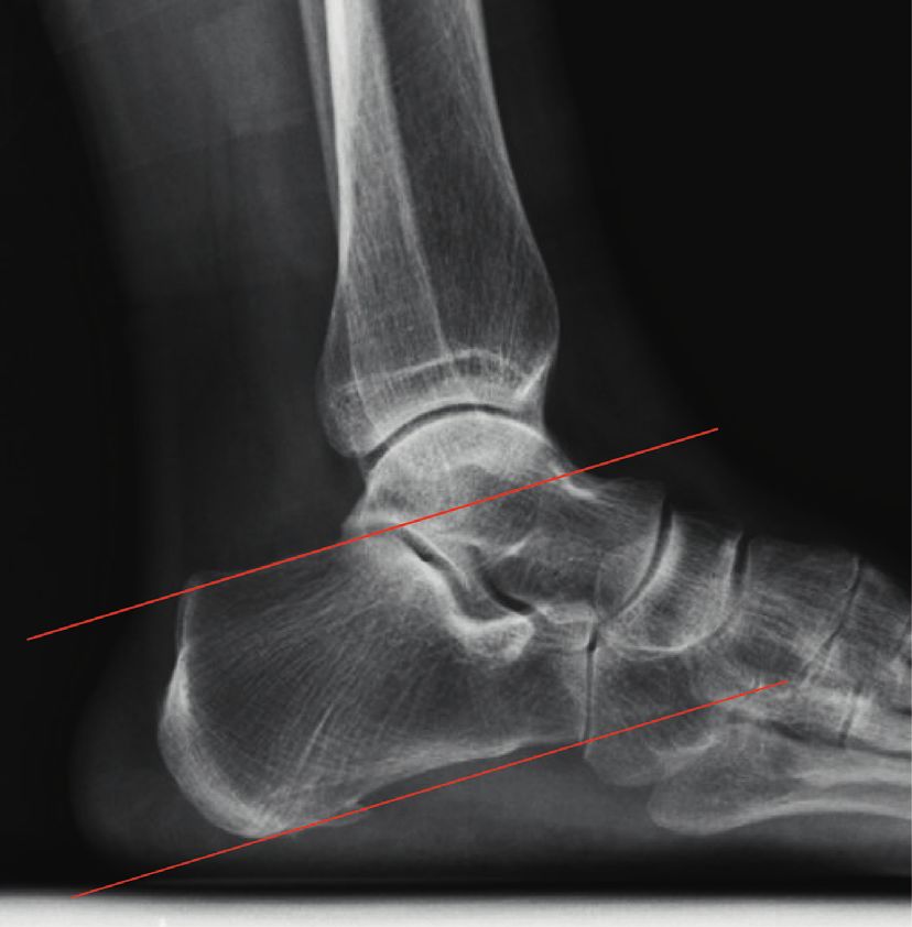
BioMed Research International 3
(a) (b) (c)
(d) (e) (f) (g)
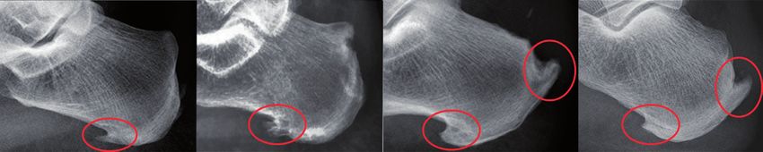
Figure 1: Examples of X-ray obtained in the study. (a) no plantar calcaneal or posterior calcaneal spur; (b, c): posterior calcaneal spurs only;
(d, e): plantar calcaneal spurs only; (f, g): spurs on bilateral sides of the calcaneus.
Figure 2: The Fowler–Philip angle was measured between an
inferior line which was tangent to the inferior margin of the cal-
caneocuboid joint and the plantar tuberosity of the calcaneus and a Figure 3: The Parallel patch lines were obtained by first drawing
superior line which was tangent to the posterior prominence at the the inferior line from the inferior margin of the calcaneocuboid
insertion of the Achilles tendon. Normal range is between 44 and 69 joint to the plantar tuberosity of the calcaneus. Then the superior
degrees, measurements greater than or equal to 75 degrees were line was drawn parallel to the inferior line beginning at the pos-
thought to be consistent with Haglund’s deformity. terior margin of the subtalar joint. If the posterior calcaneal
prominence was located above the superior line, it was considered
and posterior calcaneal spurs in heels with insertional abnormal and consistent with Haglund’s deformity.
Achilles tendinitis. With regard to plantar calcaneal spurs,
group A (posterior spurs were positive) was 5 times more probability of developing spurs on both sides of calcaneus
likely than group B (posterior spurs were negative) to have was much greater than that of developing unilateral calca-
spurs (65% compared to 12%, P < 0.05), as shown in Fig- neal spurs. Moreover, an anatomical connection between the
ure 5. Since the mechanical stimulation of plantar calcaneal Achilles tendon and the plantar fascia makes the relation-
spurs can lead to plantar fasciitis, which can cause patients to ships among the plantar fascia, calcaneus, and Achilles
develop heel pain [21], so we thought it was worthwhile for tendon biomechanically complex, so we assumed this
podiatrists to pay attention to calcaneal plantar spurs and the structure may transfer stress from one side of calcaneus to
high tension state of the plantar fascia in patients with in- another. Some studies also found that Achilles tendinopathy
sertional Achilles tendinitis in the clinical setting. In patients and heel pain can collectively benefit from similar conser-
with calcaneal plantar spurs, the positive rate of posterior vative and surgical treatments by reducing the tension on
calcaneal spurs reached 81.3%, which was far greater than both sides, such as eccentric calf stretching and gastroc-
that in patients without plantar calcaneal spurs (24.1%), as nemius recession [2, 23, 24]. Therefore, the Achilles tendon,
shown in Figure 6. These results indicated that the calcaneus, and plantar fascia should be considered as a whole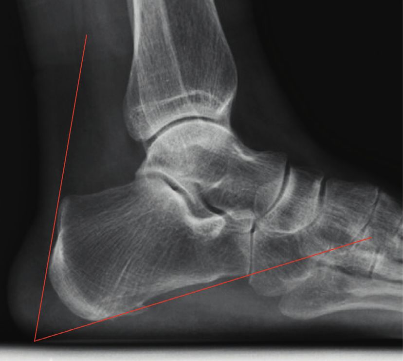
4                                                                                                                                           BioMed Research International

                                                                   Patients with insertional Achilles tendinitis

                                                                                                                              Previous Achilles tendon
                                                                                                                              surgery or noninsertional
                                                                                  Lateral view                                    Achilles tendinitis
                                                                          standing radiographs of ankle

                                                                                                                                               Excluded from
                                                                                                                                                study group
                                                 Measurements                      Study group

                                                                   Spur: present on         Spur: not present on
                            Fowler–Philip angle,
                                                                    posterior side             posterior side
                             parallel pitch lines

                                                             Group A                                                      Group B

                                                           Measurements                                                 Measurements

                                                     Presence of posterior                                          Presence of posterior
                                                        calcaneal spur                                                 calcaneal spur

                                                                                      Compare

                                                                                   Conclusion

                                                 Figure 4: A flowchart to show how the study was conducted.

                                                           Table 1: Patient profiles in group A and group B.
                                                      Patient numbers                                                                           Heel numbers
Patient group
                                Male           Female       Total     Average age (years)                                      Left         Right      Bilateral         Total
Group A                          14              5           19            46 ± 12.4                                             5           14            1              20
Group B                          23              1           24           36.9 ± 15.0                                           10           12            1              23
Total                            37              6           43           40.9 ± 14.5                                           15           26            2              43

                     20
                                                                                                                   20
                            13 (65.0%)
                     10                                                                                                        13 (81.3%)
                                                                                                                   10
                                                            3 (12.0%)                                                                                       7 (24.1%)
     Plantar spurs

                      0
                                                                                                 Posterior spurs

                                                                                                                    0

                             7 (35.0%)                                                                                          3 (18.7%)
                     10
                                                                                                                   10

                     20
                                                           22 (88.0%)                                              20
                                                                                                                                                            22 (75.9%)
                     30
                              Positive                      Negative                                               30
                                         Posterior spurs                                                                         Positive                   Negative
                                                                                                                                            Plantar spurs
                          Positive
                          Negative                                                                                          Positive
                                                                                                                            Negative
Figure 5: Plantar spurs were present in 65.0% (13 out of 20) of the
heels with calcaneal posterior spurs, while there were only 12.0% of                      Figure 6: There were 16 out of 45 heels presenting plantar spurs, of
heels presenting plantar spurs in the posterior spur negative group.                      which 13 had posterior spurs (81.3%). However, when the plantar
Plantar spurs and posterior spurs were inclined to appear at the                          spurs were negative, there were only 24.1% of heels presenting
same time (65.0%) or would not appear at all (88.0%).                                     posterior spurs.
BioMed Research International 5
Table 2: The presence of calcaneal plantar spurs in group A and diseases were not directly investigated in this study. For this
group B. Positive means that calcaneal plantar spur is observed on reason, future research can directly target the correlation
the radiograph; negative means that calcaneal plantar spur is not between the two diseases with regard to pathogenesis,
observed on the radiograph. treatment, and prognosis. Last, the study group only en-
Positive Negative Total rolled 43 patients which was a relatively small sample size.
Group A 13 7 20 Furthermore, there was only 1 female patient in group B, and
Group B 3 22 25 there were 5 female patients in group B. As far as we
Total 16 29 45 concerned, the main reason was because there were only 24
patients enrolled in group B and 19 patients in group A, so it
was the small number of patients that caused sampling error.
entity, and the pain on both sides of calcaneus must be
treated simultaneously. Therefore, the other side should 5. Conclusion
receive equal attention when podiatrists encounter pain on
either side of calcaneus in clinical work because of this First, through weight-bearing lateral ankle X-rays, a rela-
biomechanical complexity. tionship between posterior calcaneus spurs and plantar
The Fowler–Philip angle was proposed by Fowler and calcaneus spurs in insertional Achilles tendinitis patients
Philip in 1945, and the normal range is 44–69° [18]. It is was found, and a biomechanical complex involving the
believed when the angle is greater than 75°, the Haglund Achilles tendon, calcaneus, and plantar fascia was deduced.
deformity is large enough to cause swelling of the soft tissue It was suggested that insertional Achilles tendonitis and
at the end of the Achilles tendon, insertional Achilles ten- plantar fasciitis should be considered to be related and
donitis, and other diseases. However, in this survey, the treated synthetically in clinical practice. Second, the positive
author found that patients with severe insertional Achilles rate of the parallel pitch lines was higher in insertional
tendonitis whose formal conservative treatment had failed, Achilles tendonitis patients, and the Fowler–Philip angle was
the average Fowler–Philip angle was 51.39°, and only 1 not predictive of insertional Achilles tendonitis.
patient had an angle greater than 70°. This result was in-
consistent with Fowler and Philip’s theory. Fiamengo et al. Data Availability
also found no difference in the value of the Fowler–Philip
angle between the insertional Achilles tendinitis population The datasets generated during and/or analyzed during the
and a normal population [13, 14, 19]. Therefore, the author current study are available from the corresponding author
does not believe that there is a strong correlation between the on reasonable request.
Fowler-Philip angle and the occurrence of insertional
Achilles tendinitis. Conflicts of Interest
Furthermore, the author found that the positive rate of
the parallel pitch line reached 75.6% in the severe cases of The authors have no conflicts of interest to report.
insertional Achilles tendonitis requiring surgical treatment,
but other studies concluded that the false positive rate of Authors’ Contributions
parallel pitch lines in insertional Achilles tendonitis cases
Genrui Zhu, Zhifeng Wang, and Chengjie Yuan contributed
ranged from 15% to 56.8% [14, 25, 26]. The author asserts
equally to this work.
that the heterogeneity of the study populations with regard
to the severity of insertional Achilles tendonitis made it
infeasible to compare the results of the present study with Acknowledgments
previous studies. As far as we concerned, different positive This work was funded by the National Natural Science
rates of parallel pitch lines might be related to different Foundation of China (Nos. 81572176 and 8177090247)
severities of insertional Achilles tendinitis, but this concept
needs further study.
The limitations of this study are as follows: first, in this
References
study, the author only studied the correlation between spurs [1] K. Merry, M. MacPherson, E. Macdonald, M. Ryan, E. J. Park,
on two sides of the calcaneus in insertional Achilles ten- and C. Sparrey, “Differentiating sitting, standing and walking
donitis patients who received surgical treatment, and there through regional plantar pressure characteristics,” Journal of
was no case of insertional Achilles tendonitis that could be Biomechanical Engineering, vol. 142, no. 4, 2019.
alleviated by conservative treatment included as a control [2] S. Prichasuk and T. Subhadrabandhu, “The relationship of pes
group. Therefore, future studies need to explore the cor- planus and calcaneal spur to plantar heel pain,” Clinical
relation between spurs on the two sides of the calcaneus in Orthopaedics and Related Research, no. 306, pp. 192–196,
1994.
patients with less severe Achilles tendinitis who only need
[3] E. Schwartz and J. Su, “Plantar fasciitis: a concise review,” The
conservative treatment. Second, the purpose of the study was Permanente Journal, vol. 18, no. 1, pp. e105–e107, 2014.
to deduce the biomechanically complex relationships among [4] E. R. Waclawski, J. Beach, A. Milne, E. Yacyshyn, and
the Achilles tendon, calcaneus, and plantar fascia via radi- D. M. Dryden, “Systematic review: plantar fasciitis and
ography; the results suggest the necessity of jointly con- prolonged weight bearing,” Occupational Medicine, vol. 65,
sidering these two diseases in clinical practice, but the two no. 2, pp. 97–106, 2015.6 BioMed Research International
[5] J. Sullivan, E. Pappas, and J. Burns, “Role of mechanical tendinopathy,” Foot & Ankle International, vol. 38, no. 10,
factors in the clinical presentation of plantar heel pain: im- pp. 1160–1169, 2017.
plications for management,” The Foot, vol. 42, Article ID [23] P. Engkananuwat, R. Kanlayanaphotporn, and N. Purepong,
101636, 2019. “Effectiveness of the simultaneous stretching of the Achilles
[6] S. C. Wearing, J. E. Smeathers, S. R. Urry, E. M. Hennig, and tendon and plantar fascia in individuals with plantar fasciitis,”
A. P. Hills, “The pathomechanics of plantar fasciitis,” Sports Foot & Ankle International, vol. 39, no. 1, pp. 75–82, 2018.
Medicine, vol. 36, no. 7, pp. 585–611, 2006. [24] K. S. Smith, C. Jones, Z. Pinter, and A. Shah, “Isolated
[7] W. K. Smith, J. A. Noriega, and W. K. Smith, “Resection of a gastrocnemius recession for the treatment of Achilles
plantar calcaneal spur using the holmium:yttrium-aluminum- tendinopathy,” Foot & Ankle Specialist, vol. 11, no. 1,
garnet (Ho:YAG) laser,” Journal of the American Podiatric pp. 49–53, 2018.
Medical Association, vol. 91, no. 3, pp. 142–146, 2001. [25] D. Chauveaux, P. Liet, J. C. Le Huec, and D. Midy, “A new
[8] A. Barg and T. Ludwig, “Surgical strategies for the treatment radiologic measurement for the diagnosis of Haglund’s de-
of insertional Achilles tendinopathy,” Foot and Ankle Clinics, formity,” Surgical and Radiologic Anatomy, vol. 13, no. 1,
vol. 24, no. 3, pp. 533–559, 2019. pp. 39–44, 1991.
[9] P. Tu, “Heel pain: diagnosis and management,” American [26] M. A. Heneghan and H. Pavlov, “The Haglund painful heel
Family Physician, vol. 97, no. 2, pp. 86–93, 2018. syndrome. Experimental investigation of cause and thera-
[10] G. M. Caudell, “Insertional Achilles tendinopathy,” Clinics in peutic implications,” Clinical Orthopaedics and Related Re-
Podiatric Medicine and Surgery, vol. 34, no. 2, pp. 195–205, search, no. 187, pp. 228–234, 1984.
2017.
[11] H. Yesil, U. Dundar, H. Toktas, N. Eyvaz, and M. Yesil, “The
effect of high intensity laser therapy in the management of
painful calcaneal spur: a double blind, placebo-controlled
study,” Lasers in Medical Science, 2019.
[12] D. B. Wibowo, R. Harahap, A. Widodo, G. D. Haryadi, and
M. Ariyanto, “The effectiveness of raising the heel height of
shoes to reduce heel pain in patients with calcaneal spurs,”
Journal of Physical Therapy Science, vol. 29, no. 12,
pp. 2068–2074, 2017.
[13] S. A. Fiamengo, R. F. Warren, J. L. Marshall, V. T. Vigorita,
and A. Hersh, “Posterior heel pain associated with a calcaneal
step and Achilles tendon calcification,” Clinical Orthopaedics
and Related Research, vol. 167, pp. 203–211, 1982.
[14] C. C. Lu, Y. M. Cheng, Y. C. Fu, Y. C. Tien, S. K. Chen, and
P. J. Huang, “Angle analysis of Haglund syndrome and its
relationship with osseous variations and Achilles tendon
calcification,” Foot and Ankle International, vol. 28, no. 2,
pp. 181–185, 2007.
[15] H. B. Menz, G. V. Zammit, K. B. Landorf, and S. E. Munteanu,
“Plantar calcaneal spurs in older people: longitudinal traction
or vertical compression?” Journal of Foot and Ankle Research,
vol. 1, no. 1, p. 7, 2008.
[16] E. Vulcano, S. B. Mani, H. Do, W. H. Bohne, and S. J. Ellis,
“Association of Achilles tendinopathy and plantar spurs,”
Orthopedics, vol. 37, no. 10, pp. e897–e901, 2014.
[17] M. Bassiouni, “Incidence of calcaneal spurs in osteo-arthrosis
and rheumatoid arthritis, and in control patients,” Annals of
the Rheumatic Diseases, vol. 24, no. 5, pp. 490–493, 1965.
[18] A. Fowler and J. Philip, “Abnormality of the calcaneus as a
cause of painful heel: its diagnosis and operative treatment,”
British Journal of Surgery, vol. 32, no. 128, pp. 494–498, 1945.
[19] H. Pavlov, M. Heneghan, A. Hersh, A. Goldman, and
V. Vigorita, “The Haglund’s syndrome: initial and differential
diagnosis,” Radiology, vol. 144, pp. 83–88, 1982.
[20] D. Schuitema, C. Greve, K. Postema, R. Dekker, and
J. M. Hijmans, “Effectiveness of mechanical treatment for
plantar fasciitis: a systematic review,” Journal of Sport Re-
habilitation, pp. 1–18, 2019.
[21] S. Christie, G. J. Styn, G. Ford, and K. Terryberry, “Proximal
plantar intrinsic tendinopathy: anatomical and biomechanical
considerations in plantar heel pain,” Journal of the American
Podiatric Medical Association, vol. 109, no. 5, pp. 412–415,
2019.
[22] R. L. Chimenti, C. C. Cychosz, M. M. Hall, and P. Phisitkul,
“Current concepts review update: insertional AchillesYou can also read


