Abstract The Novel Use of an External Oblique Nerve Catheter After Open Cholecystectomy - Cureus
←
→
Page content transcription
If your browser does not render page correctly, please read the page content below
Open Access Case
                              Report                                               DOI: 10.7759/cureus.13580

                                             The Novel Use of an External Oblique Nerve
                                             Catheter After Open Cholecystectomy
                                             Brendan O'Donovan 1 , Brian Martin 1

                                             1. Anesthesiology, University of Massachusetts Medical School-Baystate, Springfield, USA

                                             Corresponding author: Brendan O'Donovan, brendan.o'donovan@baystatehealth.org

                                             Abstract
                                             Open cholecystectomy is a painful procedure and requires a well-rounded multimodal approach for
                                             successful postoperative analgesia. A thoracic epidural is known to provide reliable pain relief for upper
                                             abdominal surgical procedures. However, in patients for whom an epidural is contraindicated, an alternative
                                             regional technique may be sought. This case discusses the novel use of an external oblique catheter after
                                             open cholecystectomy.

                                             Categories: Anesthesiology, Pain Management, General Surgery
                                             Keywords: open cholecystectomy, external oblique nerve block, tap block, abdominal wall innervation, external
                                             oblique nerve catheter, quadratus lumborum block

                                             Introduction
                                             Open cholecystectomy is a potentially painful operation. A thoracic epidural is known to provide reliable
                                             pain relief for upper abdominal surgery [1]. When an epidural placement is contraindicated, however, pain
                                             management can be difficult. The Kocher incision is a subcostal incision used for open cholecystectomy [2].
                                             This incision encroaches on the T6-T9 dermatomes on the right subcostal abdominal region [3].

                                             A clear understanding of the innervation of the abdominal wall is necessary to utilize regional anesthesia as
                                             part of a multimodal analgesic approach for post-surgical patients. The abdominal wall is supplied by the
                                             intercostal nerves (T6-T11), the subcostal nerve (T12), and the ilioinguinal/iliohypogastric nerves (L1) [4].
                                             These nerves are derived from their respective anterior rami of the T6-L1 spinal nerves. Around the
                                             midaxillary line, the lateral cutaneous branch leaves its respective intercostal nerve and travels superficially,
                                             piercing the external intercostal muscle and/or external oblique muscle to supply sensation to the lateral
                                             abdominal wall. The lateral cutaneous nerve divides into anterior and posterior branches [5]. The anterior
                                             branch extends anteriorly as far as the rectus abdominis margin, and the posterior branch extends
                                             posteriorly to supply the skin overlying the latissimus dorsi. The intercostal nerve continues its course in the
                                             transversus abdominis plane (TAP) until it reaches the rectus abdominis muscle and divides into the anterior
                                             cutaneous branch, which supplies the skin of the midline abdomen.

Review began 12/30/2020                      The traditional lateral TAP block is performed at the midpoint between the iliac crest and the subcostal
Review ended 02/21/2021
                                             margin in the midaxillary line [4]. Cadaveric injection of dye using this approach showed involvement of T11
Published 02/26/2021
                                             and T12 100% of the time, L1 93% of the time, and T10 50% of the time [6]. A lateral TAP block may not
© Copyright 2021                             reach a dermatome high enough to benefit a patient after an open cholecystectomy.
O'Donovan et al. This is an open access
article distributed under the terms of the
Creative Commons Attribution License         The oblique subcostal TAP block is performed on the anterior abdominal wall parallel to the subcostal
CC-BY 4.0., which permits unrestricted       margin with a needle entry point near the xiphoid process [4]. Chen et al. studied the extent of analgesia
use, distribution, and reproduction in any
                                             after the oblique subcostal TAP block [7]. The study showed dermatomal coverage of T7-T12 100% of the
medium, provided the original author and
                                             time, T6 75% of the time, and L1 50% of the time. The study also shed light on the fact that the oblique
source are credited.
                                             subcostal TAP block primarily anesthetizes the mid-abdomen; however, it leaves the lateral abdomen
                                             unblocked. The subcostal TAP block would be preferred over the lateral TAP block for open cholecystectomy,
                                             however, it may not completely cover the surgical site due to the lack of lateral abdominal wall coverage.

                                             The quadratus lumborum block (QLB) is performed proximal to the branching of the lateral cutaneous nerve
                                             and near the quadratus lumborum muscle, which is closer to the anterior rami of the spinal nerves compared
                                             to the TAP block. The quadratus lumborum is encased by the thoracolumbar fascia (TLF), and local
                                             anesthetic injection into the TLF is thought to be responsible for the craniocaudal spread as well as spread to
                                             the paravertebral space [8]. The TLF contains mechanoreceptors and sympathetic fibers. Blockade of these
                                             sympathetic fibers and spread to the paravertebral space are thought to contribute to the visceral analgesia
                                             achieved with the QLB. Comparatively, the TAP block solely causes somatic analgesia and does not result in
                                             visceral analgesia [9]. The current literature describes four different variants of the QLB: QL1/lateral,
                                             QL2/posterior, QL3/anterior, and QL4/intramuscular. The QLB provides reliable dermatomal coverage of T7-
                                             L1, however, there are reports of spread as high as T4 and as low as L3 [10]. Perhaps, the QLB would be a
                                             preferred technique compared to both the lateral and subcostal TAP blocks for analgesia after an open
                                             cholecystectomy due to its extent of dermatomal coverage, the addition of visceral analgesia, and its ability

                              How to cite this article
                              O'Donovan B, Martin B (February 26, 2021) The Novel Use of an External Oblique Nerve Catheter After Open Cholecystectomy. Cureus 13(2):
                              e13580. DOI 10.7759/cureus.13580
to provide both anterior and lateral abdominal wall coverage.
Although there have been no studies comparing the TAP block versus the QLB for open cholecystectomy,
two recent studies have compared the techniques for laparoscopic cholecystectomy. Baytar et al. compared
the effectiveness of the subcostal TAP block and the posterior QLB for postoperative analgesia after
laparoscopic cholecystectomy [11]. The study concluded that both techniques produced similar reductions in
postoperative pain scores and opioid consumption. The study stated that the performance of the QLB took
eight minutes longer than the subcostal TAP block and that they recommend the subcostal TAP block due to
easier application of the block. Weheba et al. produced a similar study with different results [12]. Like the
Baytar study, this trial compared the efficacy of the subcostal TAP block and the posterior QLB in providing
postoperative analgesia after laparoscopic cholecystectomy. Although the Weheba study showed similar
postoperative pain scores between the two groups, the QLB group had a significantly longer time to the first
postoperative request for rescue analgesia and less number of patients required postoperative opioids
compared to the TAP group.
Case Presentation
A 78-year-old male with a medical history of hypertension, diabetes, gout, hyperlipidemia, chronic kidney
disease stage 2, and deep vein thrombosis (on Warfarin therapy) presented to an urgent care facility with
signs of weakness, “dark-urine,” and jaundice. In the emergency department, he was found to be in new-
onset atrial fibrillation. His initial labs were significant for leukocytosis, transaminitis, and
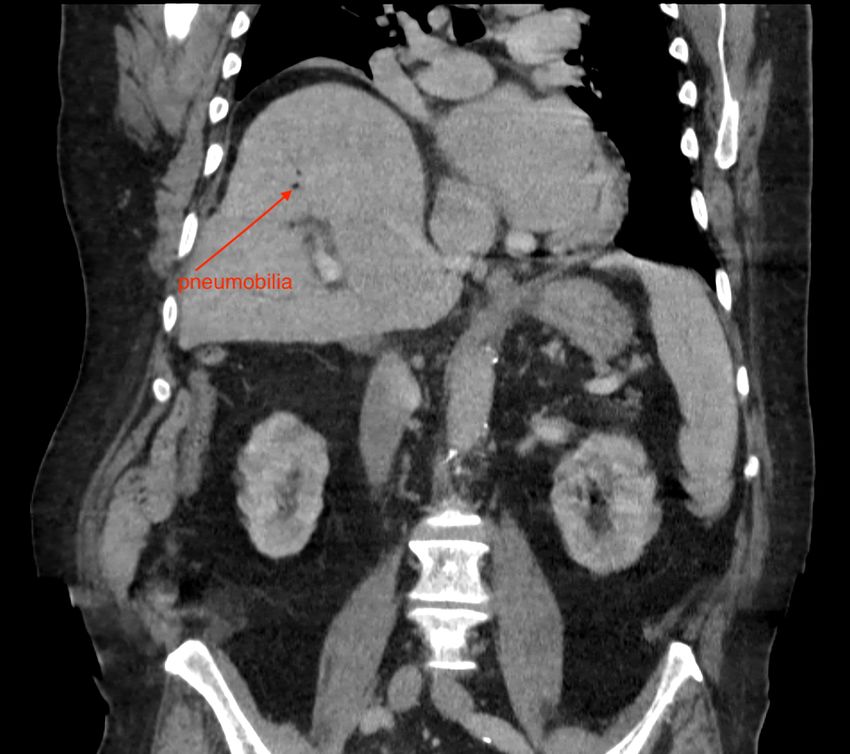
hyperbilirubinemia. An abdominal CT showed a common bile duct stone, pneumobilia, and signs of
ascending cholangitis (Figures 1, 2). He was admitted to the medicine service, started on broad-spectrum
antibiotics, and gastroenterology was consulted to perform an endoscopic retrograde
cholangiopancreatography (ERCP). Due to the patient’s new-onset atrial fibrillation, echocardiography was
performed, showing an ejection fraction of 25-35%. He was placed on a heparin infusion due to new-onset
atrial fibrillation and a history of deep venous thrombosis. An ERCP was performed on hospital day 2;
however, it was unsuccessful at removing a common bile duct gallstone. The diagnosis of ascending
cholangitis was made by visualization of a 2 cm common bile duct stone and pus draining from the papilla.
FIGURE 1: Coronal CT image showing pneumobilia
2021 O'Donovan et al. Cureus 13(2): e13580. DOI 10.7759/cureus.13580 2 of 5FIGURE 2: Coronal CT image showing a common bile duct stone
The patient was taken to the operating room on hospital day 5 for a laparoscopic cholecystectomy. The
surgical team converted to an open procedure by performing a right upper quadrant Kocher due to aberrant
anatomy and extensive adhesions. They were unable to identify the gallbladder. The common bile duct stone
was removed surgically, and a biliary T-tube was placed. As the surgical team was closing during the
operation, they inquired about the placement of a nerve catheter as an epidural was contraindicated due to
the patient’s need for anticoagulation for new-onset atrial fibrillation. At this point, the incision was mostly
closed; however, the external oblique was still exposed. The surgical team placed a B Braun Perifix®
catheter on the surface of the external oblique muscle in the right upper quadrant at the T8 dermatome. The
acute pain anesthesiology service managed the nerve catheter. The patient was extubated in the operating
room and was taken to the surgical intensive care unit for close monitoring.
The acute pain service saw the patient on postoperative day 1. His pain was managed with a bupivacaine
0.1% infusion at 7cc/hr going via the nerve catheter. He was found to have 0/10 pain at rest. He attributed
3/10 pain only when his incision site was palpated and during movement. The acute pain service followed
the patient until the catheter was removed on postoperative day 5. Each day the patient had virtually zero
pain at rest and attributed only mild pain when his incision site was palpated or during movement. The
patient’s postoperative opioid requirement was remarkably low as well. He received one dose of oxycodone
5mg on postoperative days 1 and 2, respectively. He received one dose of acetaminophen 650mg on
postoperative days 1 and 4. He received no other analgesic medication in his acute postoperative course.
Discussion
The use of an external oblique nerve catheter for postoperative analgesia has not been described yet in the
literature. Due to the low pain scores achieved and minimal narcotic use postoperatively, we hypothesize
that we successfully anesthetized the lateral cutaneous branch and the anterior cutaneous branch of the
thoracoabdominal nerves. Recently, there have been several publications describing external oblique nerve
blocks to control post-operative pain after abdominal surgery.
In 2018 Hamilton et al. proposed that injecting a local anesthetic into a thoracic fascial plane can block the
lateral cutaneous branches from T7-T11 providing adequate analgesia for lateral abdominal surgery [13].
Hamilton et al. solidified their hypothesis by a cadaver study that looked at the spread of dye after an
external oblique fascial plane block [14]. The block was performed at the sixth intercostal space in the
midclavicular line, and the needle tip was either deep or superficial to the external oblique muscle. The
study showed that injection of 20cc of dye effectively covered the anterior divisions of the lateral cutaneous
branches of the thoracoabdominal nerves from T6-T10.
In 2019, Tulgar et al. described a novel nerve block for upper abdominal wall analgesia, which they named a
TAPA block (blockage of thoracoabdominal nerves through perichondral approach) [15]. This block is
performed at the costal margin where the 9th and 10th ribs meet. A linear transducer is placed on the
costochondral angle in the sagittal plane. The authors injected 20cc of injectate between the chondrium’s
upper surface and the external oblique and 20cc of injectate between the chondrium’s lower surface and the
2021 O'Donovan et al. Cureus 13(2): e13580. DOI 10.7759/cureus.13580 3 of 5transversus abdominis muscle [16]. The authors state that the TAPA block provides analgesia from T5-T12.
They hypothesize that the TAPA block anesthetizes the lateral cutaneous branch as well as the anterior
cutaneous branch providing analgesia from the anterior axillary line to mid-abdomen from T7-T12. They
believe the block only reaches the lateral cutaneous branch of T5-T6 as these patients had pinprick
sensation 4-5cm lateral to the sternum.
We believe our nerve catheter provided excellent postoperative analgesia based on a similar principle that
Hamilton and Tulgar have recently discovered. As our nerve catheter was placed superficial to the external
oblique muscle, we believe we anesthetized the lateral cutaneous branch as well as the anterior cutaneous
branch of the thoracoabdominal nerves from T6-T9. We hypothesized that as the lateral cutaneous branch
lies above the external oblique muscle, it could be effectively blocked by a nerve catheter. Furthermore, as
the patient had no midline pain postoperatively, we believe we anesthetized the anterior cutaneous branch
as well. We presume the local anesthetic traveled in the subcutaneous tissue toward the midline blocking the
anterior cutaneous branch overlying the rectus abdominis muscle.
Although, in our case, the nerve catheter was placed under direct surgical visualization, we believe it can be
successfully placed under ultrasound guidance. Perhaps this technique would be preferred in the morbidly
obese population as the ultrasound target is more superficial and accessible compared to a TAP block or a
QLB. The QLB would be an effective technique for postoperative analgesia following an open
cholecystectomy in the majority of patients; however, it is considered a deep regional block, and its
utilization is controversial in patients on anticoagulation [8]. There is a case report describing two
incidences of hematoma formation following QLB, likely due to inadvertent lumbar artery puncture [17]. In
addition, the QLB is considered an advanced regional technique that may be difficult to perform in
inexperienced hands, whereas the superficial nature of the external oblique nerve catheter makes it easier to
perform.
Conclusions
Pain control following an open cholecystectomy can be difficult to manage, especially when an epidural is
contraindicated. A feasible option for pain control may be a subcostal external oblique catheter. We
hypothesize we were able to anesthetize the lateral cutaneous branch as well as the anterior cutaneous
branch of the thoracoabdominal nerves from T6-T9. Future studies are required to determine the
effectiveness of external oblique nerve catheters.
Additional Information
Disclosures
Human subjects: Consent was obtained or waived by all participants in this study. Conflicts of interest: In
compliance with the ICMJE uniform disclosure form, all authors declare the following: Payment/services
info: All authors have declared that no financial support was received from any organization for the
submitted work. Financial relationships: All authors have declared that they have no financial
relationships at present or within the previous three years with any organizations that might have an
interest in the submitted work. Other relationships: All authors have declared that there are no other
relationships or activities that could appear to have influenced the submitted work.
References
1. Manion SC, Brennan TJ, Riou B: Thoracic epidural analgesia and acute pain management . Anesthesiology.
2011, 115:181-188.
2. Jones MW, Deppen JG: Open cholecystectomy. StatPearls [Internet]. StatPearls Publishing, Treasure Island
(FL); 2021.
3. Fischer HB, Pinnock CA: Peripheral nerve blocks. Fundamentals of Regional Anaesthesia. Cambridge
University Press, 2004. 74.
4. Statzer N, Cummings KC 3rd: Transversus abdominis plane blocks. Adv Anesth. 2018, 36:163-180.
10.1016/j.aan.2018.07.007
5. Court C, Vialle R, Lepeintre JF, Tadié M: The thoracoabdominal intercostal nerves: an anatomical study for
their use in neurotization. Surg Radiol Anat. 2005, 27:8-14. 10.1007/s00276-004-0281-8
6. Tran TM, Ivanusic JJ, Hebbard P, Barrington MJ: Determination of spread of injectate after ultrasound-
guided transversus abdominis plane block: a cadaveric study. Br J Anaesth. 2009, 102:123-127.
10.1093/bja/aen344
7. Chen Y, Shi K, Xia Y, Zhang X, Papadimos TJ, Xu X, Wang Q: Sensory assessment and regression rate of
bilateral oblique subcostal transversus abdominis plane block in volunteers. Reg Anesth Pain Med. 2018,
43:174-179. 10.1097/AAP.0000000000000715
8. Dhanjal S, Tonder S: Quadratus lumborum block. StatPearls [Internet]. StatPearls Publishing, Treasure
Island (FL); 2020.
9. Ultrasound-Guided Transversus Abdominis Plane and Quadratus Lumborum Nerve Blocks . (2021).
Accessed: February 21, 2021: https://www.nysora.com/regional-anesthesia-for-specific-surgical-
procedures/abdomen/ultrasound-guided-transversus-abd....
10. Akerman M, Pejcic N, Velickovic I: A review of the quadratus lumborum block and ERAS . Front Med. 2018,
5:44. 10.3389/fmed.2018.00044
2021 O'Donovan et al. Cureus 13(2): e13580. DOI 10.7759/cureus.13580 4 of 511. Baytar C, Yilmaz C, Karasu D, Topal S: Comparison of ultrasound-guided subcostal transversus abdominis
plane block and quadratus lumborum block in laparoscopic cholecystectomy: a prospective, randomized,
controlled clinical study. Pain Res Manag. 2019, 2019:2815301. 10.1155/2019/2815301
12. Weheba H, Abdelsalam T, Ghareeb S, Makharita MY: Posterior quadratus lumborum block versus subcostal
transversus abdominis plane block in laparoscopic cholecystectomy. Int J Anesth Anesth. 2019, 6:093.
10.23937/2377-4630/1410093
13. Hamilton DL, Manickam BP: Is a thoracic fascial plane block the answer to upper abdominal wall analgesia? .
Reg Anesth Pain Med. 2018, 43:891-892. 10.1097/AAP.0000000000000838
14. Hamilton DL, Manickam BP, Wilson MA, Abdel Meguid E: External oblique fascial plane block. Reg Anesth
Pain Med. 2019, 44:528-529. 10.1136/rapm-2018-100256
15. Tulgar S, Senturk O, Selvi O, Balaban O, Ahiskalioğlu A, Thomas DT, Ozer Z: Perichondral approach for
blockage of thoracoabdominal nerves: anatomical basis and clinical experience in three cases. J Clin Anesth.
2019, 54:8-10. 10.1016/j.jclinane.2018.10.015
16. Tulgar S, Ahiskalioglu A, Selvi O, Thomas DT, Ozer Z: Similarities between external oblique fascial plane
block and blockage of thoracoabdominal nerves through perichondral approach (TAPA). J Clin Anesth. 2019,
57:91-92. 10.1016/j.jclinane.2019.03.027
17. Visoiu M, Pan S: Quadratus lumborum blocks: two cases of associated hematoma . Pediatr Anesth. 2019,
29:286-288. 10.1111/pan.13588
2021 O'Donovan et al. Cureus 13(2): e13580. DOI 10.7759/cureus.13580 5 of 5You can also read