Atorvastatin-induced Lichenoid Drug Eruption: A Case Report and Review of Statin-associated Cutaneous Adverse Events - Cureus
←
→
Page content transcription
If your browser does not render page correctly, please read the page content below
Open Access Case
                                          Report                                                 DOI: 10.7759/cureus.7155

                                          Atorvastatin-induced Lichenoid Drug
                                          Eruption: A Case Report and Review of
                                          Statin-associated Cutaneous Adverse
                                          Events
                                          Parnia Forouzan 1 , Ryan R. Riahi 2 , Philip R. Cohen 3

                                          1. Dermatology, University of Texas Medical School, Houston, USA 2. Dermatology, DermSurgery
                                          Associates, Sugar Land, USA 3. Dermatology, San Diego Family Dermatology, San Diego, USA

                                          Corresponding author: Parnia Forouzan, forouzanparnia@gmail.com

                                          Abstract
                                          Statin medications [3-hydroxy-3-methylglutaryl coenzyme A (HMG-CoA) reductase inhibitors]
                                          are generally used to treat hypercholesterolemia. Lichenoid drug eruptions are a potential
                                          cutaneous side effect of medications including antibiotics, antimalarials, and statins. This drug
                                          eruption can mimic features of idiopathic lichen planus in clinical presentation and pathology.
                                          We describe the case of a 73-year-old man who developed a lichenoid drug eruption secondary
                                          to atorvastatin. His clinical features, in addition to histological findings, helped to establish the
                                          diagnosis. The cutaneous eruption resolved one month after the cessation of atorvastatin and
                                          with corticosteroid therapy. Statins have been associated with adverse events including bullous
                                          dermatosis, eosinophilic fasciitis, lichenoid drug eruption, and phototoxicity. Lichenoid drug
                                          eruption associated with statin therapy requires discontinuation of the statin medication; an
                                          alternative class of medication for the treatment of hypercholesterolemia is usually necessary.

                                          Categories: Dermatology
                                          Keywords: adverse, atorvastatin, cutaneous, drug, lichen, lichenoid, eruption, planus, skin, statin

                                          Introduction
                                          Atorvastatin, a 3-hydroxy-3-methylglutaryl coenzyme A (HMG-CoA) reductase inhibitor, is
                                          commonly used to manage hypercholesterolemia. Atorvastatin usually prevents the production
                                          of cholesterol and other sterol products, including corticosteroids, vitamin D, and sex steroids,
                                          in the mevalonate pathway. However, statins can have a diverse array of effects beyond
                                          lowering the risk of cardiovascular disease [1]. Statins have been associated with various
                                          adverse cutaneous side effects including alopecia, bullous dermatosis, and lichenoid drug
Received 02/17/2020                       eruptions [1-18]. Lichenoid drug eruptions clinically mimic idiopathic lichen planus [19].
Review began 02/21/2020
Review ended 02/24/2020
Published 03/01/2020                      We report the case of a man with atorvastatin-induced lichenoid drug eruption. In addition, we
                                          describe the clinical and histopathologic characteristics of idiopathic lichen planus and
© Copyright 2020
                                          lichenoid drug eruptions as well as cutaneous adverse reactions observed with statin
Forouzan et al. This is an open
access article distributed under the      medications.
terms of the Creative Commons
Attribution License CC-BY 4.0., which
permits unrestricted use, distribution,   Case Presentation
and reproduction in any medium,
                                          A 73-year-old man presented with a pruritic rash of two months' duration on his arms, chest,
provided the original author and
source are credited.
                                          and neck. His past medical history was significant for asthma, erectile dysfunction,
                                          gastroesophageal reflux disease, and hypercholesterolemia. His current medications included

                                          How to cite this article
                                          Forouzan P, Riahi R R, Cohen P R (March 01, 2020) Atorvastatin-induced Lichenoid Drug Eruption: A
                                          Case Report and Review of Statin-associated Cutaneous Adverse Events. Cureus 12(3): e7155. DOI
                                          10.7759/cureus.7155
atorvastatin, omeprazole, ranitidine, sildenafil, and Singulair (Merck & Co, Kenilworth, NJ). He
had previously been seen by another physician who had topically treated him for eczema with
betamethasone dipropionate 0.05% cream and crisaborole 2% ointment twice daily. His
dermatitis had persisted despite therapy and he subsequently obtained a second opinion.
Cutaneous examination revealed erythematous to purple scaly plaques on the bilateral
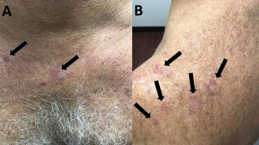
forearms, chest, upper back, and neck (Figure 1). A shave biopsy of skin eruptions on both the
left and right forearm was performed (Figure 2).
FIGURE 1: Cutaneous presentation of atorvastatin-induced
lichenoid drug eruption
Erythematous, pruritic plaques (black arrows) on the chest (A), neck, and the upper back (B)
FIGURE 2: Skin biopsy sites of statin-induced lichenoid drug
2020 Forouzan et al. Cureus 12(3): e7155. DOI 10.7759/cureus.7155 2 of 10eruption on forearms
A horizontal view of the biopsy sites (black arrows) of lichenoid drug eruption that presented as red,
planar plaques on the left (A) and the right (B) forearms are each outlined by four small purple dots
Microscopic examination revealed orthokeratosis, acanthosis, and spongiosis. A dense, band-
like inflammatory infiltrate composed predominantly of lymphocytes was present in the upper
dermis and along the dermoepidermal junction. In addition, apoptotic cells, eosinophils, and
histiocytes were observed.
Pathologic findings pointed to lichenoid dermatitis with eosinophils. Correlation of the clinical
history, lesion morphology, and pathologic findings established a diagnosis of a lichenoid drug
eruption. We suspected that the causative agent was atorvastatin, which the patient had begun
taking two months prior to the onset of his eruption.
Management included discontinuing the atorvastatin and treatment with prednisone, initially
40 mg daily with a gradual tapering of the dosage over 20 days. Additionally, a topical
betamethasone dipropionate 0.05% cream to be applied twice daily for three weeks was also
prescribed. His symptoms and skin eruption completely resolved and had not recurred at a one-
month follow-up.
Discussion
Adverse cutaneous events are a consequence of various medications including antibiotics,
anticonvulsants, and statins. Earlier studies have observed that the majority of lichenoid drug
eruptions were caused by either antimalarial agents or oral gold therapy [19].
The duration and onset of lichenoid drug eruptions are often dependent on the causative agent
and dosage. Lichenoid drug eruptions occur most often in individuals between the age of 57 to
66 years and can have an average latent period of one year between the beginning of the
medication treatment and the onset of an eruption [19]. This medication-induced eruption
should be considered when an individual receiving statin treatment develops new lesions akin
to lichen planus.
The clinical presentation and pathology of lichenoid drug eruptions can mimic those of lichen
planus (Table 1) [15-16,19-20]. Both conditions present as erythematous to purple papules and
plaques; however, lichenoid drug eruptions may be scaly, more pruritic, and resolve with
greater residual hyperpigmentation [15,19]. In addition, Wickham’s striae (a lacy, white
network of streak often located bilaterally on the buccal mucosa) and involvement of other
mucosal areas are observed less frequently in drug-induced lesions [15,19]. Compared to the
flexor surface distribution on extremities seen with idiopathic lichen planus, lichenoid drug
eruptions may present in a photodistributed or symmetric pattern [15].
2020 Forouzan et al. Cureus 12(3): e7155. DOI 10.7759/cureus.7155 3 of 10Characteristic Lichen planus Lichenoid drug eruption Reference
Similar to lichen planus but can be scaly and more
Erythematous, planar, and
pruritic; alopecia, desquamation, eczematous
Morphology polygonal papules are commonly [15,19]
papules, and greater residual hyperpigmentation may
described
also occur
A band-like lymphocyte infiltrate Similar to lichen planus but can also present with an
along the dermoepidermal junction infiltrate containing eosinophils. Focal parakeratosis,
Pathology [19,20]
is present along with apoptotic more prominent perivascular inflammation, and
keratinocytes (Civatte bodies) irregular granular layers may be present
Can appear one year after starting the causative
Onset Variable medication; onset can vary based on the medication [19]
and dosage
Dermatology
(primary lesion Extremities Arms, legs, and trunk [15,19]
location)
Distribution Flexor surface Symmetric, photodistributed pattern [15,19]
Wickham’s
Commonly present Typically not present [15]
striae
Oral/mucosal
Majority of cases Less common [19]
involvement
Diabetes mellitus, dyslipidemia,
Associated hepatitis B virus infection, hepatitis Antimalarials, beta-blockers, oral gold therapy,
[16,19]
conditions C virus infection, and thyroid penicillamine, statins, and thiazides
dysfunction
Less likely to spontaneously resolve and may not
Prognosis May spontaneously resolve regress for months even after stopping the causative [19]
agent
Can resolve spontaneously;
May resolve after discontinuing the causative drug;
however, oral and topical
Treatment however, oral and/or topical corticosteroids are [19]
corticosteroids usually expedite
usually needed to resolve the eruption
resolution
TABLE 1: Comparison between lichen planus and lichenoid drug eruption
Microscopically, both lichenoid drug eruptions and idiopathic lichen planus exhibit a band-like
lymphocytic infiltrate along the dermal-epidermal junction and apoptotic keratinocytes. Both
conditions also show acanthosis, hypergranulosis, and hyperkeratosis [20]. However, an
infiltrate with eosinophils in the dermis can help delineate lichenoid drug eruption from lichen
planus [20].
Lichenoid drug eruptions are associated with medications. In contrast, lichen planus can be
2020 Forouzan et al. Cureus 12(3): e7155. DOI 10.7759/cureus.7155 4 of 10associated with systemic conditions such as diabetes mellitus and hepatitis B or hepatitis C
viral infections. Lichenoid drug eruptions are also less likely to spontaneously resolve and may
require discontinuation of the causative agent in addition to topical and/or oral corticosteroid
therapy.
Several cutaneous adverse events have been described in patients who have received statins
(Table 2) [1-18]. Among these, bullous dermatosis, cutaneous lupus erythematosus,
dermatomyositis, eosinophilic fasciitis, and photosensitivity are the most common [1,3,5-6].
Acute generalized exanthematous pustulosis, alopecia, cheilitis, chronic actinic dermatitis,
dermatographism, eczema, erythema multiforme, pityriasis lichenoides chronica, pityriasis
rubra pilaris, porphyria cutanea tarda, purpuric lesions, and skin ulcers have also been
associated with statin use [1-2,4,7-12].
2020 Forouzan et al. Cureus 12(3): e7155. DOI 10.7759/cureus.7155 5 of 10Statin-associated adverse skin effects Reference
Acute generalized exanthematous pustulosis [1]
Alopecia [2]
Angioedema [1]
Bullous dermatosis [3]
Cheilitis [4]
Chronic actinic dermatitis [1]
Cutaneous lupus erythematosus [5]
Dermatographism [1]
Dermatomyositis [6]
Eczema [1]
Eosinophilic fasciitis [1]
Erythema multiforme [7]
Ichthyosis [1]
Lichenoid drug eruptions [13-18, CR]
Lichen planus pemphigoides [1]
Phototoxicity [1]
Pityriasis lichenoides chronica [8]
Pityriasis rubra pilaris [9]
Porphyria cutanea tarda [10]
Purpuric lesions [11]
Skin ulcers [12]
Toxic epidermal necrolysis [1]
TABLE 2: Cutaneous adverse events observed with statin medications
CR: current report
Lichenoid drug eruptions have historically been associated with antimalarials, gold, and
penicillamine. More recently, they have been observed with antineoplastics, beta-blockers, and
thiazides [16]. Our patient developed a lichenoid drug eruption secondary to atorvastatin. In
addition to atorvastatin, other statin medications have also been implicated with lichenoid
drug eruptions (Table 3) [13-18].
2020 Forouzan et al. Cureus 12(3): e7155. DOI 10.7759/cureus.7155 6 of 10Age,
Drug, race, and Location and
Morphology Pathology Treatment and result Reference
dosage sex of onset
patient
Lymphocytic infiltrate
Bilateral arms, Discontinued
73-year- along the
chest, back, and Erythematous atorvastatin;
Atorvastatin, old dermoepidermal
neck; onset after to purple, betamethasone and [CR]
40 mg/day Caucasian junction with
two months on scaly patches prednisone treatment;
male eosinophils and
atorvastatin remission in one month
histiocytes
Discontinued fluvastatin
Papules and use and treatment with
Extremities; onset plaques with A band-like mometasone-furoate
Fluvastatin, 59-year-
after four weeks on Wickham’s lymphocytic infiltrate resolved the initial
20 mg/day old
fluvastatin. striae on with apoptotic eruption in three weeks;
and woman of [13]
Redeveloped after papules. keratinocytes, later treatment with
lovastatin, 20 unknown
two weeks on Some oral hyperkeratosis, and lovastatin resulted in
mg/day ethnicity
lovastatin involvement vacuolar alteration similar eruptions.
was reported Discontinued lovastatin;
remission in three weeks
Lymphocytic
64-year- Face and upper inflammation found
Pravastatin, old back; onset three Dense along the Discontinued statin;
unknown woman of months after freckling with dermoepidermal pigmentation resolved [14]
dosage unknown beginning statin no rash junction with basal cell after nine months
ethnicity treatment damage and Civatte
bodies
Treatment with
Photodistributed,
fluocinonide 0.05% gel
symmetric fashion
and mupirocin 2%
on arms and hands;
Focal hypergranulosis, ointment was not
onset three weeks Erythematous
75-year- hyperkeratotic stratum effective. Discontinued
Pravastatin, after beginning plaques and
old Black corneum, lymphocytic statin; the eruptions [15]
10 mg/day statin treatment. papules with
man infiltrate, and vacuolar healed after four weeks;
Reappeared after shiny scales
degeneration rechallenge with
two weeks with
pravastatin led to
pravastatin
identical plaque
rechallenge
formation
A lymphocytic infiltrate Discontinued statin;
65-year- Trunk and
Flat-topped was reported in the treated with psoralen and
old extremities; onset
Rosuvastatin, and dermis with apoptotic ultraviolet A radiation
woman of three months after [16]
10 mg/day erythematous keratinocytes and focal therapy and with oral
unknown beginning statin
papules parakeratosis in the corticosteroid therapy.
ethnicity treatment
epidermis Remission in six months
Right thigh with
onset one week
2020 Forouzan et al. Cureus 12(3): e7155. DOI 10.7759/cureus.7155 7 of 10Rosuvastatin, after beginning Apoptotic Discontinued
55-year- rosuvastatin;
10 mg/day An keratinocytes, basal rosuvastatin; treatment
old South eruptions on her
and erythematous vacuolar changes, and with clobetasol [17]
Asian right thigh, back,
simvastatin, rash focal parakeratosis propionate 0.05% cream.
woman and oral mucosa
10 mg/day were present Remission in two months
were reported at
one-month follow-
up
Therapy with topical
A lymphocytic infiltrate
diflucortolone 0.1%
with eosinophils and
cream did not resolve the
histiocytes were
57-year- Red papules eruption. Discontinued
Wrists, elbows, and reported. Compact
old and simvastatin and
Simvastatin, buccal mucosa; orthokeratosis and
woman of Wickham’s bezafibrate therapy; [18]
10 mg/day onset after one focal parakeratosis in
unknown striae were eruption began to
month of statin use epidermis were found;
ethnicity noted resolve within four
Civatte bodies and
weeks, but the mucosal
vacuolar degeneration
lesions persisted at the
were also noted
six-month follow- up
TABLE 3: Characteristics of patients with statin-induced lichenoid drug eruptions
CR: current report
Lichenoid drug eruptions have been reported in one patient taking pravastatin 10 mg/day, two
patients taking rosuvastatin 10 mg/day, and two patients on simvastatin at 10 mg/day [15-18].
Our patient with the atorvastatin-induced lichenoid eruption was being treated at 40 mg/day.
Another patient developed a lichenoid drug eruption with pravastatin; however, this patient’s
dosage was not stated [14]. Another patient developed a lichenoid drug eruption on fluvastatin
20 mg daily; when she switched to lovastatin 20 mg daily, she redeveloped this drug-induced
eruption [13].
To the best of our knowledge, lichenoid drug eruptions secondary to statin medications have
been reported in two men and five women including our patient [13-18]. These individuals
ranged in age from 55 to 75 years with a median onset age of 64 years [13-18]. The median
onset age was 74 years for men and 59 years for women [13-18]. Four of the patients were of
unknown ethnicity; however, a Black man, a Caucasian man, and a South Asian woman were
described [15,17]. In the individuals who experienced a statin-induced lichenoid drug eruption,
the onset of the eruption ranged from 2 to 12 weeks after starting the statin medication with a
median of four weeks [13-18].
The cutaneous adverse event appeared on the trunk and extremities in six patients; one of the
patients had skin lesions that developed on the face [13-18]. Six patients presented with lichen
planus-like violaceous papules, and one patient demonstrated dense freckling on her face [13-
18]. Oral involvement was reported in three of the individuals, and Wickham’s striae were
observed in two patients [13,17-18].
Histologic evaluation of the statin-induced lichenoid drug eruptions demonstrated lymphocytic
infiltration of the dermoepidermal junction similar to idiopathic lichen planus [13-18]. Focal
2020 Forouzan et al. Cureus 12(3): e7155. DOI 10.7759/cureus.7155 8 of 10parakeratosis was reported in three patients [16-18]. Eosinophils were noted in two patients,
including ours [18]. Hyperkeratosis was also noted in two patients’ statin-induced lichenoid
eruptions [13,15].
Management of statin-induced lichenoid drug eruptions includes discontinuation of the
causative statin agent and treatment with topical and/or oral corticosteroids. Six of the seven
patients’ skin lesions, including ours, resolved with cessation of the statin medication and
additional therapy: an oral corticosteroid, a topical corticosteroid, or both [13,15-18]. In some
instances, the eruption persisted for several months after discontinuing the instigating agent.
Indeed, with or without additional treatment, the statin-induced drug eruptions resolved
within three weeks to nine months after the causative drug was stopped [13-18].
Conclusions
Lichenoid drug eruptions share several features with lichen planus. However, unique
characteristics of these drug-induced eruptions (including delayed onset, absence of Wickham’s
striae, and presence of eosinophils microscopically) can help distinguish lichenoid drug
eruptions from idiopathic lichen planus. Statins are generally used in the management of
hypercholesterolemia; however, several adverse cutaneous events have been observed in
patients treated with statins. Lichenoid drug eruptions are an uncommon adverse cutaneous
event associated with statin medications. The new onset of lichenoid dermatitis in an
individual receiving statin therapy should raise the concern that this skin eruption may be
associated with the medication.
Additional Information
Disclosures
Human subjects: Consent was obtained by all participants in this study. Conflicts of interest:
In compliance with the ICMJE uniform disclosure form, all authors declare the following:
Payment/services info: All authors have declared that no financial support was received from
any organization for the submitted work. Financial relationships: All authors have declared
that they have no financial relationships at present or within the previous three years with any
organizations that might have an interest in the submitted work. Other relationships: All
authors have declared that there are no other relationships or activities that could appear to
have influenced the submitted work.
References
1. Golomb BA, Evans MA: Statin adverse effects: a review of the literature and evidence for a
mitochondrial mechanism. Am J Cardiovasc Drugs. 2008, 8:373-418. 10.2165/0129784-
200808060-00004
2. Segal AS: Alopecia associated with atorvastatin . Am J Med. 2002, 113:171. 10.1016/s0002-
9343(02)01135-x
3. König C, Eickert A, Scharfetter-Kochanek K, Krieg T, Hunzelmann N: Linear IgA bullous
dermatosis induced by atorvastatin. J Am Acad Dermatol. 2001, 44:689-692.
10.1067/mjd.2001.113462
4. Mehregan DR, Mehregan DA, Pakideh S: Cheilitis due to treatment with simvastatin . Cutis.
1998, 62:197-198.
5. Laurinaviciene R, Sandholdt LH, Bygum A: Drug-induced cutaneous lupus erythematosus: 88
new cases. Eur J Dermatol. 2017, 27:28-33. 10.1684/ejd.2016.2912
6. Oztas M, Ugurlu S, Aydin O: Atorvastatin-induced dermatomyositis. Rheumatol Int. 2017,
37:1217-1219. 10.1007/s00296-017-3658-9
7. Lerch M, Mainetti C, Terziroli Beretta-Piccoli B, Harr T: Current perspectives on erythema
multiforme. Clin Rev Allergy Immunol. 2018, 54:177-184. 10.1007/s12016-017-8667-7
8. Massay RJ, Maynard AA: Pityriasis lichenoides chronica associated with the use of HMG-CoA
2020 Forouzan et al. Cureus 12(3): e7155. DOI 10.7759/cureus.7155 9 of 10reductase inhibitors. West Indian Med J. 2012, 61:743-745.
9. Gajinov ZT, Matić MB, Duran VD, Vucković N, Prcić ST, Vujanović LM: Drug-related pityriasis
rubra pilaris with acantholysis. Vojnosanit Pregl. 2013, 70:871-873. 10.2298/vsp1309871g
10. Perrot JL, Guy C, Bour Guichenez G, Amigues O, Servoz J, Cambazard F: Porphyria cutanea
tarda induced by HMG CoA reductase inhibitors: simvastatin, pravastatin. (Article in French).
Ann Dermatol Venereol. 1994, 121:817-819.
11. Kato K, Onodera K, Iwasaki Y, et al.: Pravastatin-induced rhabdomyolysis and purpura
fulminans in a patient with chronic renal failure. Int J Surg Case Rep. 2015, 8C:84-87.
Accessed: March 2, 2020: https://www.ncbi.nlm.nih.gov/pubmed/25644555/.
10.1016/j.ijscr.2015.01.042
12. Fernández-Torres R, del Pozo J, Almagro M, Yebra-Pimentel MT, Fernández-Jorge B, Mazaira
M, Fonseca E: Skin ulcers and myopathy associated with pravastatin therapy . Clin Exp
Dermatol. 2009, 34:e237-238. 10.1111/j.1365-2230.2008.03098.x
13. Sebök B, Tóth M, Anga B, Harangi F, Schneider I: Lichenoid drug eruption with HMG-CoA
reductase inhibitors (fluvastatin and lovastatin). Acta Derm Venereol. 2004, 84:229-230.
10.1080/00015550310006851
14. Pua VS, Scolyer RA, Barnetson RS: Pravastatin-induced lichenoid drug eruption. Australas J
Dermatol. 2006, 47:57-59. 10.1111/j.1440-0960.2006.00225.x
15. Keough GC, Richardson TT, Grabski WJ: Pravastatin-induced lichenoid drug eruption. Cutis.
1998, 61:98-100.
16. Vesza Z, Pires C, da Silva PM: Statin-related lichenoid dermatosis: an uncommon adverse
reaction to a common treatment. Eur J Case Rep Intern Med. 2018, 5:000844. Accessed: March
2, 2020: https://www.ncbi.nlm.nih.gov/pmc/articles/PMC6346926/. 10.12890/2018_000844
17. Wong ITY, Huang Y, Zhou Y: Drug eruption to rosuvastatin with recurrence on simvastatin: a
case report. J Cutan Med Surg. 2018, 22:359-361. 10.1177/1203475418756376
18. Roger D, Rolle F, Labrousse F, Brosset A, Bonnetblanc JM: Simvastatin-induced lichenoid
drug eruption. Clin Exp Dermatol. 1994, 19:88-89. 10.1111/j.1365-2230.1994.tb01128.x
19. Halevy S, Shai A: Lichenoid drug eruptions. J Am Acad Dermatol. 1993, 29:249-255.
10.1016/0190-9622(93)70176-t
20. Sehgal VN, Srivastava G, Sharma S, Sehgal S, Verma P: Lichenoid tissue reaction/interface
dermatitis: recognition, classification, etiology, and clinicopathological overtones. Indian J
Dermatol Venereol Leprol. 2011, 77:418-429. 10.4103/0378-6323.82389
2020 Forouzan et al. Cureus 12(3): e7155. DOI 10.7759/cureus.7155 10 of 10You can also read