Dyshidrosiform Bullous Pemphigoid: Case Reports and Review - Cureus
←
→
Page content transcription
If your browser does not render page correctly, please read the page content below
Open Access Case
                                        Report                                              DOI: 10.7759/cureus.6630

                                        Dyshidrosiform Bullous Pemphigoid: Case
                                        Reports and Review
                                        Philip R. Cohen 1

                                        1. Dermatology, San Diego Family Dermatology, San Diego, USA

                                        Corresponding author: Philip R. Cohen, mitehead@gmail.com

                                        Abstract
                                        Bullous pemphigoid is an autoimmune blistering disorder that typically presents in elderly
                                        patients as pruritic tense subepidermal blisters on the lower trunk, axilla, and groin. It is caused
                                        by circulating and tissue-bound autoantibodies directed against bullous pemphigoid antigen 1
                                        or bullous pemphigoid antigen 2 or both. Dyshidrosiform bullous pemphigoid is a rare variant
                                        of bullous pemphigoid, and it usually presents as itchy, potentially hemorrhagic, or
                                        purpuric blisters on the palms and/or soles of elderly individuals; subsequently, typical bullous
                                        lesions of bullous pemphigoid appear on other body sites. In our study, we report the features
                                        of two men with dyshidrosiform bullous pemphigoid and review the characteristics of
                                        individuals with this rare subtype of bullous pemphigoid. Including the men whose condition is
                                        described in this paper, at least 72 patients with dyshidrosiform bullous pemphigoid have been
                                        reported so far. However, complete features of the condition have not been described for all of
                                        the individuals.

                                        Based on the cases reported so far, the condition was slightly more common in women and the
                                        onset of the disease, for most of the patients, occurred between the ages of 61 and 94 years. The
                                        patients usually presented with blisters on both their palms and soles (66%) or just their soles
                                        (31%); 77% of the patients had progression of bullous pemphigoid to other areas of their body.
                                        Whether hemorrhagic blisters or purpuric lesions are associated with dyshidrosiform bullous
                                        pemphigoid remains to be determined; these features were present in 91% of the 22 patients
                                        who were described in the case reports yet were only observed in 5% of the individuals from a
                                        single larger series of 20 patients. The mainstay of therapy for dyshidrosiform bullous
                                        pemphigoid is systemic corticosteroids, with or without topical corticosteroids, and/or
                                        systemic dapsone or immunosuppressants; nearly all of the patients showed improvement after
                                        the treatment was initiated. Similar to individuals with bullous pemphigoid, at least nine of the
                                        dyshidrosiform bullous pemphigoid patients, including both patients in this report, had either a
                                        neurologic condition (seven patients) or both a neurologic condition and a psychiatric disorder
                                        (two patients). Usually, an autoimmune bullous disease, particularly dyshidrosiform bullous
                                        pemphigoid, is not initially considered in patients who present with blisters restricted to the
                                        palms and/or soles. Indeed, the lesion morphology of dyshidrosiform bullous pemphigoid
Received 01/07/2020
Review began 01/08/2020
                                        mimics several other conditions that are characterized by blisters on the hands and feet, such as
Review ended 01/09/2020                 allergic and irritant contact dermatitis, chronic bullous disease of childhood, cutaneous T-cell
Published 01/11/2020                    lymphoma, dermatophyte infection, dyshidrosis or pompholyx, epidermolysis bullosa acquisita,
© Copyright 2020
                                        erythema multiforme, herpes gestationis, lichen planus, linear IgA disease, scabies, and
Cohen. This is an open access article   systemic contact dermatitis. In conclusion, the possibility of dyshidrosiform bullous
distributed under the terms of the      pemphigoid should be considered in elderly individuals who present with the new onset of
Creative Commons Attribution License    palmar and/or plantar blisters that are either recurrent or recalcitrant to therapy or would
CC-BY 3.0., which permits
                                        subsequently also appear on other areas of the body.
unrestricted use, distribution, and
reproduction in any medium, provided
the original author and source are
credited.

                                        How to cite this article
                                        Cohen P R (January 11, 2020) Dyshidrosiform Bullous Pemphigoid: Case Reports and Review. Cureus
                                        12(1): e6630. DOI 10.7759/cureus.6630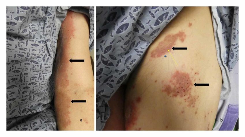
Categories: Dermatology, Family/General Practice, Internal Medicine
Keywords: blister, bullous, corticosteroid, dyshidrosiform, dyshidrosis, dyshidrotic, elderly,
pemphigoid, pompholyx, vesicle
Introduction
Bullous pemphigoid is an autoimmune blistering condition that usually occurs in elderly
individuals. The immunopathogenesis of the disease is attributed to circulating and tissue-
bound autoantibodies directed against bullous pemphigoid antigen 230 (bullous pemphigoid
antigen 1) or bullous pemphigoid antigen 180 (bullous pemphigoid antigen 2) or both [1-3]. The
condition typically presents as pruritic tense subepidermal blisters. In addition to the lower
trunk, the lesions frequently appear on the proximal flexural aspects of the arms (near the
axilla) and legs (near the groin). The lesions can be localized or widespread [1-3].
Unusual clinical variants of bullous pemphigoid have been observed in literature [1-3].
Dyshidrosiform bullous pemphigoid refers to the condition when the blisters are initially or
persistently localized to the palms and soles [4-19]. In our study, we report the features of two
men with dyshidrosiform bullous pemphigoid and review the characteristics of individuals with
this rare subtype of bullous pemphigoid.
Case Presentation
Case 1
A 61-year-old Filipino man presented with a two-month history of tender blisters on his feet.
The lesions had made it difficult for him to walk. A week before his evaluation, he had also
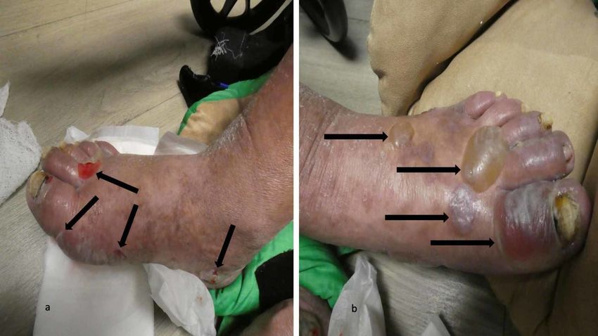
begun to develop red itchy lesions on his upper legs and central upper back. His past medical
history was only significant for hypertension for which he was receiving atenolol daily.
Cutaneous examination revealed painful, bilateral, large hemorrhagic blisters on posterior and
plantar heels (Figure 1); there were no mucosal lesions. In addition, he had a flattened blister
with a black roof on the instep of his right foot, and a blister whose roof had become detached
was also present on the dorsal left foot on the proximal fourth toe (Figure 2). Erythematous
urticarial dermal plaques were present on his central upper back (with a ruptured blister)
(Figure 3) and the proximal medial thighs (with small papules) (Figure 4).
FIGURE 1: Dyshidrosiform bullous pemphigoid presenting as
2020 Cohen et al. Cureus 12(1): e6630. DOI 10.7759/cureus.6630 2 of 11
FIGURE 1: Dyshidrosiform bullous pemphigoid presenting as
                                     plantar blisters in a 61-year-old man
                                     The posterior and plantar left heel (a) and right heel (b) show a tender large flaccid hemorrhagic
                                     blister (a) and a flattened blister (b) as the initial clinical manifestations of dyshidrosiform bullous
                                     pemphigoid; each of these blisters is outlined by black arrows. A tense blister containing clear fluid
                                     (red arrows) is present on the medial area of his left heel (a)

                                     FIGURE 2: Blisters from dyshidrosiform bullous pemphigoid on
                                     the feet of a 61-year-old man
                                     The medial view of the right foot (a) shows a black-roofed and flattened blister on the instep (blue
                                     arrows) and another flattened blister that is primarily on the posterior heel and extends to the plantar
                                     foot (black arrows). The left dorsal foot (b) has a deroofed blister on the proximal fourth toe (green
                                     arrows)

2020 Cohen et al. Cureus 12(1): e6630. DOI 10.7759/cureus.6630                                                                              3 of 11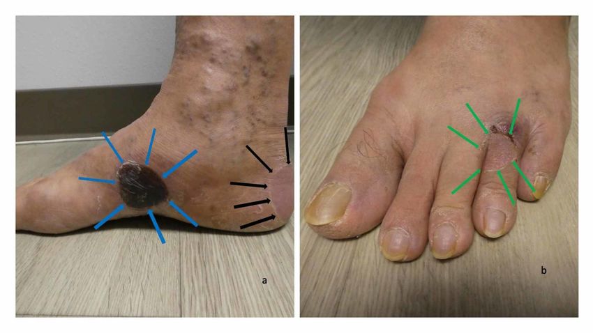
FIGURE 3: Erythematous urticarial dermal plaques and blisters
                                     on the back of a man with dyshidrosiform bullous pemphigoid
                                     Distant (a) and closer (b) views show several erythematous dermal plaques. A larger red urticaria-
                                     appearing plaque on the central upper back (black arrow), consistent with the urticarial stage of
                                     bullous pemphigoid, is present. In the center of the plaque, a blister (yellow arrows) has ruptured

                                     FIGURE 4: Erythematous urticarial dermal plaques with small
                                     papules on the right thigh of a man with dyshidrosiform
                                     bullous pemphigoid
                                     Distant (a) and closer (b) views of the right thigh show several red dermal plaques. A larger
                                     erythematous dermal plaque (black arrow) has a small papule (the tip of the yellow arrow); this was
                                     the location of the skin biopsy for hematoxylin and eosin staining which was consistent with the
                                     urticarial stage of bullous pemphigoid. The skin biopsy for direct immunofluorescence (blue star)
                                     was located adjacent to the skin lesion on the right thigh and demonstrated findings that were
                                     diagnostic for bullous pemphigoid

                                   Skin biopsies of lesions on the right thigh and left ankle were performed for routine
                                   hematoxylin and eosin staining. Microscopic examination of the right-thigh biopsy showed
                                   scattered foci of spongiosis in the epidermis; in the papillary dermis, there was a band-like
                                   infiltrate of lymphocytes and numerous eosinophils, edema, and early subepidermal
                                   vesiculation. The left-ankle biopsy demonstrated a subepidermal blister with an inflammatory
                                   infiltrate consisting of lymphocytes, histiocytes, and eosinophils in the papillary dermis.

                                   A second biopsy immediately adjacent to the right-thigh lesion was performed for direct
                                   immunofluorescence. Staining for immunoglobulin G (IgG) and C3 showed a smooth linear
                                   band of immunoreactant deposition at the dermoepidermal junction. Staining for
                                   immunoglobulin A (IgA), immunoglobulin M (IgM), and fibrinogen was negative. The
                                   hematoxylin and eosin-stained skin biopsy from the right thigh were consistent with the
                                   urticarial stage of bullous pemphigoid. Both the left- ankle (hematoxylin and eosin) and the
                                   right-thigh (direct immunofluorescence) biopsies were diagnostic for bullous pemphigoid. The

2020 Cohen et al. Cureus 12(1): e6630. DOI 10.7759/cureus.6630                                                                         4 of 11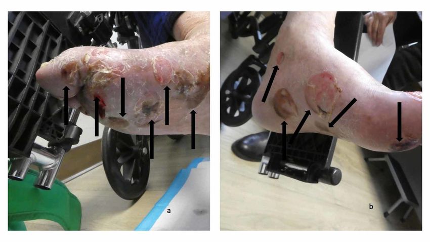
correlation of the clinical history and histopathology and immunopathology established the
                                   diagnosis of dyshidrosiform bullous pemphigoid.

                                   Serologic laboratory studies were also performed. Quantitative indirect immunofluorescence to
                                   anti-skin autoantibodies demonstrated a basement membrane zone (BMZ) staining pattern at a
                                   high titer of 1:160, confirming the diagnosis of bullous pemphigoid. The patient's eosinophil
                                   count was normal; however, his serum immunoglobulin E (IgE) level was very elevated at 7,582
                                   UI/ml (normal: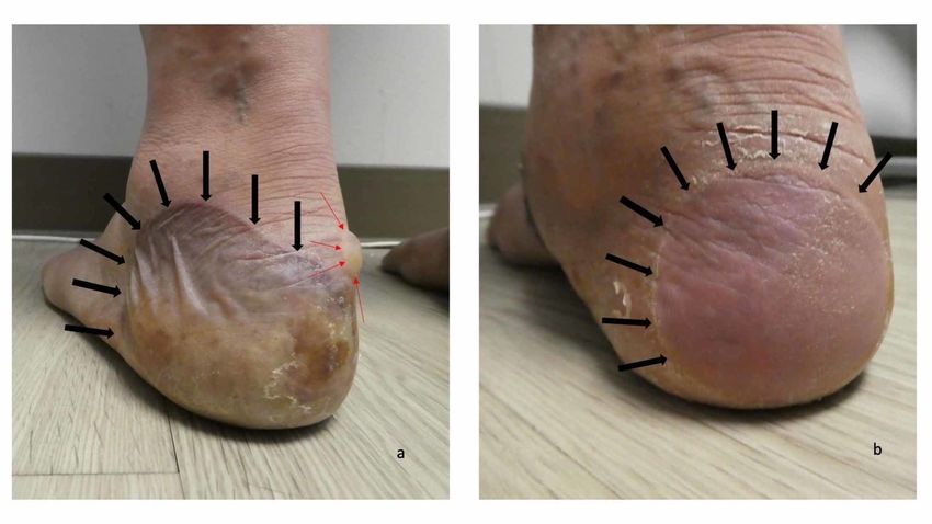
FIGURE 5: Dyshidrosiform bullous pemphigoid presenting as
                                     plantar blisters in a 65-year-old man
                                     The right foot (a) and left foot (b) show hemorrhagic and deroofed blisters (black arrows) on both the
                                     dorsal and lateral surface of his feet and toes; some of the blisters also extend to the soles of his
                                     feet

                                     FIGURE 6: Erythematous urticarial dermal plaques on the left
                                     arm and left leg of a man with dyshidrosiform bullous
                                     pemphigoid
                                     The proximal left arm (a) and the proximal left thigh (b) show several erythematous dermal plaques
                                     (black arrows). The skin biopsy on his left thigh (b) for hematoxylin and eosin staining (the tip of the
                                     yellow arrow) show pathologic changes that are consistent with the urticarial stage of bullous
                                     pemphigoid. The skin biopsy for direct immunofluorescence (blue star) is located adjacent to the
                                     skin lesion on the left thigh and demonstrate findings that are consistent with bullous pemphigoid

2020 Cohen et al. Cureus 12(1): e6630. DOI 10.7759/cureus.6630                                                                             6 of 11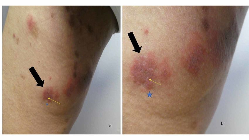
Microscopic examination of the hematoxylin and eosin-stained specimen showed eosinophilic
                                   spongiosis (showing eosinophils being present in the widened spaces between the epidermal
                                   keratinocytes), marked edema in the papillary dermis, and abundant eosinophils with some
                                   lymphocytes present in the inflammatory infiltrate in the upper dermis. These changes were
                                   consistent with those of the urticarial stage of bullous pemphigoid. The direct
                                   immunofluorescence stained specimen showed a linear band of both IgG and C3, with no
                                   staining for IgA, IgM, and fibrinogen, deposited at the dermoepidermal junction. These
                                   findings were diagnostic for bullous pemphigoid. Correlation of the clinical presentation of
                                   blisters on the feet and the pathology results (or routine-stained and immunofluorescence-
                                   stained specimens) established the diagnosis of dyshidrosiform bullous pemphigoid.

                                   The patient returned after one week. Several of the blisters on his feet had flattened (Figure 7);
                                   however, small vesicles were noted on his arm plaques (Figure 8). His topical care was
                                   continued and he was started on 60 mg of prednisolone, which was to be administered through
                                   his gastrostomy tube each morning. Follow-up, one week later, showed significant
                                   improvement of both his feet and body; there were no new blisters and the erythematous
                                   plaques on his arms and legs were nearly resolved.

                                     FIGURE 7: Blisters on the feet of a man with dyshidrosiform
                                     bullous pemphigoid
                                     Numerous flattened and deroofed blisters (black arrows) seen on the plantar and medial surfaces of
                                     the right foot (a) and left foot (b)

2020 Cohen et al. Cureus 12(1): e6630. DOI 10.7759/cureus.6630                                                                      7 of 11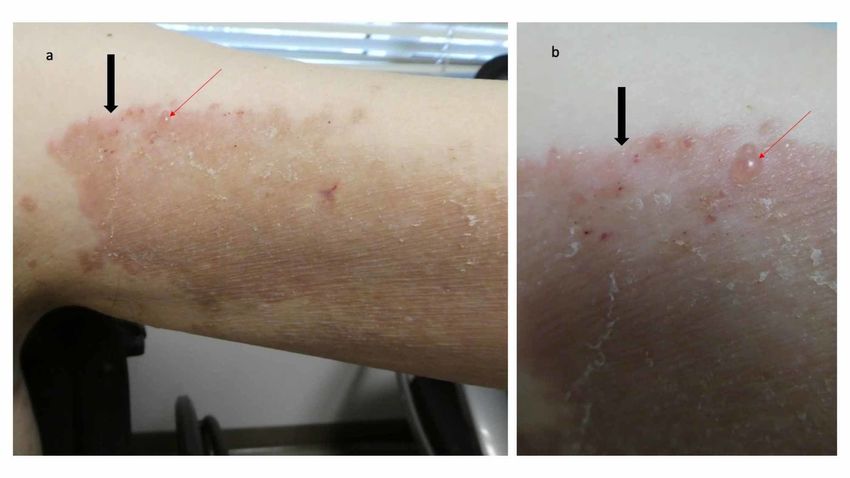
FIGURE 8: Erythematous plaque with vesicles on the left arm
                                     of a man with dyshidrosiform bullous pemphigoid
                                     The large confluent red dermal plaque on the proximal left arm (black arrow) has small vesicles;
                                     one of the vesicles is located at the tip of the red arrow

                                   Discussion
                                   Dyshidrosiform bullous pemphigoid is a unique variant of bullous pemphigoid. The
                                   morphology of the lesions mimics vesicular hand or foot dermatitis. In fact, the clinical
                                   differential diagnosis not only includes dyshidrosis or pompholyx, but also allergic and irritant
                                   contact dermatitis, chronic bullous disease of childhood, cutaneous T-cell lymphoma (vesicular
                                   palmoplantar variant), dermatophyte infection (bullous), epidermolysis bullosa acquisita,
                                   erythema multiforme, herpes gestationis, lichen planus (bullous), linear IgA disease, scabies,
                                   and systemic contact dermatitis. The possibility of an autoimmune bullous disease is usually
                                   not entertained, particularly in individuals for whom the lesions are restricted to the palms or
                                   soles or both [4-19].

                                   Dyshidrosiform bullous pemphigoid was initially described by Levine et al. in 1979 [4]. They
                                   described a 72-year-old man who initially presented with a vesicular eruption of the hands
                                   (that resolved in two weeks after treatment with tar soaks and topical corticosteroid
                                   application) and subsequently developed large tense bullae on his feet which cleared after two
                                   weeks of oral prednisone 40 mg daily. Blisters continued to periodically appear on his feet; in
                                   addition, one episode was also associated with truncal lesions and an erosion on his pharynx.
                                   Prior to skin biopsy confirmation of the diagnosis of bullous pemphigoid, he had also been
                                   treated with erythromycin, intramuscular corticosteroid, and griseofulvin. After establishing
                                   the diagnosis, he was eventually treated with dapsone 50 mg thrice daily for seven months; all
                                   of his lesions resolved after five days and there was no recurrence at follow-up four months
                                   after stopping the treatment [4].

                                   Since the publication of Levine et al.’s paper, at least 71 additional patients, including the
                                   individuals in this report, with dyshidrosiform bullous pemphigoid have been reported in the
                                   literature. Most of the reports (24 papers) only described a single patient. However, there were
                                   two reports with two patients, four reports with three patients, one report with four patients,

2020 Cohen et al. Cureus 12(1): e6630. DOI 10.7759/cureus.6630                                                                          8 of 11
one report with nine patients, and one report with 20 patients (one of whom was previously
described) [4-19]. Also, three larger studies of dyshidrosiform bullous pemphigoid have been
performed [17-19]. One group of investigators only identified three patients with
dyshidrosiform bullous pemphigoid in 86 individuals with bullous pemphigoid, who were
treated in the largest teaching hospital in Taiwan from 1977 to 1994. The three patients
predominantly developed vesicles and bullae on their palms and soles that were difficult to
differentiate from pompholyx. All of the palm and sole lesions responded to treatment with
prednisolone 30-40 mg per day [19].
However, the other two research groups observed 28% (20 of 71 patients) and 45% (nine of 20
patients) of individuals with palmar and/or plantar lesions in their series of bullous pemphigoid
patients [17,18]. The palm and/or plantar lesions were the presenting feature in four of the 20
patients [18]. Also, only one of the 20 patients had hemorrhagic bullae [6,18].
The third study took place during a period of three years and included 20 bullous pemphigoid
patients from Stockholm, Sweden; there were nine dyshidrosiform bullous pemphigoid
patients, two of whom only had palm and/or sole lesions. The investigators emphasized that all
nine of the dyshidrosiform bullous pemphigoid patients had prodromal symptoms, such as an
eczematous eruption (three patients), a papular eruption (three patients), an eczematous and
papular eruption (one patient), an intertriginous eruption (one patient), or an urticarial and
papular eruption (one patient) for greater than (five patients) or less than (four patients) three
months [17]. Based on the increased number of bullous pemphigoid patients with palm and/or
sole lesions observed in the latter two studies, it is conjectured that dyshidrosiform bullous
pemphigoid occurs more commonly than implied by the publication of patient descriptions in
the individual case reports.
Based on the studies, epidemiology information was available for 38 of the 72 dyshidrosiform
bullous pemphigoid patients. The onset age of dyshidrosiform bullous pemphigoid ranged from
20 years to 94 years (median: 76 years); however, only three of the patients were in their
twenties. The age of the other 35 individuals ranged from 61 to 94 years [4-19]. Dyshidrosiform
bullous pemphigoid was only slightly more common in women (20 patients) than men (18
patients). The onset age in women ranged from 23 years to 94 years (median: 77 years).
Similarly, the onset age in men ranged from 20 years to 92 years (median: 75 years) [4-
19]. Many of the patients initially had blisters on both their palms and soles (19 of 29
individuals, 66%) [4-19]. However, some of the patients only presented with lesions on either
their palms (one of 29 individuals, 3%) or their soles (nine of 29 individuals, 31%).
Some of the researchers emphasized the association of hemorrhagic blisters or purpuric lesion
with dyshidrosiform bullous pemphigoid [6,7]. The blisters were hemorrhagic and purpura was
present in 91% of the patients (20 of 22 individuals) whose lesions were described in their
individual case reports [4-16]. In contrast, only one of the 20 patients (5%) from a large series
of patients had hemorrhagic lesions [18]. Similar to both of the patients described in this report,
77% of the patients (23 of 30 individuals) had progression of bullous pemphigoid to other areas
of their body [4-19]; however, oral lesions were only described in three patients [6,11,16]. The
duration of time between the onset of lesions on the palms and/or soles and new blisters on
other body sites ranged from one week to seven months (median: seven weeks). In common
with nine dyshidrosiform bullous pemphigoid patients who had prodromal symptoms prior to
the onset of their blisters [17], one man also developed pruritic papules on his arms and upper
back prior to the appearance of blisters on his palms [15].
All of the patients had histopathologic confirmation of their bullous pemphigoid diagnosis [4-
19]. This not only included hematoxylin and eosin-stained sections of formalin-fixed lesional
skin tissue specimens, but also immunofluorescence (direct, indirect or both) studies. Only a
2020 Cohen et al. Cureus 12(1): e6630. DOI 10.7759/cureus.6630 9 of 11small number of patients had enzyme-linked immunosorbent assay or Western blot testing for
autoantibodies against bullous pemphigoid antigen 1 or bullous pemphigoid antigen 2 or both
[11,14]. Treatment of dyshidrosiform bullous pemphigoid was described for 32 of the patients
[4-19]. Systemic corticosteroids-starting dosage ranging from 10-80 mg daily (median: 30 mg
daily) was used in the management of most (26 individuals, 81%) of the patients. In fact, one
group of investigators was able to successfully treat dyshidrosiform bullous pemphigoid
patients with a lower daily dosage of prednisone [6].
Other interventional agents were also used in the treatment of dyshidrosiform bullous
pemphigoid patients [4-19]. These included topical corticosteroids (12 individuals), dapsone
(seven individuals whose dosage ranged from 50-200 mg daily; median: 150 mg daily), oral
antibiotics such as erythromycin, doxycycline, or tetracycline (three individuals), and
nicotinamide (one individual). Immunosuppressant agents were also used either alone or as a
corticosteroid agent: azathioprine (three individuals whose dosage ranged from 100-150 mg
daily; median: 100 mg daily) and cyclophosphamide (one individual whose daily dosage was
100 mg). Nearly all of the patients improved with the treatment [4-19]. Resolution of lesions
occurred within one week to one month; however, recurrent episodes of palm and/or sole
lesions were not uncommon either during tapering or after stopping of the systemic treatment.
Indeed, many patients were still receiving therapy when they were reported. Two of the patients
died from conditions deemed to be unrelated to bullous pemphigoid or its treatment, one from
respiratory failure and the other from cardiac arrest [11,16].
An association between bullous pemphigoid and neurologic or psychiatric disorders has been
identified [1-3]. Both of the men in this report had neurologic disorders, either epilepsy or
Parkinsonism. In addition, at least seven of the other dyshidrosiform bullous pemphigoid
patients had a neurologic disorder: cerebrovascular accidents (two men and one woman)
[6,11,15], Parkinsonism (two women) [14,16], peripheral neuropathy (one woman) [16], and
senile dementia (one woman) [11]. One of the men in this report also had manic depression
syndrome. Similarly, one of the women patients suffered from depression [14]. Whether there is
an increased incidence of neurologic and psychiatric conditions in dyshidrosiform bullous
pemphigoid patients as compared to individuals with bullous pemphigoid without
dyshidrosiform-like lesions remains to be determined.
Conclusions
Dyshidrosiform bullous pemphigoid is a rarely described variant of bullous pemphigoid. Similar
to idiopathic bullous pemphigoid, dyshidrosiform bullous pemphigoid typically presents with
pruritic lesions in elderly individuals; the hemorrhagic or purpuric blisters on the palms and/or
soles are often followed by the development of typical bullous lesions on other body sites.
Nearly all dyshidrosiform bullous pemphigoid patients improve after the diagnosis is
established and treatment is initiated. The mainstay of therapy is systemic corticosteroids,
with or without topical corticosteroids, and/or systemic dapsone or immunosuppressants.
Additional Information
Disclosures
Human subjects: Consent was obtained by all participants in this study. Conflicts of interest:
In compliance with the ICMJE uniform disclosure form, all authors declare the following:
Payment/services info: All authors have declared that no financial support was received from
any organization for the submitted work. Financial relationships: All authors have declared
that they have no financial relationships at present or within the previous three years with any
organizations that might have an interest in the submitted work. Other relationships: All
authors have declared that there are no other relationships or activities that could appear to
have influenced the submitted work.
2020 Cohen et al. Cureus 12(1): e6630. DOI 10.7759/cureus.6630 10 of 11References
1. Bernard P, Antonicelli F: Bullous pemphigoid: a review of its diagnosis, associations and
treatment. Am J Clin Dermatol. 2017, 18:513-528. 10.1007/s40257-017-0264-2
2. Genovese G, Di Zenzo G, Cozzani E, Berti E, Cugno M, Marzano AV: New insights into the
pathogenesis of bullous pemphigoid: 2019 update. Front Immunol. 2019, 10:1506. Accessed:
January 11, 2020: https://www.frontiersin.org/articles/10.3389/fimmu.2019.01506/full.
10.3389/fimmu.2019.01506
3. Miyamoto D, Santi CG, Aoki V, Maruta CW: Bullous pemphigoid. An Bras Dermatol. 2019,
94:133-146. 10.1590/abd1806-4841.20199007
4. Levine N, Freilich A, Barland P: Localized pemphigoid simulating dyshidrosiform dermatitis.
Arch Dermatol. 1979, 115:320-321. 10.1001/archderm.1979.04010030028010
5. Liu HN, Su WP, Rogers RS 3rd: Clinical variants of pemphigoid. Int J Dermatol. 1986, 25:17-
27. 10.1111/j.1365-4362.1986.tb03397.x
6. Barth JH, Fairris GM, Wojnarowska F, White JE: Haemorrhagic pompholyx is a sign of bullous
pemphigoid and an indication for low-dose prednisolone therapy. Clin Exp Dermatol. 1986,
11:409-412. 10.1111/j.1365-2230.1986.tb00483.x
7. Duhra P, Ryatt KS: Haemorrhagic pompholyx in bullous pemphigoid. Clin Exp Dermatol. 1988,
13:342-343. 10.1111/j.1365-2230.1988.tb00719.x
8. Mohr C, Duschet P, Bonsmann G, Luger TA, Gschnait F, Schwarz T: Dyshidrosiform bullous
pemphigoid. (Article in German). Hautarzt. 1993, 44:785-788.
9. Beylot-Barry M, Doutre MS, Beylot C: Dyshidrotic pemphigoid. (Article in French) . Ann
Dermatol Venereol. 1995, 122:81-83.
10. Braun B, Baima B, Sticherling M: Bullous pemphigoid manifesting as dyshidrotic eczema and
prurigo nodularis. (Article in German). Hautarzt. 2002, 53:739-743. 10.1007/s00105-002-0348-
6
11. Sugimura C, Katsuura J, Moriue T, Matsuoka Y, Kubota Y: Dyshidrosiform pemphigoid: report
of a case. J Dermatol. 2003, 30:525-529. 10.1111/j.1346-8138.2003.tb00426.x
12. Patrizi A, Rizzoli L, Benassi L, Neri I: Another case of dyshidrosiform pemphigoid. J Eur Acad
Dermatol Venereol. 2003, 17:370. 10.1046/j.1468-3083.2003.00792_17.x
13. Kim YJ, Kim MY, Kim HO, Park YM: Dyshidrosiform bullous pemphigoid. Acta Derm
Venereol. 2004, 84:253-254. 10.1080/00015550410025200
14. Forschner A, Fierlbeck G: Localized pemphigoid on the soles of both feet . Int J Dermatol. 2005,
44:312-314. 10.1111/j.1365-4632.2004.01932.x
15. Seike M, Nakajima K, Ikeda M, Kodama H: Coexistence of nodular and dyshidrosiform
pemphigoid. J Dermatol. 2006, 33:375-376. 10.1111/j.1346-8138.2006.00086.x
16. Basseri S, Ly TY, Hull PR: Dyshidrotic bullous pemphigoid: case report and review of
literature. J Cutan Med Surg. 2018, 22:614-617. 10.1177/1203475418763544
17. Asbrink E, Hovmark A: Clinical variations in bullous pemphigoid with respect to early
symptoms. Acta Derm Venereol. 1981, 61:417-421.
18. Barth JH, Venning VA, Wojnarowska F: Palmo-plantar involvement in auto-immune blistering
disorders—pemphigoid, linear IgA disease and herpes gestationis. Clin Exp Dermatol. 1988,
13:85-86. 10.1111/j.1365-2230.1988.tb00664.x
19. Chang YT, Liu HN, Wong CK: Bullous pemphigoid—a report of 86 cases from Taiwan . Clin Exp
Dermatol. 1996, 21:20-22. 10.1111/j.1365-2230.1996.tb00005.x
2020 Cohen et al. Cureus 12(1): e6630. DOI 10.7759/cureus.6630 11 of 11You can also read