Risk factors of left atrial appendage thrombus in patients with non-valvular atrial fibrillation
←
→
Page content transcription
If your browser does not render page correctly, please read the page content below
Open Medicine 2021; 16: 361–366
Research Article
Yaowu Liu, Didi Zhu, Yunyun Xiao, Yeqian Zhu, Qianxing Zhou, Liqun Ren, Long Chen*
Risk factors of left atrial appendage thrombus
in patients with non-valvular atrial fibrillation
https://doi.org/10.1515/med-2021-0009 diameter ≥49.5 mm), non-paroxysmal AF and both larger LA
received March 17, 2020; accepted September 26, 2020 and non-paroxysmal AF were 30% (12/40), 15.2% (17/112)
Abstract and 39.1% (9/23), respectively.
Objective ‒ To investigate the risk factors of left atrial Conclusion ‒ Enlarged LA (anteroposterior diameter
appendage thrombus (LAAT) in patients with non-valvular ≥49.5 mm) and non-paroxysmal AF were independent
atrial fibrillation (AF). risk factors of LAAT in non-valvular AF patients.
Methods ‒ We collected the clinical data of patients Keywords: atrial fibrillation, left atrial appendage thrombus,
with non-valvular AF who underwent transesophageal risk factors
echocardiography (TEE) at the Zhongda Hospital of
Southeast University between January 2016 and June
2019. The patients were divided into two groups, LAAT
and non-LAAT. We performed comparative analysis, receiver 1 Introduction
operating characteristic (ROC) curve analysis and logistic
regression analysis to estimate the risk factors of LAAT. Atrial fibrillation (AF) is the most common sustained
Results ‒ A total of 442 patients with non-valvular AF arrhythmia with a prevalence of approximately 3% in
were enrolled in the study. LAAT was detected by TEE in adults [1,2], accounting for one-third of hospitalizations
20 cases (4.7%). Compared with patients without LAAT, for cardiac rhythm disorders [3]. AF is independently
patients with LAAT had higher CHA2DS2-VASc scores (3 associated with increased risks of mortality and
vs 2, p = 0.001), higher values of D-dimer (180.0 vs morbidity, partly due to increased risk of stroke caused
90.0 μg/L, p = 0.003), larger LA anteroposterior dia- by the arrhythmia [4,5]. AF-related ischemic stroke is the
meters (50.5 vs 41.0 mm, p < 0.001) and higher ratios of result of detachment of left atrial thrombus. Studies
non-paroxysmal AF (85.0% vs 23.6%, p < 0.001). ROC revealed that 90% of left atrial thrombi originate from
curve analysis revealed that the cutoff value of LA ante- left atrial appendage (LAA) [6], and thrombus of LAA
roposterior diameter was 49.5 mm. After adjusting for was associated with its structure and morphology [7].
other confounders, logistic regression analysis showed In spite of the anatomical factors of LAA, other risk
that enlarged LA (anteroposterior diameter ≥49.5 mm) factors associated with left atrial appendage thrombus
and non-paroxysmal AF were independently associated (LAAT) were rarely reported. Early detection and inter-
with higher risks of LAAT (OR = 7.28, 95% CI: 2.36–22.47; vention of LAAT-related risk factors are important for
OR = 8.89, 95% CI: 2.33–33.99, respectively). The propor- reducing the incidence of thromboembolism in AF
tions of LAAT in patients with larger LA (anteroposterior patients. Herein, we collected and analyzed the clinical
data of patients with non-valvular AF who underwent
transesophageal echocardiography (TEE) to explore
the potential risk factors of LAAT.
* Corresponding author: Long Chen, Department of Cardiology,
Zhongda Hospital of Southeast University, Nanjing, 210009, China,
e-mail: longchen.crown@163.com
Yaowu Liu, Didi Zhu, Qianxing Zhou, Liqun Ren: Department of 2 Methods
Cardiology, Zhongda Hospital of Southeast University, Nanjing,
210009, China
Yunyun Xiao: Department of Geriatrics, Affiliated Drum Tower 2.1 Study population
Hospital, Nanjing University Medical School, Nanjing, 210008,
China
Yeqian Zhu: Department of Cardiology, The First Affiliated Hospital This retrospective cohort study included consecutive
of Nanjing Medical University, Nanjing, 210029, China patients with non-valvular AF who underwent TEE at
Open Access. © 2021 Yaowu Liu et al., published by De Gruyter. This work is licensed under the Creative Commons Attribution 4.0
International License.
362 Yaowu Liu et al.
the Zhongda Hospital of Southeast University between odds ratios (ORs) with 95% confidence intervals (CIs). All
January 2016 and June 2019. Evaluation of AF was based statistical analyses were performed using STATA 12.0
on diagnostic criteria of the latest guidelines for the software (Stata Corporation, College Station, TX, USA).
management of AF [8]. According to clinical characteris- All tests were two-sided and p < 0.05 was considered
tics, AF can be classified into paroxysmal, persistent, or significant.
long-standing persistent AF. Paroxysmal AF is defined as
AF that terminates spontaneously or with intervention
within 7 days. Persistent AF is defined as continuous
AF that is sustained beyond 7 days. Long-standing 3 Results
persistent AF is defined as continuous AF of >12 months
duration. Since the exactly continuous time of some
3.1 Clinical characteristics of study
patients with persistent AF was difficult to ensure, we
were unable to distinguish persistent AF from long- population
standing persistent AF for these cases. In this study,
we classified AF into two subtypes, namely, paroxy- A total of 422 patients [234 males and 188 females;
smal AF (episodes that last for ≤7 days) and non- median age, 65 years (range, 33–86 years)] with non-
paroxysmal AF (episodes that sustain for >7 days). valvular AF who underwent TEE, were enrolled in the
We excluded patients with valvular AF (AF patients with study. TEE revealed LAAT in 20 out of 422 cases (4.7%).
moderate to severe mitral stenosis or mechanical heart Compared with the non-LAAT group, patients in the
valves), with congenital heart disease, and who received LAAT group had higher score of CHA2DS2-VASc (3 vs 2,
anticoagulant therapy >3 weeks before TEE tests. This p = 0.001), higher value of D-dimer (180.0 vs 90.0 μg/L,
study was approved by the ethics committee review board p = 0.003), greater anteroposterior diameter of left atrium
of Zhongda Hospital of Southeast University, China. (LA) (50.5 vs 41.0 mm, p < 0.001), and higher proportion
of non-paroxysmal AF (85.0% vs 23.6%, p < 0.001).
However, there were no significant differences in gender,
age, smoking, drinking, history of diseases (hyperten-
2.2 Data collection sion, diabetes, coronary heart disease), values of plate-
lets, prothrombin time (PT), activated partial thrombo-
We collected general and clinical information, including plastin time (APTT), serum creatinine, left ventricular
age, gender, history of smoking, drinking and chronic end diastolic dimension (LVEDD) and left ventricular ejec-
diseases, CHA2DS2-VASc score, AF types, results of blood tion fraction (LVEF) between the two groups. The baseline
tests, measured parameters of transthoracic echocardio- characteristics of patients are presented in Table 1.
graphy (TE) and TEE, from medical records in the hospital
system. LAAT was defined as abnormal lumpy echogenic
masses of LAA seen in ≥2 sections with clear boundaries
using TEE [9]. 3.2 Results of ROC curve analysis
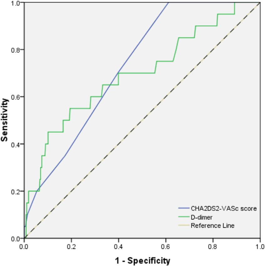
Table 2 shows the results of ROC curve analysis. The cutoff
points of CHA2DS2-VASc score, D-dimer and anteropos-
2.3 Statistical analysis terior diameter of LA were 1.5, 255.5 μg/L and 49.5 mm,
respectively, to predict LAAT. ROC curves are shown in
Included patients were divided into two groups (LAAT Figures 1 and 2.
and non-LAAT). Continuous variables were presented
as median and quartiles, and non-parametric test was
used for inter-group comparisons. Categorical variables
were presented as percentages, and χ2 test was used for 3.3 Logistic regression analysis
comparisons. The receiver operating characteristic (ROC)
curve was used to analyze and determine the appropriate According to the cutoff values, the normal D-dimer group
cutoff point of risk factors to predict LAAT. Logistic (normal group) was defined as the value of D-dimer
regression analyses were performed to find independentRisk factors of left atrial appendage thrombus in patients with non-valvular atrial fibrillation 363 Table 1: Comparisons of baseline characteristics between LAAT and non-LAAT groups Clinical characteristics LAAT (n = 20) Non-LAAT (n = 402) P value Male, n (%) 8 (40) 226 (56.2) 0.154 Age, median (IQR) – years 63 (53–73) 65 (58–72) 0.088 Smoking, n (%) 4 (20) 85 (21.1) 0.903 Drinking, n (%) 3 (15) 32 (8) 0.265 Hypertension, n (%) 16 (80) 246 (61.2) 0.093 Diabetes, n (%) 5 (25) 54 (13.4) 0.147 Coronary artery disease, n (%) 10 (50) 142 (35.3) 0.185 CHA2DS2-VASc score, median (IQR) 3 (2–4) 2 (1–3) 0.001 Platelet count, median (IQR), 109/L 174 (152–217) 184 (148–220) 0.641 PT, median (IQR), s 12.2 (11.4–15.5) 12.0 (11.3–13.6) 0.360 APTT, median (IQR), s 32.9 (30.3–41.85) 32.4 (29.6–37.1) 0.324 D-dimer, median (IQR), μg/L 180.0 (69.5–330.1) 90.0 (49.0–142.8) 0.003 SCr, median (IQR), μmol/L 75.0 (61.3–92.5) 77.0 (65.0–88.0) 0.703 LA anteroposterior diameter, median (IQR), mm 50.5 (44.0–54.4) 41.0 (37.4–44.7)
364 Yaowu Liu et al.
CHA2DS2-VASc score for each patient. They found that
the risk of LAAT increased with increasing CHA2DS2-
VASc score. The CHA2DS2-VASc score was an indepen-
dent risk factor for LAAT (OR 3.26, 95% CI: 2.3–4.65;
p = 0.001) in multivariate logistic analysis [17]. We also
found that patients with LAAT had higher CHA2DS2-
VASc scores compared with patients without LAAT.
However, no exact cutoff point of CHA2DS2-VASc score
was used to predict the risk of LAAT because of the low
specificity (0.39) in this study.
Blood biomarkers were found to be associated with
the presence of LAAT. For example, a study by Habara
showed diagnostic discrimination with D-dimer, with an
OR of 97.6 (95% CI: 17.3–595.8) for LAAT and a 97%
negative predictive value [18]. In our study, patients in
the LAAT group had higher value of D-dimer (180.0
vs 90.0 μg/L, p = 0.003) compared with the non-LAAT
group. However, no exact cutoff point was used to predict
the risk of LAAT because of low sensitivity (0.46). The
difference was that the proportion of recent embolic
Figure 1: ROC analysis of LA anteroposterior diameter in predicting events 2 weeks before TEE was high (23%) in the previous
LAAT. LA = left atrium, LAAT = left atrial appendage thrombus. study.
By analyzing the clinical data of patients with non-
valvular AF who underwent TEE, we found that enlarged
LA anteroposterior diameter was a risk factor of LAAT in
non-valvular AF patients. Scherr et al. included 732 cases
referred for catheter ablation of AF. All patients were
anti-coagulated for ≥4 weeks prior to the procedure.
TEE was performed in all patients within 24 h prior to
ablation. A total of 12 patients had LA thrombus (1.6%),
and larger LA diameter was found to be associated with
LA thrombus (OR = 1.6, 95% CI: 1.1–2.3) [19]. To investi-
gate predictors of LAAT formation in patients with AF,
Nishikii-Tachibana studied 543 AF patients who under-
went TEE before pulmonary vein isolation. All patients
were anti-coagulated with warfarin before ablation and
LAATs were observed in 2.1% of patients. Multivariate
analysis showed that increased LA volume (>50 mL)
was significantly associated with increased prevalence
of LAATs [20]. The two studies included patients treated
with anticoagulants for ≥3 weeks before the ablation. In
this study, we excluded patients who had received
Figure 2: ROC analysis of CHA2DS2-VASc score and D-dimer in
anticoagulant therapy more than 3 weeks before TEE
predicting LAAT. LAAT = left atrial appendage thrombus. tests to avoid the effects of anticoagulants on LAAT.
However, results of the above studies were similar to
our study, and we also found that enlarged LA increased
diabetes mellitus, prior stroke or transient ischemic LAAT risk. Our data revealed that enlarged LA (antero-
attack [doubled], vascular disease, age 65–74, female) posterior diameter ≥49.5 mm) was an independent risk
is the most powerful scoring system for the prediction factor of LAAT (OR = 7.28, p < 0.001). The exact me-
of stroke [16]. Uz et al. reviewed 309 non-valvular chanism by which enlarged LA increases LAAT risk
patients who had undergone TEE, and calculated the remains unclear. Generally, enlargement of the LARisk factors of left atrial appendage thrombus in patients with non-valvular atrial fibrillation 365
Table 3: Logistic regression analysis to evaluate risk factors of LAAT
Variables B SE Wald Adjusted OR (95% CI) P value
D-dimer (elevated/normal) 1.01 0.59 2.92 2.75 (0.86–8.75) 0.088
LA diameter (larger/smaller) 1.99 0.58 11.91 7.28 (2.36–22.47) 0.001
AF type (non-paroxysmal/paroxysmal) 1.55 0.46 10.21 8.89 (2.33–33.99) 0.001
B, regression coefficient; SE, standard error; OR, odds ratio; CI, confidence interval; LA, left atrium; AF, atrial fibrillation.
represents remodeling of the atrial structure, and 5 Conclusion
severe LA remodeling can lead to deterioration of
atrial mechanical function (loss of atrial contractile Enlarged LA (anteroposterior diameter ≥49.5 mm) and
force) resulting in blood flow stagnation and throm- non-paroxysmal AF were independent risk factors of
bosis in the atrium. LAAT in non-valvular AF patients.
In addition, our study revealed that patients with
non-paroxysmal AF were associated with a higher risk Acknowledgments: This study was supported by the
of LAAT compared with patients with paroxysmal AF Project of Nanjing Medical Science and Technology
(OR = 8.89, p < 0.001). This result was different from Development (YKK18258).
traditional clinical research in observing the risk of TE
events in AF patients. In 2000, Hart et al. published a Disclosure: The manuscript was read and approved by all
cohort study comparing 460 subjects with intermittent authors, the requirements for authorship have been met,
AF with 1,552 sustained AF patients treated with aspirin, and each author believes that the manuscript represents
and followed-up for a mean of 2 years. Analysis showed original work, if that information is not provided in
that the annualized rate of ischemic stroke was similar another form.
between those with intermittent (3.2%) and sustained AF
(3.3%) [21]. Diagnosis of intermittent AF required at least Conflict of interest: The authors declare no conflict of
two electrocardiogram-documented episodes before entry, interest.
and no dynamic electrocardiogram (Holter) was used. This
design was easier to include intermittent patients with
relatively high burden of AF, and may be the source of
inconsistent results. Similar to our finding, Scherr et al. References
observed that patients who were in AF at the time of TEE
were more likely to have thrombus compared with all other [1] Björck S, Palaszewski B, Friberg L, Bergfeldt L. Atrial fibrilla-
patients (2.9% vs 0.7%; p = 0.03) [19]. The atrium shrinks tion, stroke risk, and warfarin therapy revisited: a population-
350–600 beats per minute every day during AF, and pro- based study. Stroke. 2013;44(11):3103–8. doi: 10.1161/
STROKEAHA.113.002329.
longed rapid shrinkage may lead to loss of LA function,
[2] Haim M, Hoshen M, Reges O, Rabi Y, Balicer R, Leibowitz
resulting in LAAT. Therefore, patients with persistent AF M. Prospective national study of the prevalence, incidence,
can easily form LAAT compared with patients with paroxy- management and outcome of a large contemporary cohort of
smal AF because of the different durations [22]. patients with incident non-valvular atrial fibrillation. J Am
Finding the risk factors for LAAT is of great signifi- Heart Assoc. 2015;4(1):e001486. doi: 10.1161/
JAHA.114.001486.
cance to identify the high-risk patients. The proportions
[3] Kirchhof P, Benussi S, Kotecha D, Ahlsson A, Atar D, Casadei B,
of LAAT in patients with larger LA (LA anteroposterior dia- et al. ESC Guidelines for the management of atrial fibrillation
meter ≥49.5 mm), non-paroxysmal AF, and both larger LA developed in collaboration with EACTS. Eur Heart J.
and non-paroxysmal AF were 30% (12/40), 15.2% (17/112) 2016;37(38):2893–962. doi: 10.1093/eurheartj/ehw210.
and 39.1% (9/23), respectively, before anticoagulant [4] Andersson T, Magnuson A, Bryngelsson I-L, Frøbert O,
therapy. Therefore, we recommend that TEE should be Henriksson KM, Edvardsson N, et al. All-cause mortality in
2,72,186 patients hospitalized with incident atrial fibrillation
performed if risk factors are detected (LA anteropos-
1995–2008: a Swedish nationwide long-term case-control
terior diameter ≥49.5 mm or/and non-paroxysmal AF) study. Eur Heart J. 2013;34(14):1061–7. doi: 10.1093/
for evaluation of LAAT. eurheartj/ehs469.366 Yaowu Liu et al.
[5] Hahne K, Mönnig G, Samol A. Atrial fibrillation and silent cohort of patients with atrial fibrillation. Eur Heart J.
stroke: links, risks, and challenges. Vasc Health Risk Manage. 2016;37(42):3203–10. doi: 10.1093/eurheartj/ehw077.
2016;12:65–74. doi: 10.2147/VHRM.S81807. [15] Kim T-H, Yang P-S, Uhm J-S, Kim J-Y, Pak H-N, Lee M-H, et al.
[6] Mahajan R, Brooks AG, Sullivan T, Lim HS, Alasady M, CHADS-VASc score (congestive heart failure, hypertension,
Abed HS, et al. Importance of the underlying substrate in age ≥ 75 [doubled], Diabetes mellitus, prior stroke or transient
determining thrombus location in atrial fibrillation: implica- ischemic attack [doubled], vascular disease, age 65–74,
tions for left atrial appendage closure. Heart. female) for stroke in asian patients with atrial fibrillation: a
2012;98(15):1120–6. doi: 10.1136/heartjnl-2012-301799. Korean nationwide sample cohort study. Stroke.
[7] Beigel R, Wunderlich NC, Ho SY, Arsanjani R, Siegel RJ. The left 2017;48(6):1524–30. doi: 10.1161/STROKEAHA.117.016926.
atrial appendage: anatomy, function, and noninvasive eva- [16] Lip GYH, Nieuwlaat R, Pisters R, Lane DA, Crijns HJGM. Refining
luation. JACC Cardiovasc Imaging. 2014;7(12):1251–65. clinical risk stratification for predicting stroke and throm-
doi: 10.1016/j.jcmg.2014.08.009. boembolism in atrial fibrillation using a novel risk factor-
[8] Calkins H, Hindricks G, Cappato R, Kim YH, Saad EB, based approach: the euro heart survey on atrial fibrillation.
Aguinaga L, et al. HRS/EHRA/ECAS/APHRS/SOLAECE expert Chest. 2010;137(2):263–72. doi: 10.1378/chest.09-1584.
consensus statement on catheter and surgical ablation of [17] Uz O, Atalay M, Dogan M, Isilak Z, Yalcin M, Uzun M, et al. The
atrial fibrillation. Heart Rhythm. 2017;14(10):e275–444. CHA2DS2-VASc score as a predictor of left atrial thrombus in
doi: 10.1016/j.hrthm.2017.05.012. patients with non-valvular atrial fibrillation. Med Principles
[9] Ikezawa K, Shigekawa M, Sengoku K, Yoshioka T, Sakamori R, Pract Int J Kuwait Univ Health Sci Cent. 2014;23(3):234–8.
Sakata Y, et al. Left atrial appendage thrombus detected by doi: 10.1159/000361028.
transesophageal examination with linear endoscopic ultra- [18] Habara S, Dote K, Kato M, Sasaki S, Goto K, Takemoto H, et al.
sound. Clin Case Rep. 2019;7(7):1327–30. doi: 10.1002/ Prediction of left atrial appendage thrombi in non-valvular
ccr3.2223. atrial fibrillation. Eur Heart J. 2007;28(18):2217–22.
[10] Lupercio F, Carlos Ruiz J, Briceno DF, Romero J, Villablanca PA, doi: 10.1093/eurheartj/ehm356.
Berardi C, et al. Left atrial appendage morphology assessment [19] Scherr D, Dalal D, Chilukuri K, Dong J, Spragg D, Henrikson CA,
for risk stratification of embolic stroke in patients with atrial et al. Incidence and predictors of left atrial thrombus prior to
fibrillation: a meta-analysis. Heart Rhythm. catheter ablation of atrial fibrillation. J Cardiovasc
2016;13(7):1402–9. doi: 10.1016/j.hrthm.2016.03.042. Electrophysiol. 2009;20(4):379–84. doi: 10.1111/j.1540-
[11] Zimetbaum P. Atrial fibrillation. Ann Intern Med. 8167.2008.01336.x.
2017;166(5):ITC33–48. doi: 10.7326/AITC201703070. [20] Nishikii-Tachibana M, Murakoshi N, Seo Y, Xu D, Yamamoto M,
[12] Hijazi Z, Lindbäck J, Alexander JH, Hanna M, Held C, Hylek EM, Ishizu T, et al. Prevalence and clinical determinants of left
et al. The ABC (age, biomarkers, clinical history) stroke risk atrial appendage thrombus in patients with atrial fibrillation
score: a biomarker-based risk score for predicting stroke in before pulmonary vein isolation. Am J Cardiol.
atrial fibrillation. Eur Heart J. 2016;37(20):1582–90. 2015;116(9):1368–73. doi: 10.1016/j.amjcard.2015.07.055
doi: 10.1093/eurheartj/ehw054. [21] Hart RG, Pearce LA, Rothbart RM, McAnulty JH, Asinger RW,
[13] Zhu W, Fu L, Ding Y, Huang L, Xu Z, Hu J, et al. Meta-analysis of Halperin JL. Stroke with intermittent atrial fibrillation:
ATRIA versus CHADS-VASc for predicting stroke and throm- incidence and predictors during aspirin therapy. Stroke pre-
boembolism in patients with atrial fibrillation. Int J Cardiol. vention in atrial fibrillation investigators. J Am Coll Cardiol.
2017;227:436–42. doi: 10.1016/j.ijcard.2016.11.015. 2000;35(1):183–7. doi: 10.1016/s0735-1097(99)00489-1.
[14] Aspberg S, Chang Y, Atterman A, Bottai M, Go AS, Singer DE. [22] Al-Saady NM, Obel OA, Camm AJ. Left atrial appendage:
Comparison of the ATRIA, CHADS2, and CHA2DS2-VASc stroke structure, function, and role in thromboembolism. Heart.
risk scores in predicting ischaemic stroke in a large Swedish 1999;82(5):547–54. doi: 10.1136/hrt.82.5.547.You can also read