Annals of Clinical Medicine and Research
←
→
Page content transcription
If your browser does not render page correctly, please read the page content below
Annals of Clinical Medicine and Research                                                                                                        Research Article
                                                                                                                                         Published: 06 Aug, 2021

  Demographic Profiles, Distribution of Lesion on CT Brain
  and Outcome in Patients with Spontaneous Intracerebral
   Hemorrhage (ICH) in a Tertiary Care Hospital: A Cross
                      Sectional Study
                                              Md Shoriful Islam1, Richmond Ronald Gomes*2, Monjur Hasan FM3
                                              1
                                                  DNCC dedicated COVID Hospital Dhaka , Dhaka, Bangladesh
                                              2
                                                  Ad-Din Women’s Medical College Hospital, Dhaka, Bangladesh
                                              3
                                                  Ad-Din Sakina Women’s Medical College Hospital, Jashore, Bangladesh

                                              Abstract
                                              Spontaneous Intracerebral Hemorrhage (ICH) has remained is the least treatable form of stroke
                                              despite recent improvements in medical treatment. Treatment usually supportive and medical
                                              such as ventilatory support, blood pressure reduction, osmotherapy, fever control, seizure control
                                              and nutritional support and treatment of comorbid conditions. This study was carried out to see
                                              demographic variability, clinical presentation, causes and outcome of spontaneous intracerebral
                                              hemorrhage.
                                              Methods and Materials: This were a cross sectional observational prospective in study on 50
                                              spontaneous ICH patients admitted in Medicine department of Khulna Medical College Hospital
                                              from November 2020 to April 2021.
                                              Result: The study showed that spontaneous ICH was most common in between 41 to 70 years.
                                              Their age frequency was 14 (28%) in 41 to 50 years, 15 (30%) in 51 to 60 years, 12 (24%) in 61 to 70
                    OPEN ACCESS               years, 5 (10%) in 71 to 80 years and 4 (8%) in more than 81 years age group. Among the patients,
                     *Correspondence:         64% (32) were male and 36% (18) were female. Hemorrhage in the right cerebral hemisphere was
     Richmond Ronald Gomes, Ad-Din            in 52% (26) & in left cerebral hemisphere in 48% (24) patients. Hemorrhage was more common in
    Women’s Medical College Hospital,         the basal ganglia 60% (30), thalamus 14% (7), lobar in 14% (7), pontine in 6% (3), internal capsule
                     Dhaka, Bangladesh,       in 6% (3) patients. Ventricular hemorrhage was present in 36% (18) & absent in 64% (32) patients.
     E-mail: rrichi.dmc.k56@gmail.com         Among 50 cases 88% (44) survived and died 12% (6) and good recovery was in 24% (12), moderately
          Received Date: 12 Jul 2021
                                              disable 22% (11) severely disable 26% (13), vegetative 16% (8) and death 12% (6) patients. Among
                                              the cases GCS Score 8 or less 30% were alive and 12% died and. Among those cases Glasgow coma
         Accepted Date: 03 Aug 2021
                                              scale score 9 or more all were alive. Among patients with Glasgow coma scale score 8 or less good
        Published Date: 06 Aug 2021
                                              recovery was (00), moderately disable (03), severely disable (04), vegetative (08), dead (06). There
                                 Citation:    were statistically significant association (p-value =0.002) between low (8 or less) Glasgow coma scale
  Shoriful Islam Md, Gomes RR, Monjur         Score and outcome of patients with spontaneous ICH. Among patients with Glasgow coma scale
      Hasan FM. Demographic Pro iles,         score 9 or more good recovery (12), moderately disable (08), severely disable (09), vegetative (00),
 Distribution of Lesion on CT Brain and       dead (00). Among 36% (18) patients of ventricular hemorrhage 26% (13) were alive and 10% (05)
 Outcome in Patients with Spontaneous         died. Among 64% (32) patients without ventricular hemorrhage 62% (31) were alive and 2% (01)
   Intracerebral Hemorrhage (ICH) in a        died. There is statistically significant association (p-value =0.01) between ventricular hemorrhage
         Tertiary Care Hospital: A Cross      and outcome of patients with spontaneous ICH.
    Sectional Study. Ann Clin Med Res.
                                              Conclusion: Spontaneous ICH is common in Indian subcontinent. As death occur due to ICH
                       2021; 2(4): 1034.
                                              itself, associated co morbidities or due to complications, management in stroke care unit, High
   Copyright © 2021 Richmond Ronald           dependency unit and Intensive care unit is required.
          Gomes. This is an open access
                                              Keywords: Spontaneous; Intracerebral Hemorrhage; Osmotherapy; Seizure; Glasgow coma
    article distributed under the Creative
                                              scale
   Commons Attribution License, which
   permits unrestricted use, distribution,
                                              Introduction
        and reproduction in any medium,
   provided the original work is properly        Cerebrovascular diseases are the third leading cause of death after heart disease and cancer in
                                     cited.   developed countries. They also come first in terms of causing death and disability in neurologic

Remedy Publications LLC.                                                      1                                         2021 | Volume 2 | Issue 4 | Article 1034
Richmond Ronald Gomes, et al.,                                                                                   Annals of Clinical Medicine and Research

diseases in adults [2]. Non-traumatic intracerebral hemorrhage is            Social Science) 17.0.
bleeding into the parenchyma of the brain that may extend into the
                                                                                 Inclusion criteria:
ventricles and, in rare cases, the subarachnoid space. Spontaneous
intracerebral hemorrhage is second most common causes of stroke                 1. All patients with Spontaneous intracerebral hemorrhage
following ischemic stroke. Depending on the underlying cause of              admitted in Medicine wards of KMCH.
bleeding, intracerebral hemorrhage is classified as either primary or
                                                                                 2. Voluntarily given consent.
secondary. Primary intracerebral hemorrhage accounting for 78% to
88% of all cases, originates from the spontaneous rupture of small               Exclusion criteria:
vessels damaged by chronic hypertension or amyloid angiopathy [5].
                                                                                 1. Patients of traumatic intracerebral hemorrhage
The world-wide incidence of intracerebral hemorrhage from 10 to
20 cases per 100,000 population, [6,7] and increases with age [6,8].             2. Recurrent stroke.
Intracerebral hemorrhage is more common in men than women,
                                                                                 3. Not willing to give informed consent.
particularly those older than 55 years of age [8,9], and in certain
populations, including blacks and Japanese [6,10].                           Results
    Hypertension is the most important risk factor for spontaneous               Table 1 showing age distribution of patients with spontaneous
intracerebral hemorrhage [11]. Intracerebral hemorrhage commonly             ICH. It was most common in between 41 to 70 years. Their age
affects cerebral lobes, basal ganglia, the thalamus, the brain stem          frequency 14 (28%) were in 41 to 50 years, 15 (30%) were in 51 to 60
(predominantly the pons), and the cerebellum as a result of ruptured         years, 12 (24%) were in 61 to 70 years, 5 (10%) were in 71 to 80 years,
vessels [14]. Extension into the ventricles occurs in association with       4 (8%) were more than 81 years old.
deep, large hematomas. The classic presentation of intracerebral
                                                                                 Chi-square (χ2) test was employed to analyses the data. P-value
hemorrhage is sudden onset of a focal neurological deficit that
Richmond Ronald Gomes, et al.,                                                                                                  Annals of Clinical Medicine and Research

Table 3: Hemorrhage site distribution of the participants.                                   Table 8: Relation between GCS Score and outcome of the patients.
         Hemorrhage site                        Frequency                    Percent            Outcome of patients                            GCS score
                 Lobar                              7                          14                                           8 or less      9 or more      Total       P-Value
                 Pontine                            3                          6                    Good recovery               0              12          12
          Internal capsule                          3                          6                   Moderately disable           3              8           11
              Basal ganglia                        30                          60                   Severely disable            4              9           13
                Thalamus                            7                          14            Table 8 represents those relations between GCS score and outcome of
                                                                                             spontaneous intracerebral hemorrhage. Where GCS Score was 8 or less
                  Total                            50                         100            good recovery (00), moderately disable (03), severely disable (04), vegetative
Table 3 shows that hemorrhage occur commonly in the basal ganglia.                           (08), death (06). Hemorrhage with GCS Score 9 or more good recovery (12),
Hemorrhage site distribution - basal ganglia 60% (30), thalamus 14% (7), lobar               moderately disable (08), severely disable (09), vegetative (00), dead (00). There
14% (7), pontine 6% (3) and internal capsule 6% (3).                                         is statistically significant association (p-value =0.000) between GCS score and
                                                                                             outcome of patients with spontaneous intracerebral hemorrhage.
Table 4: Ventricular hemorrhage of the participants.
         Ventricular hemorrhage                    Frequency                  Percent

                   Present                                      18              36

                    Absent                                      32              64

                     Total                                      50              100
Table 4 shows that ventricular hemorrhage was present in 36% (18) & absent in
64% (32) patients.
Table 5: Patient’s condition with spontaneous intracerebral hemorrhage.
         Patient’s condition                     Frequency                   Percent

                   Alive                            44                         88

                  Dead                                  6                      12

                   Total                            50                         100
Table 5 shows patient’s condition with spontaneous intracerebral hemorrhage.                   Figure 1: Sex distribution of the patients.
Among 50 cases more were alive 88% (44) and died 12% (6).                                      Figure 1 showing spontaneous ICH is more common in male than female.
                                                                                               Among 50 cases, 64% (32) were male whereas 36% (18) were female.
Table 6: Relation between GCS^ score and outcome of the patients.
                          Outcome of patient

   GCS Score                    Alive               Death            Total     P-Value        20                                                [](36%)
    8 or less                    15                         6          21
                                                                                              15         [](26%)            [](24%)
   9 or more                     29                         0          29       0.002
                                                                                              10                                                                     [](14%)
      Total                      44                         6          50
Table 6 shows relation between GCS score and outcome of the patients. Among                    5
21 patients with GCS Score 8 or less survived 15 and died 06. Rests of the
                                                                                               0
29 patients with GCS Score 9 or more all were alive. There were statistically
                                                                                                      Farmer/ Day        Businessman         Housemaker           Service-holder
significant association (p-value =0.002) between low (8 or less) GCS Score and
outcome of patients with spontaneous intracerebral hemorrhage.
                                                                                                         Labour
                                                                                               Figure 2: Occupation of the patients.
Table 7: Relation between ventricular hemorrhage and outcome of the patients.
                                                                                               Figure 2 demonstrates that spontaneous intracerebral hemorrhage was
                                 Outcome of patient                                            found in farmer/day labor 26% (13), businessman 24% (12), and housemaker
                                                                                               36% (18), and service holder 14% (7).
     Ventricular hemorrhage             Alive      Death             Total     P-Value

                Present                  13             5             18
                                                                                             16 to 60 years. Study by Adnan et al. [21] showed that compared with
                Absent                   31             1             32        0.01
                                                                                             woman, men had a younger age of onset. All studies have shown a
                 Total                   44             6             50
                                                                                             steep rise in incidence with increasing age.
Table 7 presents relation between ventricular hemorrhage and outcome of the
patients. Among 36% (18) patients with ventricular hemorrhage 26% (13) were                      In this study, spontaneous ICH is more common in male 64%
alive and 10% (05) died. Among 64% (32) patients without ventricular hemorrhage              (32) than female 36% (18). In study by Ong [19] showed that male
62% (31) were alive and 2% (01) died. There is statistically significant association
                                                                                             to female ratio was 1:0.77. Adnan [21], in a study showed that
(p-value =0.01) between ventricular hemorrhage and outcome of patients with
spontaneous intracerebral hemorrhage.                                                        compared with woman, men had a younger age of onset (54 vs. 60
                                                                                             years; p
Richmond Ronald Gomes, et al., Annals of Clinical Medicine and Research
14 [](26%)
[](24%)
12 [](22%)
10
[](16%)
8
[](12%)
6
4
2
0
Good Recovery Moderately Disable Severly Disable Vegetative Death
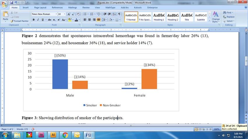
Figure 3: Showing distribution of smoker of the participants.
Figure 3 represents spontaneous ICH more common in smoker. Among 50 Figure 5: Outcome of treatment of spontaneous intracerebral hemorrhage.
cases male smoker 25 (50%) and female 1 (2%) and nonsmoker male 7 Figure 5 shows patients outcome after treatment of spontaneous intracerebral
(14%) and female 17 (34%). hemorrhage. Among 50 cases good recovery 24% (12), moderately disable
22% (11) severely disable 26% (13), vegetative 16% (8) and death 12% (6).
scan was done in all the patients at the time of admission. 46% of
the patients had primarily putaminal bleeding. Lobar bleeding was
present in 28% of patients. Thalamic bleeding in 13% of the patients,
cerebellar bleeding in 3% and pontine bleeding in 4%of patients [15].
Hemorrhage found in ganglio-thalamic 23 (46%), ganglio-capsular
10 (20%), lobar 14 (28%) cerebellar 1(2%) and pontine 2(4%). In
study by Bhatia et al. [17] showed the sites of hematoma included
ganglionic (70.6%), thalamic (16.8%), lobar (4.2), brainstem (7%) and
cerebellar (1.4%).
We found ventricular hemorrhage were present in 36% (18) &
absent in 64% (32) patients. In the study, Kafle [16] showed that 26%
cases had intraventricular extension.
Figure 4: Hemorrhage side of the participants.
Figure 4 presents hemorrhage side distribution which was 52% (26) in the In our study we found outcome of patients with spontaneous
right side and 48% (24) in the left side. intracerebral hemorrhage, 88% (44) were alive and 12% (6) died.
Kafle [16] showed 90% of the patients who were admitted improved
Giulia [22] found men with low SEP (Socioeconomic Position) with and were discharged from the hospital. The mortality rate was 6%.
an ischemic event were more likely to be hospitalized for a new stroke Condition of 4% patients deteriorated and were taken to home on
than men with high SEP. Women with low SEP with hemorrhagic family member’s request. In a study by Yonghong et al. [23] concluded
stroke were more likely to be hospitalized for cardiovascular disease that increased systolic and diastolic blood pressures were significantly
compared with women with high SEP. and positively associated with death and disability among patient
with acute hemorrhagic stroke, but not acute ischemic stroke. Ong
In our study, we found that spontaneous ICH was common in
and Raymond [19] found the mortality at one month was 20.3%. A
poor and middle-class family. Among the patients, poor was 40%
recent study by Zia et al., found that for those younger than 75 years
(20), solvent 58% (29), very good 2% (1). In a study by Giulia [22]
of age, male sex predicted a poor outcome.
showed that stroke incidence strongly differs between socioeconomic
groups reflecting a heterogeneous distribution of lifestyle and clinical In our study we found patients with GCS Score 9 or more all were
risk factors. Strategies for primary prevention should target less alive. Among 42% patients with GCS Score 8 or less, 30% (15) were
affluent people. alive and 12% (06) died. All death was among those having GCS score
were 8 or less. Among patients with GCS Score 8 or less were good
In this study, we found that spontaneous ICH was more common
recovery in 0% (00), moderately disable in 6% (03), severely disable
in smoker. Among 50 cases, male smoker was 25 (50%) and female
in 8% (04), vegetative in 16% (08) and death in 12% (06). In rest
1 (2%) and nonsmoker male was 7 (14%) and female was 17 (34%).
of the cases with GCS Score 9 or more were good recovery in 24%
Kafle [16] showed that 21 percent of patients were smoker. In study
(12), moderately disable in 16% (08), severely disable in 18% (09),
by Zaharia et al. [11] found that, cigarette smoker (13.1%). Doctor
vegetative in 0% (00), death in 0% (00). There is statistically significant
et al. [15] showed in their study, history of smoking was present in
association (p-value =0.000) between GCS score and outcome of
24 cases (48%), all were male and 17 patients (34%) were currently
patients with spontaneous intracerebral hemorrhage. There was
smoking. Craig S Anderson reported history of smoking in 29%
statistically significant association (p-value =0.002) between low
of patients and ex-smoking in 19% of patients out of 60% cases of
(8 or less) GCS Score and outcome of patients with spontaneous
spontaneous intracerebral hemorrhage.
intracerebral hemorrhage.
In our study we found that right sided hemorrhage was in 52%
Most of all deaths in our study occurred among patients with
(26) & left sided was in 48% (24) cases. Hemorrhages were more
ventricular hemorrhage. Among 36% patients of ventricular
common in the basal ganglia. Hemorrhage site distributions were
hemorrhage 10% (05) died and 26% (13) were alive. Among those
basal ganglia 60% (30), thalamus 14% (7), lobar 14% (7), pontine
patients without ventricular hemorrhage 62% (31) were alive and
6% (3) and internal capsule 6% (3). Kafle [16], in their study CT
Remedy Publications LLC. 4 2021 | Volume 2 | Issue 4 | Article 1034Richmond Ronald Gomes, et al., Annals of Clinical Medicine and Research
only 2% (01) died. There is statistically significant association (p-value 10. Broderick JP, Adams HP, Barsan W, Feinberg W, Feldmann E, Grotta
=0.01) between ventricular hemorrhage and outcome of patients with J, et al. Guidelines for the management of spontaneous intracerebral
spontaneous intracerebral hemorrhage. hemorrhage: A statement for healthcare professionals from a special
writing group of the Stroke Council, American Heart Association. Stroke.
Conclusion 1999;30(4):905-15.
Spontaneous ICH is a major cause of morbidity and mortality 11. Zaharia B, Plesea IE, Georgescu CC, Camenta A, Georgescu CV,
Enache SD, et al. morphoclinical study of intracerabral hemorrhage
among stroke patients. Hypertension is the most common cause of
with intraventricular extention. Romanian J Morphol and Embryol.
spontaneous ICH; others are smoking, dyslipidemia, diabetes mellitus 2005;46(3):199-206.
and family history of stroke. Mainstay of treatment is supportive,
including airway maintenance, diabetes control, blood pressure 12. Adams HP Jr, Brott TG, Furlan AJ, Gomez CR, Grotta J, Helgason CM,
et al. Guidelines for thrombolytic therapy for acute stroke: A supplement
control, treatment and prophylaxis of convulsion, temperature
to the guidelines for the management of patients with acute ischemic
control, nutritional support, careful fluid therapy and rehabilitation stroke: A statement for healthcare professionals from a special writing
is also needed for improved mortality and morbidity. group of the Stroke Council, American Heart Association. Circulation.
1996;94(5):1167-74.
Limitation of Study
13. Qureshi AI, Safdar K, Weil J, Barch C, Bliwise DL, Colohan AR, et al.
The present study did not represent the actual scenario of Predictors of early deterioration and mortality in black Americans with
spontaneous ICH in Bangladesh because the study was conducted spontaneous intracerebral hemorrhage. Stroke. 1995;26(10):1764-7.
in one tertiary level hospital (Khulna Medical College and Hospital
14. Mayer SA, Sacco RL, Shi T, Mohr JP. Neurologic deterioration in
(KMCH). Sample size and duration of the study was short. Actual noncomatose patients with supratentorial intracerebral hemorrhage.
measurement of intracranial pressure was not possible. Advanced Neurology. 1994;44(8):1379-84.
investigation facilities (Cerebral angiogram, MRI of brain) were
15. Doctor NM, Pandya RB, Vaghani CV, Marwadi MR, Gheewala GK,
limited. There was no advanced life support available.
Barfiwala VA. A study on clinical profile, risk factors and mortality in
References hypertensive intracerebral hemorrhage in a tertiary care hospital in Surat
city. National J Med Res. 2013;4(3):381-4.
1. Sacco RL. Risk factors, outcomes, and stroke subtypes for ischemic stroke.
Neurology. 1997;49(5 suppl 4):S39-44. 16. Kafle DR. Outcome of patients with spontaneous intracerebral hemorrhage
at a tertiary care hospital in Nepal. J Nobel Med College. 2013;4(2):35-43.
2. Foulkes MA, Wolf PA, Price TR, Mohr JP, Hier DB. The Stroke Data Bank:
Design, methods, and baseline characteristics. Stroke. 1988;19(5):547-54. 17. Bhatia R, Singh H, Singh S, Padma MV, Prasad K, Tripathi M, et al. A
prospective study of in-hospital mortality and discharge outcome in
3. Broderick JP, Brott T, Tomsick T, Huster G, Miller R. The risk of spontaneous intracerebral hemorrhage. Neurol India. 2013;61(3):244-8.
subarachnoid and intracerebral hemorrhages in blacks as compared with
whites. N Engl J Med. 1992;326:733-6. 18. Hsiang J, Zhu X, Wong L, Kay R, Poom W. Putaminal and thalamic
hemorrhage in ethnic Chinese living in Hong Kong. Surg Neurol.
4. Furlan AJ, Whisnant JP, Elveback LR. The decreasing incidence of 2009;46(5):441-5.
primary intracerebral hemorrhage: A population study. Ann Neurol.
1979;5(4):367-73. 19. Ong TZ and Raymond AA. Risk factors for stroke and predictors of one-
month morality. Singapore Med J. 2002;43(10):517-21.
5. Giroud M, Gras P, Chadan N, Beuriat P, Milan C, Arveux P, et al. Cerebral
hemorrhage in a French prospective population study. J Neurol Neurosurg 20. Juvela S. Risk factors for impaired outcome after spontaneous intracerebral
Psychiatry. 1991;54:595-598. hemorrhage. Arch Neutol. 1995;52(12):1193-200.
6. Sacco RL, Mayer SA. Epidemiology of intracerebral hemorrhage. In: 21. Adnan IQ, Suri MA, Safdar K, Jeffery RO, Robert SJ, Michael RF.
Feldmann E, editor. Intracerebral hemorrhage. Armonk, N.Y.: Futura Intracerebral hemorrhage in blacks: Risk factors, subtypes, and outcome.
Publishing. 1994:3-23. Stroke. 1997;28(5):961-4.
7. Suzuki K, Kutsuzawa T, Takita K, Ito M, Sakamoto T, Hirayama A, 22. Giulia C, Nera A, Francesco F, Alberto PC. Socioeconomic differences
et al. Clinico-epidemiologic study of stroke in Akita, Japan. Stroke in stroke incidence and prognosis under a universal healthcare system.
1987;18(2):402-6. Stroke. 2009;40(8):2812-9.
8. Brott T, Thalinger K, Hertzberg V. Hypertension as a risk factor for 23. Yonghong Z, Reilly KH, Tong W, Tan Xu, Chen J, Bazzano LA. Blood
spontaneous intracerebral hemorrhage. Stroke 1986;17(6):1078-83. pressure and clinical outcome among patients with acute stroke in linner
Mongolia, China. J hypertens. 2008;26(7):1446-52.
9. Mutlu N, Berry RG, Alpers BJ. Massive cerebral hemorrhage: Clinical and
pathological correlations. Arch Neurol. 1963;8:644-61.
Remedy Publications LLC. 5 2021 | Volume 2 | Issue 4 | Article 1034You can also read