Clinical Features of Ulnar Tunnel Syndrome and the Diagnostic Value of Nerve Conduction Measurements
←
→
Page content transcription
If your browser does not render page correctly, please read the page content below
doi: 10.2490/prm.20210010                                               Progress in Rehabilitation Medicine 2021; Vol. 6, 20210010

ORIGINAL ARTICLE
      Clinical Features of Ulnar Tunnel Syndrome and the
     Diagnostic Value of Nerve Conduction Measurements
   Shingo Nobuta, MD, PhD a Hiroshi Okuno, MD, PhD a Taku Hatta, MD, PhD a Ryo Sato, MD, PhD a
                                     and Eiji Itoi, MD, PhD b

              Objectives: The purposes of this study were to assess the clinical features of ulnar tunnel
              syndrome (UTS) and to investigate the diagnostic value of nerve conduction measurements for
              UTS. Methods: Eighteen patients with UTS were reviewed retrospectively. Fifteen patients had
              intrinsic muscle atrophy and motor weakness, and 15 had numbness with hypesthesia. The com-
              pound muscle action potentials (CMAPs) from the first dorsal interosseous (FDI) muscle and
              the abductor digiti minimi (ADM) muscle and the sensory nerve action potential (SNAP) from
              the little finger were recorded and analyzed. All patients underwent ulnar tunnel release surgery
              and neurolysis. Static two-point discrimination test results and pinch strengths were assessed
              before and after surgery. Results: Before surgery, FDI-CMAP was recorded in 17 patients, and
              ADM-CMAP in 16, and all showed delayed latency and/or low amplitude. SNAP was recorded in
              eight patients and two showed delayed latency. The causes of ulnar nerve lesions were ganglion in
              five patients, traumatic adhesion in four, ulnar artery aberrancy in four, pisohamate arch in three,
              anomalous muscle in one, and ulnar vein varix in one. The sites of the lesions were in zone 1 of
              the ulnar tunnel anatomy in 12 patients, in zone 2 in 2, and in zones 1 and 2 in 4. After surgery, all
              patients obtained recovery of motor function and sensation; however, postoperative FDI-CMAP
              and ADM-CMAP did not improve to the normal range. Conclusions: The causes of UTS were
              ganglion, traumatic adhesion, ulnar artery aberrancy, and pisohamate arch. Both FDI-CMAP and
              ADM-CMAP were valuable for electrophysiological diagnosis of UTS.

            Key Words: abductor digiti minimi; first dorsal interosseous; nerve conduction measurement;
                       ulnar nerve lesion; ulnar tunnel syndrome

                     INTRODUCTION                                       reported five cases of UTS caused by ganglion.16) In 1861,
                                                                        Guyon19) reported the anatomy of the ulnar area and predict-
   Ulnar tunnel syndrome (UTS) is an uncommon form of                   ed that problems could occur with entrapment of the ulnar
ulnar entrapment neuropathy at the wrist that can be caused             nerve, and in 1908 Hunt20) described three patients with oc-
by several intrinsic or extrinsic factors.1–4) A review of the          cupational neuritis. Seddon5) and Richmond6) reported ulnar
literature indicated the following possible causes of UTS:              nerve palsy caused by a carpal ganglion. In 1965, Dupont
ganglions,5–16) traumatic neuropathies,8) anomalous muscle              et al.7) used the term ulnar tunnel syndrome and reported
or fibrous bands,10,17,18) ulnar artery thromboses or aber-             four cases. An exact clinical diagnosis of UTS and detection
rancy,7,10) wrist fracture,5) carpal osteoarthritis,8) pisohamate       of the location of the causative lesion are difficult, and elec-
arch,11) and idiopathic.3) Most previous reports have dealt             trophysiological diagnosis may help to confirm the diagno-
with a small number of cases,5–7,11–18) and we previously               sis.2–4,11,13–16,21–25) The purposes of this study were to assess

Received: October 16, 2020, Accepted: January 27, 2021, Published online: February 13, 2021
a Department of Orthopaedic Surgery, Tohoku Rosai Hospital, Sendai, Japan
b Department of Orthopaedic Surgery, Tohoku University School of Medicine, Sendai, Japan

Correspondence: Shingo Nobuta, MD, PhD, Department of Orthopaedic Surgery, Tohoku Rosai Hospital, 4-3-21 Dainohara, Aoba-ku,
Sendai, Miyagi 981-8563, Japan, E-mail: s-nobuta@tohokuh.johas.go.jp
Copyright © 2021 The Japanese Association of Rehabilitation Medicine
            This is an open-access article distributed under the terms of the Creative Commons Attribution Non-Commercial No
            Derivatives (CC BY-NC-ND) 4.0 License. http://creativecommons.org/licenses/by-nc-nd/4.0/

                                                                    1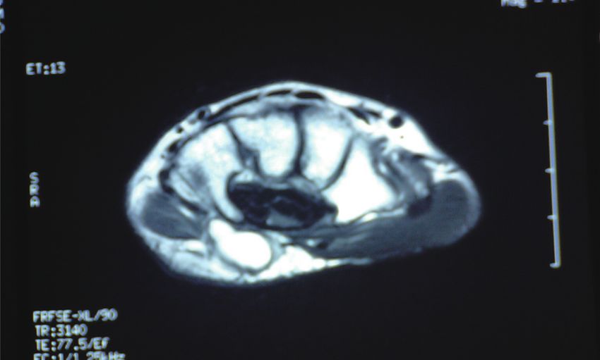
2                                                                 Nobuta S, et al: Clinical Features of Ulnar Tunnel Syndrome

Table 1. Details and test results for eighteen ulnar tunnel syndrome patients
                                                                                                       Follow up
   Case     Age       Sex        Side     Duration of   Hypesthesia      TPD        Pinch       m         TPD       Pinch
                                           symptoms                     (mm)         (kg)                 (mm)       (kg)
     1.      34        F           L          7m           p, d          40          0           6         35         0.3
     2.      38        M           R          1m           p, d           15         0           6          5         2.0
     3.      36        M           R          2m           p, d          20          2.8         6          5         4.5
     4.      41        F           R         84 m          none            5         2.5       13           5         3.3
     5.      80        F           R          5m           p, d           45         1.8       17          10         3.2
     6.      33        F           R          8m           p, d           15         2.6         9          7         3.4
     7.      76        F           R          2m           p, d           30         0.8         6         10         3.4
     8.      68        M           L          5m           p, d           30         1.5         8          7         3.5
     9.      57        M           R          5m           p, d           10         2.4       54           7         4.2
    10.      44        F           R         24 m            p            15         2.3         7          7         3.5
    11.      58        M           R          1m             p           40          2.8       10          15         4.5
    12.      54        F           R          1m             p            10         4.2       12           5         4.5
    13.      61        M           R          5m           p, d          25          4.2       48          10         4.5
    14.      45        F           R          5m             p            10         2.0         5          5         4.0
    15.      56        F           L          3m             p             5         0.5         7          5         3.6
    16.      66        F           L          2m          none             7         1.3       10           5         4.2
    17.      64        M           R          3m             p           25          4.0         7         20         7.0
    18.      56        M           R          1m          none             5         2.1         6          5         7.4
 F, female; M, male; L, left; R, right; m, months; p, palmar side; d, dorsal side; TPD, two-point discrimination; Pinch, pulp
pinch strength.

the clinical features of UTS and to investigate the diagnostic     and on both the palmar and dorsal sides in nine (Table 1).
value of nerve conduction measurements for UTS.                    A Tinel-like sign at the ulnar tunnel was not seen in any of
                                                                   these patients. The intrinsic muscles included the first dorsal
              PATIENTS AND METHODS                                 interosseous (FDI) and the abductor digiti minimi (ADM).
                                                                   The results of the static two-point discrimination (TPD) test
  Eighteen hands from 18 patients with UTS treated between         on the little finger ranged from 5 to 45 mm, with a mean of
May 2008 and July 2016 were reviewed after a mean follow-          19.6 mm. The pulp pinch strength ranged from 0 to 4.2 kg,
up of 11 months (range, 5–54 months). Details of the 18 cases      with a mean of 2.1 kg (Table 2). T1-weighted MRI of the
are shown in Table 1. The ages of the patients (8 men and 10       wrist in 17 patients demonstrated a soft tissue mass in 5
women) at surgery ranged from 33 to 80 years, with a mean          patients16) (cases 12,14–16, and 18), a high-signal area in 8
age of 53 years. The right side was affected in 14 patients,       (cases 1–4, 6, 8, 9, and 17), and normal findings in 4 (cases
and the dominant extremity was involved in 12 patients.            5, 7, 10, and 13).
The mean duration of symptoms was 9 months (range, 1–84               Nerve conduction measurements were performed before
months). UTS was diagnosed based on clinical symptoms,             and after surgery. The compound muscle action potentials
electrophysiological evaluations, and magnetic resonance           (CMAPs) from the FDI and ADM and the sensory nerve
imaging (MRI) findings. Written informed consent was               action potential (SNAP) from the little finger were recorded
obtained from each patient. All patients except for three          and analyzed. We used a Nicolet Viking electromyography
(cases 12, 13, and 17) had motor weakness and atrophy of           system (Nicolet Instruments, Madison, WI, USA) and a 10-
the intrinsic muscles with a positive Froment’s sign and a         mm silver disc. The palmar skin temperature was not allowed
claw finger deformity of the little finger. Fifteen patients had   to fall below 32°C. FDI-CMAP and ADM-CMAP were
numbness and hypesthesia in the ulnar nerve distribution;          recorded by supramaximal stimulation of the ulnar nerve at
hypesthesia was seen only on the palmar side in six patients       the wrist. The stimulus duration was 0.2–0.5 ms. SNAP was

                              Copyright © 2021 The Japanese Association of Rehabilitation Medicine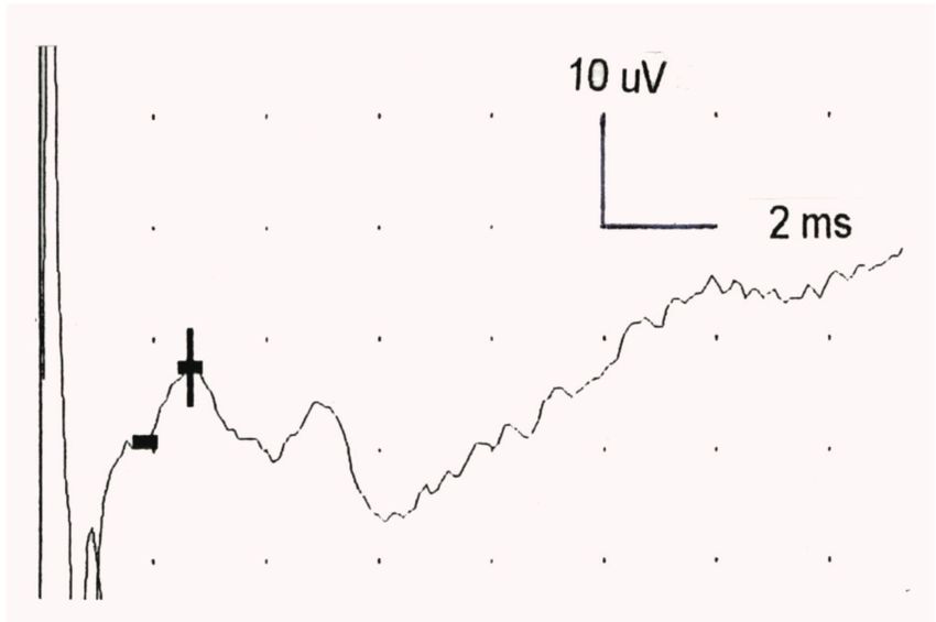
Prog. Rehabil. Med. 2021; Vol.6, 20210010                                                                                     3

Table 2. Overall results before and after surgery for UTS
                                                          Preoperative                Final follow-up         P value
 Static TPD test on little finger (mm) (SD)                 19.6 (13.0)                   9.3 (7.6)
4 Nobuta S, et al: Clinical Features of Ulnar Tunnel Syndrome
Table 3. Details of causes, zone, and nerve conduction measurements in UTS patients
Parameter measurements before / after surgery
FDI-CMAP ADM-CMAP SNAP
Case Cause Zone Lat. (ms) Amp. (mV) Lat. (ms) Amp. (mV) Lat. (ms) Amp (μV)
1. ua 1 3.4/2.6 0.9/1.7 2.4/2.6 1.5/2.3 nr
2. am 1 3.8/4.7 1.0/1.5 3.9/4.6 0.3/5.0 4.2/nr 2.0/nr
3. uv 1 6.0/3.9 1.3/2.1 6.6/4.5 0.2/2.3 8.9/nr 1/nr
4. ta 2 3.9/4.0 4.4/2.9 3.0/2.4 3.8/6.2 nr
5. ta 1 17.0/3.7 0.05/0.2 ur/3.6 ur/0.5 nr
6. ua 1 4.5/4.4 3.5/6.5 nr 2.0/nr 5.0/nr
7. ua 1 ur/6.3 ur/0.5 12.7/4.1 0.1/0.6 nr
8. pa 1, 2 6.5/5.1 0.1/1.3 3.1/3.2 0.6/0.4 nr
9. pa 1, 2 7.5/5.7 0.5/0.6 3.5/5.4 0.6/1.0 nr
10. pa 1, 2 5.1/4.2 0.7/0.7 4.9/3.7 0.3/1.2 nr
11. ta 1 10.3/6.1 0.1/1.5 6.2/4.2 0.1/4.1 3.1/5.0 2.0/5.0
12. gl 1 5.6/5.0 10.7/10.3 6.0/4.1 2.1/1.8 2.5/1.7 25/48
13. ta 1 5.2/4.7 0.8/0.9 3.8/3.1 5.0/3.3 nr
14. gl 1, 2 12.6/5.3 0.1/4.0 11.8/3.9 0.2/4.4 2.6/2.8 10/10
15. gl 2 6.5/4.9 0.6/6.8 3.1/3.1 3.6/5.6 nr
16. gl 1 4.4/3.7 0.1/0.8 3.5/2.9 0.5/0.4 2.0/nr 8.0/nr
17. ua 1 4.8/3.7 1.3/1.1 4.5/4.5 0.7/1.7 nr
18. gl 1 6.4/4.2 1.5/12.2 2.6/2.7 0.5/6.6 2.6/nr 20/nr
Underlined data are within the normal range.
ua, ulnar artery aberrancy; am, anomalous muscle; uv, ulnar vein varix; ta, traumatic adhesion; pa, pisohamate arch; gl,
ganglion; Lat., latency; Amp., amplitude; nr, normal range.
patients. Case Presentation
After surgery, all patients recovered motor function and A 45-year-old right-hand-dominant woman (case 14)
sensation. The mean TPD improved from 19.6 to 9.3 mm (P presented with a 5-month history of onset and intrinsicProg. Rehabil. Med. 2021; Vol.6, 20210010 5
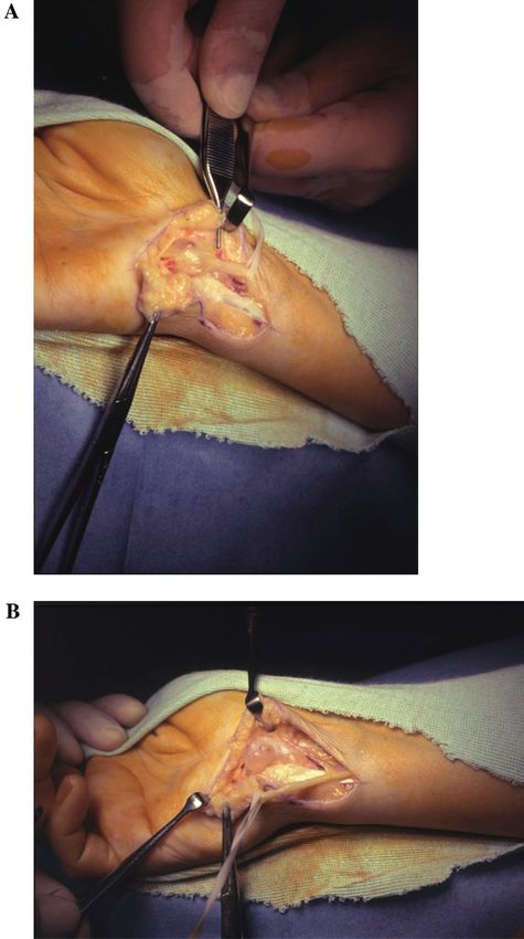
Fig. 1. Axial T1-weighted MRI showed a cystic mass lesion
at the ulnar tunnel (arrow) in case 14.
Fig. 3. ADM data for case 14. (A) Preoperatively, ADM-
CMAP latency was 11.8 ms with an amplitude of 0.1 mV.
(B) Five months after surgery, latency was 3.9 ms with an
amplitude of 4.4 mV.
Fig. 4. SNAP data for case 14 showed normal latency (2.6
Fig. 2. FDI data for case 14. (A) Preoperatively, FDI- ms) and amplitude (8 μV).
CMAP latency was 12.6 ms with an amplitude of 0.1 mV.
(B) Five months after surgery, latency was 5.3 ms with an
bifurcated, and zone 3 surrounds the superficial or sensory
amplitude of 4.0 mV.
branch of the ulnar nerve. Depending on the site of compres-
sion, clinically, the lesion may be in the motor, sensory, or
CMAP and ADM-CMAP revealed a shortened latency and mixed branch,15) whereas the sensory branch on the dorsal
an increased amplitude (Figs. 2B, 3B). ulnar side is normal in UTS.4) However, if paresthesia is seen
on the dorsal ulnar side of the hand, the likely lesion site
DISCUSSION is the cubital tunnel.4) In our series, compression involved
zone 1 in 16 cases (89%; 12 cases in zone 1 only and 4 cases
The ulnar tunnel is anatomically classified into three in zones 1 and 2) and zone 2 in 6 cases (2 cases in zone 2
zones12): zone 1 is the area proximal to the bifurcation of only and 4 cases in zones 1 and 2). For the two cases with
the ulnar nerve, zone 2 encompasses the motor branch of compression in zone 2 only, case 4 showed no sensory loss
the ulnar nerve (except the branch to the ADM) after it has and case 15 showed palmar side hypesthesia. Hypesthesia
Copyright © 2021 The Japanese Association of Rehabilitation Medicine6 Nobuta S, et al: Clinical Features of Ulnar Tunnel Syndrome
Fig. 6. Photomicrograph (hematoxylin-eosin stain, original
magnification ×20) showing the ganglion cyst with a thick-
walled cystic space and focal myxoid change in the sur-
rounding matrix in case 14.
that ulnar nerve lesion in zones 1 and 2 are likely caused by
ganglions or fractures of the hamate, and that lesions in zone
Fig. 5. (A) Intraoperative photograph of a ganglion which 3 are caused by vascular lesions resulting from thrombosis
was mainly compressing the motor branch of the ulnar nerve or aneurysm.12) However, in our series, the causes of zone
at zones 1 and 2 (arrow) in case 14. (B) The ganglion arose 1 lesions were ganglions, ulnar artery aberrancy, traumatic
from triquetrohamate joint. adhesion, anomalous muscle, and ulnar vein varix. Murata et
al.3) stated that surgical exploration is the only reliable way
to clarify the site of compression.
was seen only on the palmar side in six patients, and both The expected theoretical symptoms of compressions in the
on the palmar and dorsal sides in nine patients. These het- three anatomical zones are: zone 1 lesions – delayed ADM
erogeneous findings underline the fact that an exact clinical latency and FDI latency and diminished SNAP amplitude;
diagnosis of UTS and detection of the location of the lesion zone 2 compression – delayed FDI latency according to the
are often difficult. A possible explanation for the nine pa- site of compression, although ADM latency may be normal;
tients who had hypesthesia both on the palmar and dorsal and zone 3 lesions – diminished SNAP amplitude and
sides is the presence of an aberrant dorsal sensory branch normal ADM-CMAP and FDI-CMAP.4) In our series of 18
of the ulnar nerve which diverged with the palmar sensory cases, there were 16 cases of zone 1 compression; of these,
branch in the ulnar tunnel. Murata et al.3) reported that 90% 14 showed delayed FDI latency, 15 showed low FDI ampli-
of cases with UTS were in zone 1. A previous report stated tude, 14 had delayed ADM latency, and 14 had low ADM
Copyright © 2021 The Japanese Association of Rehabilitation MedicineProg. Rehabil. Med. 2021; Vol.6, 20210010 7
amplitude. Of the eight patients who underwent SNAP, the and ADM-CMAP did not improve to the normal range,
results were normal in six (Table 3). Consequently, both and residual delayed latency and low amplitude were seen
FDI-CMAP and ADM-CMAP were valuable for a definite despite recovery of the intrinsic muscles (Tables 2 and 3).
electrodiagnosis of UTS; however, SNAP was not useful for In these cases, from the viewpoint of neurophysiology, my-
confirming the diagnosis. Cases 12, 13, and 17 showed ad- elinization and axonal regeneration of fibers in the FDI and
equate pinch strengths (4.2, 4.2, and 4.0 kg, respectively), but ADM branches were insufficient, notwithstanding a mean
cases 13 and 17 had low FDI-CMAP amplitudes. These two follow-up of 11 months.
cases originally had adequate pinch strengths on the normal There were several limitations to this study. First, we did
side (5.4 and 5.2 kg, respectively); therefore, the decrease in not investigate the relationship between MRI findings and
pinch strength before surgery and rehabilitation was small. the causes of UTS. Second, we could not clarify the relation-
Surgery was indicated for these cases with severe numbness ship between the electrophysiological data and the recovery
and pain in the ulnar nerve distribution. time for intrinsic muscles. Third, to detect further improve-
Lumbrical-interossei motor studies21) and short segment ment of FDI-CMAP and ADM-CMAP, longer follow-up is
incremental studies (SSIS, inching method) of FDI-CMAP needed.
have been reported and indicated that SSIS was valuable for
diagnosis of the precise localization of UTS.22–25) Neverthe- CONCLUSIONS
less, SSIS is somewhat time-consuming and technically
difficult,24) particularly stimulating a site on the palmar side. The causes of UTS in our series were ganglion, traumatic
Accordingly, we performed traditional nerve conduction adhesion, ulnar artery aberrancy, or pisohamate arch. Both
measurements of CMAPs and SNAP. Murata et al.3) reported FDI-CMAP and ADM-CMAP were valuable for electro-
normal values for ADM-CMAP latency of 2.5 mV, and a SNAP latency of 15 μV. In the current series, based on our crite- covery of intrinsic muscles.
ria, we identified delayed latencies and low amplitudes for
CMAPs and SNAPs. ACKNOWLEDGMENTS
Nerve conduction measurements for UTS have been re-
ported,2,3,10,11,13–16) and they all indicated delayed conduction The authors are grateful to Ms. Yumi Watabe, Ms. Hiromi
at the wrist. However, few studies have examined nerve Takeda, Ms. Yoko Kusakari, Pathologist Noriyuki Iwama,
conduction before and after surgery.2,11,14) Uriburu et al.11) MD, Fumie Nakayama, MD, and Honorary Director Kat-
reported three cases of UTS and found that FDI-CMAP sumi Sato, MD, Tohoku Rosai Hospital, for assisting in the
was recordable in one case after surgery. Moreover, post- manuscript preparation.
operatively, FDI latency was shortened from 7 to 4 ms in
one patient and from 24 to 4 ms in another. Ebeling et al.2) CONFLICTS OF INTEREST
described nine cases of UTS and found that FDI latency was
shortened postoperatively. Erkin et al.14) reported a patient The authors declare that there are no conflicts of interest.
with a ganglion and found that the FDI latency was shortened
REFERENCES
from 3.5 to 3.2 ms and the FDI amplitude increased from 2.1
to 5.4 mV postoperatively. Inaparthy et al.15) reported that the
time for hypothenar muscles to recover to the normal range 1. Shea JD, McCLAIN EJ: Ulnar-nerve compression
was 12 to 14 weeks in their patients. In our series of 18 pa- syndromes at and below the wrist. J Bone Joint Surg
tients, after rehabilitation, the mean pinch strength increased Am 1969;51:1095–1103. DOI:10.2106/00004623-
from 2.1 to 3.1 kg at 2 months after surgery. In a previous 196951060-00004, PMID:5805411
report, we described five cases of UTS caused by ganglion 2. Ebeling P, Gilliatt RW, Thomas PK: A clinical and elec-
and found that both FDI-CMAP and ADM-CMAP were trical study of ulnar nerve lesions in the hand. J Neurol
valuable for electrophysiological diagnosis 16); however, the Neurosurg Psychiatry 1960;23:1–9. DOI:10.1136/
current study describes 18 cases of UTS with various causes, jnnp.23.1.1, PMID:13819155
including ganglion, and the results were incidentally similar
to those of the past report. In our current series, FDI-CMAP
Copyright © 2021 The Japanese Association of Rehabilitation Medicine8 Nobuta S, et al: Clinical Features of Ulnar Tunnel Syndrome
3. Murata K, Shih JT, Tsai TM: Causes of ulnar tun- 15. Inaparthy PK, Anwar F, Botchu R, Jähnich H, Katchbu-
nel syndrome: a retrospective study of 31 subjects. J rian MV: Compression of the deep branch of the ulnar
Hand Surg Am 2003;28:647–651. DOI:10.1016/S0363- nerve in Guyon’s canal by a ganglion: two cases. Arch
5023(03)00147-3, PMID:12877855 Orthop Trauma Surg 2008;128:641–643. DOI:10.1007/
4. Chen SH, Tsai TM: Ulnar tunnel syndrome. J Hand Surg s00402-008-0636-4, PMID:18509691
Am 2014;39:571–579. DOI:10.1016/j.jhsa.2013.08.102, 16. Nobuta S, Sonofuchi K, Itoi E: Electrophysiological
PMID:24559635 features of ulnar tunnel syndrome caused by gan-
5. Seddon HJ: Carpal ganglion as a cause of paralysis glion − a descriptive study. Int J Phys Med Rehabil
of the deep branch of the ulnar nerve. J Bone Joint 2018;06:496. DOI:10.4172/2329-9096.1000494
Surg Br 1952;34-B:386–390. DOI:10.1302/0301- 17. Hirooka T, Hashizume H, Nagoshi M, Shigeyama Y,
620X.34B3.386, PMID:12999919 Inoue H: Guyon’s canal syndrome. A different clini-
6. Richmond DA: Carpal ganglion with ulnar nerve cal presentation caused by an atypical fibrous band.
compression. J Bone Joint Surg Br 1963;45-B:513–515. J Hand Surg Am 1997;22:52–53. DOI:10.1016/S0266-
DOI:10.1302/0301-620X.45B3.513, PMID:14058327 7681(97)80016-2, PMID:9061525
7. Dupont C, Cloutier G, Prévost Y, Dion MA: Ulnar- 18. Spinner RJ, Lins RE, Spinner M: Compression of
tunnel syndrome at the wrist. J Bone Joint Surg Am the medial half of the deep branch of the ulnar nerve
1965;47:757–761. DOI:10.2106/00004623-196547040- by an anomalous origin of the flexor digiti minimi.
00010, PMID:14299666 A case report. J Bone Joint Surg Am 1996;78:427–
8. Vanderpool DW, Chalmers J, Lamb DW, Whiston 430. DOI:10.2106/00004623-199603000-00015,
TB: Peripheral compression lesions of the ulnar PMID:8613451
nerve. J Bone Joint Surg Br 1968;50-B:792–803. 19. Guyon F: Note sur une disposition anatomique propre a
DOI:10.1302/0301-620X.50B4.792, PMID:4303276 la face anterieure de la region du poignet et non encore
9. Hayes JR, Mulholland RC, O’Connor BT: Compres- decrite. Bull Soc Anat Paris 1861;6:184–186.
sion of the deep palmar branch of the ulnar nerve. 20. Hunt JR: Occupation neuritis of the deep palmar branch
Case report and anatomical study. J Bone Joint of the ulnar nerve. J OF Nerv Ment Dis 1908;35:673–
Surg Br 1969;51-B:469–472. DOI:10.1302/0301- 689. DOI:10.1097/00005053-190811000-00001
620X.51B3.469, PMID:5820788 21. Kothari MJ, Preston DC, Logigian EL: Lumbrical-
10. Kleinert H, Hayes JE: The ulnar tunnel syndrome. Plast interossei motor studies localize ulnar neuropa-
Reconstr Surg 1971;47:21–24. DOI:10.1097/00006534- thy at the wrist. Muscle Nerve 1996;19:170–174.
197101000-00005, PMID:4320782 DOI:10.1002/(SICI)1097-4598(199602)19:23.0.CO;2-B, PMID:8559165
drome of the deep motor branch of the ulnar nerve. 22. Hatori M, Sakurai M, Miyasaka Y, Nobuta S: Elec-
(Piso-Hamate Hiatus syndrome). J Bone Joint Surg Am trodiagnosis of ulnar tunnel syndrome by inching
1976;58:145–147. DOI:10.2106/00004623-197658010- technique. J Jpn Soc Surg Hand 1989;6:346–350.
00032, PMID:1249106 23. McIntosh KA, Preston DC, Logigian EL: Short-
12. Gross MS, Gelberman RH: The anatomy of the distal segment incremental studies to localize ulnar nerve
ulnar tunnel. Clin Orthop Relat Res 1985;&NA;238– entrapment at the wrist. Neurology 1998;50:303–306.
247. DOI:10.1097/00003086-198506000-00033, DOI:10.1212/WNL.50.1.303, PMID:9443503
PMID:3995823 24. Cowdery SR, Preston DC, Herrmann DN, Logigian
13. Kuschner SH, Gelberman RH, Jennings C: Ulnar EL: Electrodiagnosis of ulnar neuropathy at the wrist:
nerve compression at the wrist. J Hand Surg Am conduction block versus traditional tests. Neurol-
1988;13:577–580. DOI:10.1016/S0363-5023(88)80100- ogy 2002;59:420–427. DOI:10.1212/WNL.59.3.420,
X, PMID:3418064 PMID:12177377
14. Erkin G, Uysal H, Keleş I, Aybay C, Özel S: Acute 25. Seror P: Electrophysiological pattern of 53 cases of
ulnar neuropathy at the wrist: a case report and review ulnar nerve lesion at the wrist. Neurophysiol Clin
of the literature. Rheumatol Int 2006;27:191–196. 2013;43:95–103. DOI:10.1016/j.neucli.2012.11.037,
DOI:10.1007/s00296-006-0166-8, PMID:16896989 PMID:23540258
Copyright © 2021 The Japanese Association of Rehabilitation MedicineYou can also read