Laparoscopic surgery for urachal remnants in pubescent children: a case series
←
→
Page content transcription
If your browser does not render page correctly, please read the page content below
Hashizume et al. Surgical Case Reports     (2020) 6:120
https://doi.org/10.1186/s40792-020-00884-z

 CASE REPORT                                                                                                                                       Open Access

Laparoscopic surgery for urachal remnants
in pubescent children: a case series
Naoki Hashizume1,2,3* , Masahiro Ohtaki1, Kouei Nihei4, Kaoru Sakamoto2, Yasuhiro Shirahata2, Tetsuya Shimada2,
Eriko Ohta2, Daisuke Yamai2, Akihiro Takeshi2, Kaito Sato2, Satoshi Suzuki2 and Minoru Yagi3

  Abstract
  Background: Various techniques are applied in laparoscopic surgery for the treatment of urachal remnants, which
  are less invasive and associated with lower morbidity. We herein report a case series in which we treated urachal
  remnants and medial umbilical ligaments using a laparoscopic approach.
  Case presentation: From 2015 to 2019, seven patients (male, n = 5; female, n = 2) with a urachal remnant were
  treated by laparoscopic surgery in our institute. Five boys and two girls with a median age of 11 years (range 10–
  15 years) were enrolled in this series. The clinical results of laparoscopic treatment, the perioperative records, and
  the pathologic results were evaluated. The operation was performed with the use of three ports and an EZ access®
  (Hakko Medical, Nagano, Japan), which is a silicon cap for the wound retractor (Lap Protector®, Hakko Medical,
  Nagano, Japan). The removal of the urachal remnant and medial umbilical ligaments was completed with a median
  operative time of 92 min (range 69–128). The median hospital stay after surgery was 4 days (range 2–5). No patients
  developed intra-postoperative complications or recurrence.
  Conclusions: Although our data are preliminary, complete laparoscopic removal of symptomatic urachal remnants
  and medial umbilical ligaments was a safe and effective minimally invasive approach, with better cosmetic
  outcomes.
  Keywords: Laparoscopic surgery, Urachal remnant, Children

Introduction                                                                          urachal remnants have been reported, which are less in-
Urachal remnants occasionally require intervention                                    vasive and associated with lower morbidity [3–5]. We
when they become infected and symptomatic [1, 2].                                     herein report our surgical techniques and the devices
Intervention is recommended over drainage of the ab-                                  that we used in performing laparoscopic surgery for the
scess cavity and antibiotic therapy in order to reduce the                            treatment of the urachal remnant and the medial umbil-
risk of recurrence and the potential for malignant                                    ical ligaments in pubescent children.
change of the urachal remnant. The traditional approach
for removing a urachal remnant has been open surgery
                                                                                      Case presentation
with a hypogastric transverse or midline infra-umbilical
                                                                                      Patients
incision, which is associated with increased morbidity
                                                                                      Seven patients of 10 to 15 years of age (male, n = 5; female,
and longer convalescence. Recently, various techniques
                                                                                      n = 2) with a urachal remnant were treated by laparoscopic
for applying laparoscopic surgery in the treatment of
                                                                                      surgery from January 2015 to December 2019. The patients’
                                                                                      medical records were reviewed retrospectively. All patients
* Correspondence: hashidume_naoki@med.kurume-u.ac.jp                                  presented with a low abdominal infra-umbilical infection
1
 Department of Pediatric Surgery, Tsuruoka Municipal Shonai Hospital, 4-20            and umbilical discharge. Preoperative evaluations included
Izumi-machi, Tsuruoka-shi, Yamagata 997-0033, Japan
2
 Department of Surgery, Tsuruoka Municipal Shonai Hospital, Tsuruoka, Japan           ultrasonography and computerized tomography of the ab-
Full list of author information is available at the end of the article                domen. Antibiotics were administered, and drainage was
                                      © The Author(s). 2020 Open Access This article is licensed under a Creative Commons Attribution 4.0 International License,
                                      which permits use, sharing, adaptation, distribution and reproduction in any medium or format, as long as you give
                                      appropriate credit to the original author(s) and the source, provide a link to the Creative Commons licence, and indicate if
                                      changes were made. The images or other third party material in this article are included in the article's Creative Commons
                                      licence, unless indicated otherwise in a credit line to the material. If material is not included in the article's Creative Commons
                                      licence and your intended use is not permitted by statutory regulation or exceeds the permitted use, you will need to obtain
                                      permission directly from the copyright holder. To view a copy of this licence, visit http://creativecommons.org/licenses/by/4.0/.
Hashizume et al. Surgical Case Reports (2020) 6:120 Page 2 of 5
performed as an initial treatment. After several months of vision on the left side of the abdomen (Fig. 1). A 30° lap-
acute symptoms, each patient underwent laparoscopic exci- aroscope was used in all procedures. A wound retractor
sion of the urachal remnant. All values are expressed as the separated the dropped cephalic side of the urachus and
median (range, minimum to maximum). the medial umbilical ligaments (Fig. 2a). Any bowel or
omental adhesions from prior operations or inflamma-
Surgical procedures tory reactions of the infected urachal remnant and med-
All patients were resected urachal remnant and the med- ial umbilical ligaments were lysed off using ultrasonic
ial umbilical ligaments. Under general anesthesia, after scissors (Fig. 2b). A single ENDOLOOP ligature® (ETHI-
making a zigzag skin incision at the umbilicus, the fascial CON; Bridgewater, NJ, USA) was placed at the caudal
opening cephalic side of the urachus was wholly dis- stump of the medial umbilical ligaments, which was tied
sected from the umbilicus. As medial umbilical liga- and then transected with ultrasonic scissors (Fig. 2c).
ments can develop chronic infection in the event of The caudal stump of the urachus was tied with a single
omphalitis [6], the medial umbilical ligaments were ENDOLOOP ligature® and transected just above the
resected continuously. After the cephalic side of the ura- bladder dome with ultrasonic scissors (Fig. 2d). If pre-
chus and the medial umbilical ligaments were dropped operative or intraoperative investigations revealed com-
into the abdominal cavity, an intraperitoneal wound re- munication or adhesion between the urachus and the
tractor (Lap Protector®, Hakko Medical, Nagano, Japan) bladder, we performed bladder cuff resection and then
was placed in the open umbilicus. An EZ access® (Hakko transverse suturing using a linear stapler. The defected
Medical, Nagano, Japan), which is a silicon cap that can peritoneum was not repaired. The excised specimen was
be used as a wound retractor with an additional 12-mm exteriorized via a wound retractor and sent for a histo-
port (EZ trocar®, Hakko Medical, Japan), was placed in pathological examination. The clinical results of the lap-
the cap, and insufflation was performed using CO2 to aroscopic excision of urachal remnants and medial
maintain an intraabdominal pressure of 8 mmHg. An- umbilical ligaments, the perioperative records, and the
other three 5-mm ports were inserted under direct pathologic results were evaluated.
Fig. 1 Skin incision and port placement for laparoscopic removal of a urachal remnant. After making a zigzag skin incision in the umbilicus, the
fascial opening on the cephalic side of the urachus was dissected from the umbilicus. Then, on the cephalic side of the urachus and the medial
umbilical ligaments, an EZ access®, which is a silicon cap for a wound retractor (Lap Protector®), was inserted intraperitoneally. Another three 5-
mm ports were inserted under direct vision on the left side of the abdomenHashizume et al. Surgical Case Reports (2020) 6:120 Page 3 of 5
Outcome
The patient demographic and perioperative data are
shown in Table 1. The median patient age was 11 years
(range 10–15). The median body mass index was 18.6
kg/m2 (range 17.1–19.1). The median interval of pre-
operative umbilical infection was 4 months (range 2–6).
The median operative time was 98 min (range 69–128).
The median hospital stay was 4 days (range 2–5). The
results of the pathological evaluations were as follows:
infected urachal sinus (n = 3), infected patent urachus
and partially sealed off (n = 2), 1 case of a normal ura-
chal cyst, and 1 case of a normal patent urachus that
was partially sealed off. All patients started to eat an oral
diet and began ambulating on postoperative day 1. In
our study, there were no intraoperative complications.
In addition, we found no postoperative complications,
including recurrence of symptoms in patients with or
without infection and intestinal obstruction for adhesion
formation.
Discussion
The urachus is an embryonic connection between the
bladder and allantois, which elongates as the bladder de-
scends into the pelvis causing it to obliterate and form
the medial umbilical ligament [1]. When the process of
obliteration is incomplete, a remnant of the urachus will
remain in either the form of a urachal cyst, patent ura-
chus, urachal sinus, or vesicourachal diverticulum [1, 2].
An infected urachal remnant can cause abdominal pain,
abdominal tenderness, fever, nausea, vomiting, dysuria,
voiding difficulty, urethritis, epididymitis, and orchitis at
presentation [7]. Intravenous antibiotic treatment is
mandatory for infected urachal cysts. After intravenous
antibiotic treatment and subsequent follow-up, surgical
management has remained the standard of care, as it
avoids infection and prevents malignant degeneration in
later life [2].
The principal treatment for a urachal remnant is
the complete excision of the whole tract. This re-
quires a long midline skin incision in the lower abdo-
men, which is inevitably associated with a cosmetic
disadvantage due to the conspicuous scar. To alleviate
this drawback, laparoscopic excision of the urachal
remnant was first demonstrated in 1993 by Trondsen
et al. [8]. Since that report, there have been several
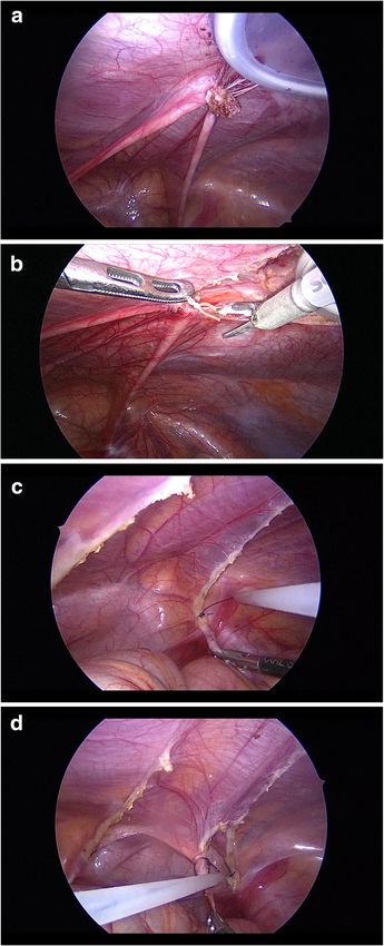
Fig. 2 Laparoscopic view of the urachal remnant. a A wrap trials of laparoscopic surgery to correct urachal
protector separated the cephalic side of the urachal remnant and anomalies [3–5]. It has also been claimed that a lap-
the medial umbilical ligaments. b The urachal remnant was lysed off aroscopic procedure provides better cosmesis. When
using ultrasonic scissors. c The caudal stump of the medial umbilical
the distance between the umbilicus and the dome of
ligaments was tied with a single ENDOLOOP ligature®. d The caudal
stump of the urachus was tied with a single ENDOLOOP ligature® the urinary bladder is short, as it is in infants, the
umbilical approach may be suitable for resecting a
urachal remnant. The umbilical approach is appropri-
ate for infants, and laparoscopic surgery is recom-
mended for older children [5].Hashizume et al. Surgical Case Reports (2020) 6:120 Page 4 of 5
Table 1 Patient characteristics and perioperative data
Case Age Sex BMI After infection or abscess Operation time Hospital stay Pathologic diagnosis of the urachal
(year) (month) (min) (days) remnant
1 10 M 19.1 5 128 4 Infected urachal sinus
2 10 F 18.7 3 120 3 Infected patent urachus and partially seal
off
3 11 M 17.4 3 92 2 Infected urachal sinus
4 11 F 18.6 4 81 3 Infected urachal sinus
5 14 M 19.1 2 93 5 Normal patent urachus and partially seal
off
6 14 M 17.1 6 69 4 Normal urachal cyst
7 15 M 17.1 4 89 4 Infected patent urachus and partially seal
off
In contrast with other laparoscopic approaches to the mm single port was placed for the wound retractor. If
treatment of urachal anomalies, our technique had some severe communication or adhesion between the urachus
distinct features, including devices and trocar sites. First, and the bladder occurs, a wound retractor may facilitate
the luminal wall of a urachal remnant is composed of multiple port insertion.
transitional epithelium, and infection may occur due to In our method, the defected peritoneum was not
the accumulation of materials within the cyst. In our repaired. There was a controversy about suturing the de-
technique, the cephalic side of the urachal remnant fection of peritoneal layers or not. In the cesarean sec-
could be completely resected. Omphalitis secondary to tion, non-closure of the visceral and parietal peritoneum
symptomatic urachal remnants often necessitates simul- at lower segment cesarean section is associated with
taneous resection of the umbilicus. Since its incomplete fewer postoperative complications, is more cost-
resection can result in recurrence, adequate debridement effective, and is simpler than the traditional operative
of the infected tissue is mandatory. Many cases of lap- technique of closing both peritoneal layers [14]. In our
aroscopic surgery do not include simultaneous umbilical study, there was no case of intestinal obstruction for ad-
resection for symptomatic urachal remnants with open hesion formation.
urachal resection [8, 9]. The reason for this is that umbi- The present study was associated with some limita-
licoplasty is very complicated for laparoscopic surgery. tions. This study was retrospective in nature and only in-
This is because umbilicoplasty is very complicated for cluded a small number of cases. Furthermore, there
laparoscopic surgery. Our method involving en bloc re- were no patients under 10 years of age.
section of the umbilicus followed by umbilicoplasty was
deemed the best method for achieving the complete Conclusion
resolution of symptoms and ensuring superior cosmesis In conclusion, laparoscopic excision seems to be a safe and
in pubescent children. Second, another three 5-mm less invasive method for the treatment of urachal remnants.
working camera ports were inserted under direct vision However, a prospective, large, multi-institutional random-
on the right side of the abdomen with the patient in a ized study is needed to validate the advantages of a laparo-
supine position. These ports provided a good view of the scopic approach to an open approach.
full length of the urachus and allowed adequate access
Acknowledgements
to both the umbilicus and the bladder dome, without
The authors thank Brian Quinn for his critical reading of the manuscript.
any hindrance or discomfort. Lateral side ports and EZ
access® placements could reduce the risk of incomplete Authors’ contributions
excision of the urachal remnant. Compared with other NH and MO drafted the manuscript. NH carried out the acquisition of the
data. MY participated in the critical revision of the manuscript. The authors
reports [10–13], the mean total operative time of this read and approved the final manuscript.
technique was shorter than that of other techniques
such as multi-port technique that the first port was Funding
No funding
placed in the side of the abdomen or umbilical portion
as the camera port and 2 or 3 ports inserted [10, 11] and Availability of data and materials
single-site surgery technique used only EZ access® place- All data generated during this study are included in this published article.
ments [12].
Ethics approval and consent to participate
Third, for bladder cuff resection and subsequent trans- This study was reviewed and approved by the Ethics Committee of Tsuruoka
verse suturing using a linear stapler, an additional 12- Municipal Shonai Hospital.Hashizume et al. Surgical Case Reports (2020) 6:120 Page 5 of 5
Consent for publication
Informed consent was obtained from the patient for the publication of this
case report, including their medical data and images.
Competing interests
The authors declare that they have no competing interests.
Author details
1
Department of Pediatric Surgery, Tsuruoka Municipal Shonai Hospital, 4-20
Izumi-machi, Tsuruoka-shi, Yamagata 997-0033, Japan. 2Department of
Surgery, Tsuruoka Municipal Shonai Hospital, Tsuruoka, Japan. 3Department
of Pediatric Surgery, Kurume University School of Medicine, Kurume, Japan.
4
Department of Surgery, Niigata Prefectural Tsubame Rosai Hospital,
Tsubame, Japan.
Received: 25 March 2020 Accepted: 24 May 2020
References
1. Aylward P, Samson K, Raynor S, Cusick R. Operative management of urachal
remnants: an NSQIP based study of postoperative complications. J Pediatr
Surg. 2020;55(5):873–7.
2. Stopak JK, Azarow KS, Abdessalam SF, Raynor SC, Perry DA, Cusick RA.
Trends in surgical management of urachal anomalies. J Pediatr Surg. 2015;
50:1334–7.
3. Sukhotnik I, Aranovich I, Mansur B, Matter I, Kandelis Y, Halachmi S.
Laparoscopic surgery of urachal anomalies: a single-center experience. Isr
Med Assoc J. 2016;18:673–6.
4. Tatekawa Y. Surgical strategy of urachal remnants in children. J Surg Case
Rep. 2019;2019:rjz222.
5. Sato H, Furuta S, Tsuji S, Kawase H, Kitagawa H. The current strategy for
urachal remnants. Pediatr Surg Int. 2015;31:581–7.
6. Yoo KH, Lee SJ, Chang SG. Treatment of infected urachal cysts. Yonsei Med
J. 2006;47:423–7.
7. Keingsberg K. Letter: Infection of umbilical artery stimulating paret urachus.
J Pediatr. 1975;86:151–2.
8. Trondsen E, Reiertsen O, Rosseland AR. Laparoscopic excision of urachal
sinus. Eur J Surg. 1993;159:127–8.
9. Jeong HJ, Han DY, Kwon WA. Laparoscopic management of complicated
urachal remnants. Chonnam medical journal. 2013;49(1):43–7.
10. Tanaka K, Misawa T, Baba Y, Ohashi S, Suwa K, Ashizuka S, Yoshizawa J, Ohki
T. Surgical management of urachal remnants in children: open versus
laparoscopic approach: a STROBE-compliant retrospective study. Medicine.
2019;98(40):e17480.
11. Maemoto R, Matsuo S, Sugimoto S, Tokuka A. Umbilical resection during
laparoscopic surgery for urachal remnants. Asian journal of endoscopic
surgery. 2019;12(1):101–6.
12. Yanishi M, Kinoshita H, Matsuzaki T, Yoshida T, Ohsugi H, Taniguchi H, Sugi
M, Matsuda T. Laparoendoscopic single-site surgery for urachal remnants: a
single-center experience. Urologia internationalis. 2020;104(1-2):70–4.
13. Fujiogi M, Michihata N, Matsui H, Fushimi K, Yasunaga H, Fujishiro J. Early
outcomes of laparoscopic versus open surgery for urachal remnant
resection in children: a retrospective analysis using a nationwide inpatient
database in Japan. Journal of laparoendoscopic & advanced surgical
techniques. Part A. 2019;29(8):1067–72.
14. Grundsell HS, Rizk DE, Kumar RM. Randomized study of non-closure of
peritoneum in lower segment cesarean section. Acta obstetricia et
gynecologica Scandinavica. 1998;77(1):110–5.
Publisher’s Note
Springer Nature remains neutral with regard to jurisdictional claims in
published maps and institutional affiliations.You can also read