Colchicine poisoning complicated by medulla oblongata myelinolysis: a case report
←
→
Page content transcription
If your browser does not render page correctly, please read the page content below
Case Report

Colchicine poisoning complicated by medulla oblongata
myelinolysis: a case report
Wei Jiang1#, Xuan-Yu Tan2#, Jia-Ai Li1, Kang Qu1, Peng Yu3, Ming Dong1
1
Department of Neurology and Neuroscience Center, The First Hospital of Jilin University, Changchun, China; 2Department of Neurosurgery, The
First Hospital of Jilin University, Changchun, China; 3Department of Ophthalmology, The Second Hospital of Jilin University, Changchun, China
#
These authors contributed equally to this work.
Correspondence to: Ming Dong, MD, PhD. Department of Neurology and Neuroscience Center, The First Hospital of Jilin University, Xinmin Street
#71, Changchun 130021, China. Email: neuromdong@163.com; Peng Yu, MD, PhD. Department of Ophthalmology, The Second Hospital of Jilin
University, Ziqiang Street #218, Changchun 130041, China. Email: 895222700@qq.com.

                 Abstract: Medulla oblongata myelinolysis is an extremely rare manifestation of extrapontine myelinolysis
                 (EPM). Herein, we report a case of a 34-year-old man with a history of gout who presented repeated
                 vomiting and diarrhea after ingesting 15 colchicine pills. A hyponatremia diagnosis was given and after
                 an intensive treatment, his serum sodium level increased from 118 to 129 mmol/L within 24 hours.
                 Brain magnetic resonance imaging (MRI) revealed a lesion in the medulla oblongata that appeared as a
                 hypointense area in T1-weighted images and a hyperintense area in T2-weighted images. A diagnosis of
                 medulla oblongata myelinolysis and colchicine poisoning was then given, and methylprednisolone therapy
                 was initiated. Seventeen days later, the patient achieved a good outcome with methylprednisolone therapy.
                 However, his medulla oblongata lesion remained detectable with MRI. Medulla oblongata myelinolysis is
                 an extremely rare manifestation of EPM, and unique for being colchicine-induced. This case shows that
                 colchicine poisoning can lead to hyponatremia, which in turn can induce myelinolysis if not treated correctly.
                 As exemplified by our patient’s case, desirable treatment outcomes are possible in such cases, although these
                 outcomes may not be associated with a visible reduction of the brain lesions in MRI scans.

                 Keywords: Colchicine; myelin sheath; medulla oblongata; demyelinating diseases; case report

                 Submitted Dec 17, 2019. Accepted for publication Mar 04, 2020.
                 doi: 10.21037/apm-19-627
                 View this article at: http://dx.doi.org/10.21037/apm-19-627

Introduction                                                               and EPM are associated with rapid osmotic changes (3).
                                                                           To our knowledge; only two cases of medulla oblongata
Colchicine, a drug used primarily to treat and prevent gout,
                                                                           myelinolysis has been reported in literature (4,5). Moreover,
has a narrow therapeutic index, with no clear distinction
                                                                           to date, this is the first case of colchicine-induced medulla
between non-toxic and toxic dosages (1). Patients who
ingest therapeutic oral doses of colchicine may experience                 oblongata myelinolysis. We report herein a case of medulla
abdominal pain, diarrhea, nausea, and vomiting. These                      oblongata myelinolysis caused by colchicine poisoning in
symptoms may lead to hyponatremia, a condition generally                   which the patient’s rapid recovery was not reflected by the
defined as a plasma sodium level of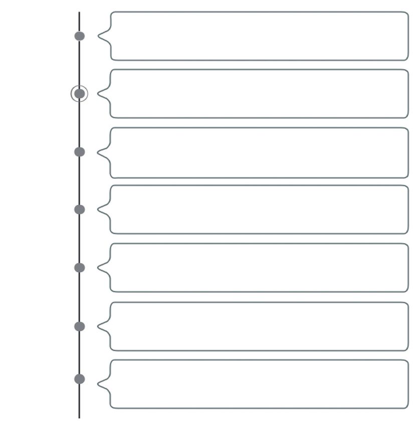
2350 Jiang et al. Colchicine poisoning and medulla oblongata myelinolysis
145 A B
Na+ concentration (mmol/L)
140
135
T1
130
125
120
115
0 5 10 15 20
Day post-admission
Figure 1 Changes in serum sodium concentrations after admission.
The red dashed line represents the lowest serum sodium T2
concentration that is considered acceptable under treatment
protocols.
with nausea, vomiting, diarrhea, and dizziness that
arose 3 days after he ingested 15 colchicine pills (total
colchicine dose, 0.5 mg). He had a history of gout, and
T2
he had felt a terrible pain in his right toe on the day of Sagittal
colchicine ingestion. Based on his previous experience
with colchicine, he initially took 3 tablets, which provided
no relief; he then took another 3 tablets. The pain eased
somewhat but was not eliminated. To further ameliorate
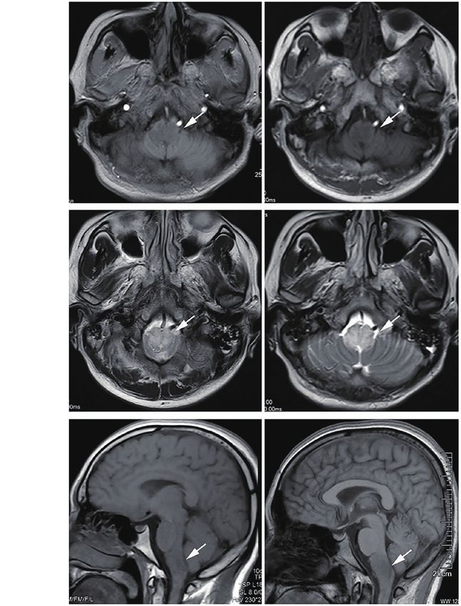
Figure 2 Time-dependent MRI observations. (A) Three days
the pain, he consumed 9 additional tablets over the next
after symptom onset, MRI scans revealed a lesion in the medulla
6 hours. After taking 15 tablets, his pain was substantially
oblongata that appeared as a hypointense area in T1-weighted
eased, but he began to experience dizziness, nausea, and
images and a hyperintense area in T2-weighted images (white
vomiting. These symptoms persisted for 3 days, during
arrows); (B) follow-up MRI performed 17 days later revealed that
which the patient felt increasingly languid. His wife became
the lesion was persistent in both T1- and T2-weighted images
concerned and brought him to the emergency department.
(white arrows). MRI, magnetic resonance imaging.
Upon examination, his vital signs and hepatic and renal
functions were normal. However, he was very lethargic.
His serum sodium concentration of 118 mmol/L indicated bilaterally, and his patellar tendon reflexes were active.
hyponatremia. We immediately initiated sodium correction His serum creatine kinase, aspartate aminotransferase, and
treatment and administered an antiemetic. His sodium level lactate dehydrogenase levels and white blood cell counts
consequently increased to 129 mmol/L within 24 hours were normal. However, a Medical Research Council scale
(Figure 1). evaluation indicated a limb strength score of 0/5, leading to a
Despite the improved sodium level, he gradually developed diagnosis of tetraparesis.
weakness in his limbs, which was accompanied by dyspnea, On the third day after admission, brain MRI revealed
tachycardia, and hypotension. The following vital signs a lesion in the medulla oblongata that appeared as a
were recorded at that time: respiratory rate, 30 breaths/min; hypointense area in T1-weighted images and a hyperintense
body temperature, 36.7 ℃; heart rate, 120 beats/min; and area in T2-weighted images (Figure 2A). We considered
blood pressure, 80/55 mmHg. A blood gas analysis revealed these findings suggestive of medulla oblongata myelinolysis.
a partial oxygen pressure of 58 mmHg, indicating respiratory We therefore administrated intravenous methylprednisolone
failure. We subsequently initiated mechanical ventilation (dose, 80 mg/d for the first 10 days and 40 mg/d for the next
and a dopamine infusion at a rate of 5 mL/h. His pupils 10 days). After the third day of treatment, the patient’s vital
had diameters of 3.0 mm. The Babinski sign was intact signs normalized. By treatment day 10, his muscle strength
© Annals of Palliative Medicine. All rights reserved. Ann Palliat Med 2021;10(2):2349-2353 | http://dx.doi.org/10.21037/apm-19-627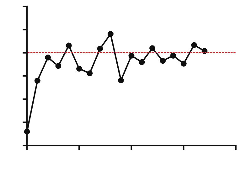
Annals of Palliative Medicine, Vol 10, No 2 February 2021                                                                                  2351

                                   3 days before
                                                        Taking to much colchicine caused poisoning
                                     admission

                                       at               Patient’s serum indicates hyponatremia and initiated sodium
                                    admission           correction treatment

                                    1 day after         Sodium level increased 11 mmol/L within 24 hours.
                                    admission           Patient’s muscle strength score of 0/5 on the Medical
                                                        Research Council accompanied with abnormal vital signs

                                    3 days after        MRI showed medulla oblongata myelinolysis and
                                     admission          initiated treatment of methylprednisolone

                                    6 days after
                                     admission          Patient’s vital signs normalized

                                   10 days after        Muscle strength improved to a score of
                                    admission           3/5 on the Medical Research Council

                                   17 days after
                                    admission           Muscle strength continued to improved and a repeat
                                                        MRI revealed that the lesion persisted

Figure 3 Timeline about historical and current information from this case of care organized. MRI, magnetic resonance imaging.

had improved to a score of 3/5 on the Medical Research                         of hyponatremia then exacerbated the respiratory and
Council scale, and it continued to improve thereafter to a                     circulatory failure and muscle weakening.
score of 4/5 on day 17. No adverse or unexpected events                           Generally, acute flare-ups in adults with gout are treated
were observed during treatment. He did not exhibit hepatic                     by administering oral colchicine doses of 1.2 mg/d three to
or renal insufficiency during this period. Despite these                       four times per week. The standard oral dose for prophylaxis
clinical improvements, a follow-up MRI scan revealed                           is 0.5–0.6 mg/d administered three to four times per week.
that his medulla oblongata lesion persisted (Figure 2B). A                     However, even the recommended dose for acute pain
summary of the timeline is shown in Figure 3.                                  can induce gastrointestinal side effects before it provides
                                                                               relief from gout symptoms (9). The initial gastrointestinal
                                                                               symptoms of acute toxicity occur within the first 24 hours,
Discussion
                                                                               whereas multiorgan failure develops 24–72 hours after
The excessive administration of colchicine or accidental                       ingestion. Additionally, hyponatremia, hypocalcemia, and
ingestion of colchicine-containing plants can lead to                          metabolic acidosis may occur (10). Our patient had no
acute colchicine poisoning, which initially manifests as                       medical history of hyponatremia, had not recently used
gastrointestinal effects but may progress to multiorgan                        diuretic agents, and lacked evidence of adrenal, thyroid,
dysfunction (6). This latter stage of colchicine poisoning                     pituitary, or renal insufficiency. His medical history
may involve respiratory and circulatory failure and muscle                     implicated colchicine poisoning as the most probable
weakness (1,7,8). However, our patient did not present                         contributor to the development of severe hyponatremia.
with any clinical manifestations of multiorgan failure or                         As variants of ODS, both CPM and EPM can occur
respiratory and circulatory failure upon admission to the                      when hyponatremia is not treated properly. EPM generally
hospital. Therefore, we attribute the hyponatremia solely                      occurs as a comorbidity of CPM, but some case reports
to his initial gastrointestinal reaction to colchicine. The                    indicate that it can precede CPM or occur in isolation
medulla oblongata lesion induced by the rapid correction                       of CPM (3). CPM, the classic presentation, reflects the

© Annals of Palliative Medicine. All rights reserved.               Ann Palliat Med 2021;10(2):2349-2353 | http://dx.doi.org/10.21037/apm-19-627
2352 Jiang et al. Colchicine poisoning and medulla oblongata myelinolysis
greater susceptibility of the pontine white matter tracts to lesion in MRI scans was inconsistent with the patient’s good
this condition, whereas EPM affects other brain regions response to methylprednisolone therapy and subsequent
(e.g., the cerebellum, thalamus, basal ganglia, or subcortical clinical recovery. The lack of subsequent dynamic evolution
white matter). Although the latter condition is not as of MRI was a limitation in this study. We attempted to
rare as previously believed (3,11,12), medulla oblongata advise the patient to review the MRI, but unfortunately, the
myelinolysis remains an extremely rare manifestation of patient refused due to long-distance travel.
EPM (13). In brain MRI scans, ODS typically appears
as hyperintense lesions in the central pons or associated
Conclusions
extrapontine structures in T2-weighted and fluid-
attenuated inversion recovery images, with corresponding We report a rare case of medulla oblongata myelinolysis
hypointensities in T1-weighted images (11). The findings induced by a rapid recovery from colchicine toxicity-
from our patient’s case were consistent with previously induced hyponatremia. This case clarifies the clinical and
reported observations. neuroradiological characteristics of medulla oblongata
It has been shown that demyelination in patients with myelinolysis. Moreover, the patient’s good outcome
ODS can be reversible, with near-complete restoration following methylprednisolone treatment was not
being possible even in patients whose myelin has entirely accompanied by a resolution of the medulla oblongata
disappeared in conventional MRI images (14-17). However, lesion, thus confirming that in such cases, MRI findings
even after the restoration of myelin sheaths, these patients may not be indicative of the actual clinical situation.
exhibit residual MRI findings that are thought to represent
permanent damage (14).
Acknowledgments
Although the exact pathogenesis of ODS remains unclear,
the rapid correction of chronic hyponatremia has been Funding: This work was supported by a grant from the
shown to induce pathological changes in animal models National Natural Science Foundation of China to MD
(i.e., dogs and rats) (18,19). Currently, most neurologists (Grant No. 31872772).
who treat patients with ODS have reached a consensus that
to ensure patient safety, serum sodium levels should not be
Footnote
increased by more than 10 mmol/L/d, although the most
recent recommendation suggests a maximum correction Conflicts of Interest: All authors have completed the ICMJE
rate of 8 mmol/L/d (13). In our patient’s case, the vomiting uniform disclosure form (available at http://dx.doi.
and diarrhea induced by the ingestion of a high dose of org/10.21037/apm-19-627). The authors have no conflicts
colchicine led to hyponatremia, and the subsequent rapid of interest to declare.
correction of hyponatremia induced medulla oblongata
myelinolysis. We noted that the 11-mmol/L increase in our Ethical Statement: The authors are accountable for all
patient’s serum sodium level on the first day of treatment aspects of the work in ensuring that questions related
exceeded the recommended serum sodium correction rate. to the accuracy or integrity of any part of the work are
Hyponatremia is a relatively common manifestation of appropriately investigated and resolved. All procedures
colchicine poisoning. Although increasing attention has performed in studies involving human participants were in
been given to the diagnosis and treatment of colchicine accordance with the ethical standards of the institutional
poisoning and the associated serious complications, this and/or national research committee(s) and with the Helsinki
report is, to our knowledge, the first to describe medulla Declaration (as revised in 2013). Written informed consent
oblongata myelinolysis caused by colchicine poisoning. Our was obtained from the patient for publication of this
findings from this case have enriched our understanding of manuscript and any accompanying images.
the clinical and neuroradiological characteristics of medulla
oblongata myelinolysis. Our observations demonstrate the Open Access Statement: This is an Open Access article
risks associated with rapid sodium correction and proves distributed in accordance with the Creative Commons
that MRI findings and clinical symptoms are not always well Attribution-NonCommercial-NoDerivs 4.0 International
correlated. In this case, the persistent medulla oblongata License (CC BY-NC-ND 4.0), which permits the non-
© Annals of Palliative Medicine. All rights reserved. Ann Palliat Med 2021;10(2):2349-2353 | http://dx.doi.org/10.21037/apm-19-627Annals of Palliative Medicine, Vol 10, No 2 February 2021 2353
commercial replication and distribution of the article with polyradiculoneuropathy. J Clin Neurosci 2014;21:331-2.
the strict proviso that no changes or edits are made and the 9. Varughese GI, Varghese AI, Tahrani AA. Colchicine: time
original work is properly cited (including links to both the to rethink. N Z Med J 2007;120:U2429.
formal publication through the relevant DOI and the license). 10. Güven AG, Bahat E, Akman S, et al. Late diagnosis of
See: https://creativecommons.org/licenses/by-nc-nd/4.0/. severe colchicine intoxication. Pediatrics 2002;109:971-3.
11. Singh TD, Fugate JE, Rabinstein AA. Central pontine
and extrapontine myelinolysis: a systematic review. Eur J
References
Neurol 2014;21:1443-50.
1. Finkelstein Y, Aks SE, Hutson JR, et al. Colchicine 12. Zhuang L, Xu Z, Li Y, et al. Extrapontine myelinolysis
poisoning: the dark side of an ancient drug. Clin Toxicol associated with pituitrin: case report and literature review.
(Phila) 2010;48:407-14. BMC Neurol 2014;14:189.
2. Spasovski G, Vanholder R, Allolio B, et al. Clinical practice 13. Martin RJ. Central pontine and extrapontine myelinolysis:
guideline on diagnosis and treatment of hyponatraemia. the osmotic demyelination syndromes. J Neurol
Eur J Endocrinol 2014;170:G1-47. Neurosurg Psychiatry 2004;75 Suppl 3:iii22-8.
3. Kumar S, Fowler M, Gonzalez-Toledo E, et al. 14. Yuh WT, Simonson TM, D'Alessandro MP, et al.
Central pontine myelinolysis, an update. Neurol Res Temporal changes of MR findings in central pontine
2006;28:360-6. myelinolysis. AJNR Am J Neuroradiol 1995;16:975-7.
4. Kulczycki J, Kozlowski P, Iwinska-Buksowicz B. Central 15. Menger H, Jorg J. Outcome of central pontine
myelinolysis of medulla oblongata. Case report. Neurol and extrapontine myelinolysis (n = 44). J Neurol
Neurochir Pol 1994;28:757-61. 1999;246:700-5.
5. Bhagavan BS, Wagner JA, Juanteguy J. Central pontine 16. Floris G, Di Stefano F, Melis R, et al. Isolated bipallidal
myelinolysis and medullary myelinolysis. Arch Pathol Lab lesions caused by extrapontine myelinolysis. Neurology
Med 1976;100:246-52. 2013;81:1722-3.
6. Horioka K, Tanaka H, Isozaki S, et al. Acute colchicine 17. Kallakatta RN, Radhakrishnan A, Fayaz RK, et al. Clinical
poisoning causes endotoxemia via the destruction and functional outcome and factors predicting prognosis
of intestinal barrier function: the curative effect of in osmotic demyelination syndrome (central pontine and/
endotoxin prevention in a murine model. Dig Dis Sci or extrapontine myelinolysis) in 25 patients. J Neurol
2020;65:132-40. Neurosurg Psychiatry 2011;82:326-31.
7. Kuncl RW, Duncan G, Watson D, et al. Colchicine 18. Laureno R. Central pontine myelinolysis following rapid
myopathy and neuropathy. N Engl J Med correction of hyponatremia. Ann Neurol 1983;13:232-42.
1987;316:1562-8. 19. Kleinschmidt-DeMasters BK, Norenberg MD. Rapid
8. Ghosh PS, Emslie-Smith AM, Dimberg EL. correction of hyponatremia causes demyelination: relation
Colchicine-induced myoneuropathy mimicking to central pontine myelinolysis. Science 1981;211:1068-70.
Cite this article as: Jiang W, Tan XY, Li JA, Qu K, Yu P, Dong
M. Colchicine poisoning complicated by medulla oblongata
myelinolysis: a case report. Ann Palliat Med 2021;10(2):2349-2353.
doi: 10.21037/apm-19-627
© Annals of Palliative Medicine. All rights reserved. Ann Palliat Med 2021;10(2):2349-2353 | http://dx.doi.org/10.21037/apm-19-627You can also read